

The treatment of small animal wounds will commonly require a staged approach. Definitive treatment will often be delayed while emergency treatment of the patient and its wound, and initial stabilisation is performed. Analgesia and effective restraint is often required, until the animal is stable enough for sedation or a general anaesthetic (GA). Wounds can be classified by their cause and the type of tissue damage caused: incisional, abrasion, avulsion (or degloving), shearing, puncture or perforated, and burns. Regardless of the aetiology of the wound, the factor which has the single biggest impact on future healing is the presence of contamination and necrotic tissue.
Once the patient is stable a more thorough evaluation may be carried out. Appropriate chemical restraint may be required for examination. Diagnostic imaging may be used to check for foreign material, penetrating injuries, associated fractures, dislocations and tendon or ligament damage. A management plan should take into account the wound's location, size, damage to local structures and the amount of tissue loss.
Wound healing phases
There are three phases in wound healing
- The inflammatory phase (immediate)
- The proliferative (repair) phase (days 3 to 7 post injury)
- The remodelling phase (days 5 to 7 onwards post injury) (Anderson, 1996).
Each of the above phases rather than occurring at completely separate times as completely separate processes, in fact all overlap as they progress and hence influence the development and duration of the next phase, meaning each phase is crucial to the end result.
Early inflammatory phase
The initial injury triggers coagulation and stimulation of the inflammatory cascade resulting in chemotaxis and an influx of inflammatory cells. The magnitude of the inflammatory response will be dependent on the severity of the trauma (O'Dwyer, 2007).
During the early inflammatory stage, wound management is commonly aimed at the debridement of the wound, in order to remove necrotic tissue, and debris/foreign material, from the wound bed. The mainstays of debridement in acute wounds are surgical and mechanical debridement. Dressings at this time may include mechanical debridement dressings such as wet-to-dry dressings, or the use of Debrisoft (supplied by Pioneer Vet Products).
Mechanical debridement
Debrisoft
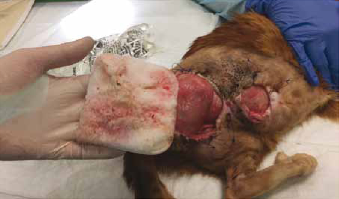
Debrisoft is a monofilament debridment pad that is made up of more than 18 million specially angled fibres. It is used to remove sloughy tissue, debris and necrotic/non-viable tissue from the wound bed. Debrisoft can be used in conscious, as well as unconscious patients, and works rapidly (Figure 1). Its gentle action means viable tissue within the wound bed, i.e. granulation tissue and epithelial cells, remain intact. Its simplicity, low cost and rapid results makes it a very viable option for debridement. Case study 1, shows the product being used on initial presentation and for the first few dressing changes to remove necrotic tissue and sloughy material.
Wet-to-dry dressing
Wet-to-dry dressings are a useful tool in wound management in veterinary practice. Their effectiveness relies on the adherence of the dressing to the wound bed, which on removal lifts the debris and necrotic tissue that becomes trapped in the mesh of the dressing (Figure 2).
The application of wet-to-dry dressings involves moistening a sterile gauze swab using sterile saline or Hartmann's solution, which is in contact with the wound bed; this is then protected by standard secondary (cotton wool or dry swabs) and tertiary bandage layers. The moisture from the swab dilutes the exudates in the wound, which is absorbed into the secondary layer and the swab dries and adheres to the wound surface. The dressing needs to be changed at least daily before any strike through is noted; any delay leads to a delay in healing and increased risk of infection. Removal is often painful and sedation or anaesthesia is usually required.
Surgical debridement
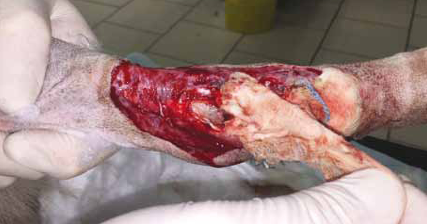
Surgical (or ‘Sharp’) debridment will often be involved. This involves the cutting away of devitalised tissue from the wound, which is a rapid and effective debridement technique (Figure 3), as well as allowing the surgeon the opportunity to thoroughly explore, and assess the wound and underlying structures. As with any surgical procedure, debridement should be performed using strict aseptic technique, to prevent introducing further bacterial contamination into the wound, so the surgical site should be draped, and the clinician gowned and gloved. Pioneer Vet Products have a comprehensive range of surgical protective wear including variable fenestration drapes, ideal as the aperture can be adjusted to fit around individual wounds.
Generally two approaches to surgical debridement can be used; ‘en bloc’ or layered debridement. The choice depends on the availability of surrounding tissue for closure, and therefore the need to preserve as much skin as possible.
En bloc debridement
In areas where there is adequate normal tissue to allow closure afterwards, the whole affected tissue can be excised with a border of healthy tissue — in a similar fashion to removing a tumour with healthy margins in all planes. This is termed en-bloc debridement, ideal areas for this technique include the trunk or proximal limbs where there is plenty of available skin to close the deficit. The wound is usually packed with surgical swabs and sutured closed prior to excision of the entire wound.
Layered debridement
More commonly when debriding a wound, devitalised tissue is removed gradually in layers, allowing conservation of tissue where possible; this may be important in areas such as the lower limbs and feet where there is inadequate skin to allow en-bloc debridement.
Superficial tissue is removed first, followed by debridement of deeper tissues. As debridement progresses, instruments can be changed or disinfected and rinsed to prevent contaminating areas that have already been debrided.
Other forms of debridement
Autolytic
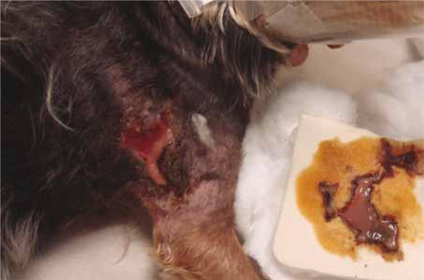
Autolytic debridement is a selective debridement brought about by the release of the patient's own proteolytic enzymes (e.g. collagenase, elastase) and the activation of phagocytes. These enzymes soften and breakdown necrotic tissue, and are mostly produced by leucocytes. This is a natural process that occurs in all wounds, but its action can be enhanced by using products that promote and maintain an ideal moist wound environment, which promotes the activity of leucocytes and macrophages. The moist environment also promotes the swelling of necrotic tissue, which loosens it from the wound bed. Assistance with autolytic debridement may also be considered, which can include the use of manuka honey, e.g. Manuka Fill as with Case study 2, Manuka IG Impregnated Gauze dressing, hydrogels in combination with absorbent dressings such as Activheal Foam Non-adhesive, Activheal Adhesive or Alleviate dressings (all available from Pioneer Vet Products), to absorb the exudate and sloughy material produced at the wound bed (Figure 4).
Enzymatic
Enzymatic agents are relatively selective in loosening and removing necrotic tissue (Gregory, 1999). No veterinary product exists in the UK, but in the USA an ointment derived from bacteria (Bacillus subtillus), which contains the enzyme sutilain, is available and can be used in wounds with minimal necrotic tissue.
A much wider range of proteolytic enzyme preparations are available for wound management in humans, including enzymes derived from plants (e.g. pineapple), bacteria and animals (e.g. Antarctic krill).
Larval
The use of maggots in wounds is an idea that has been around for hundreds of years. In human medicine the most commonly used maggot is that of the greenbottle fly (Lucillia sericata) (Gregory, 1999). Maggots are able to selectively debride necrotic tissue while sparing healthy structures; this technique is particularly advantageous when debriding wounds where vital structures exist and therefore surgical debridement is contraindicated.
Hydrosurgery
Hydrosurgical units emit a very high-pressure stream of saline that cuts tissues. The Versajet (Smith and Nephew) is the most commonly used unit in human hospitals where they are used for debridement of wounds, chronic ulcers, and burns. Human reports suggest that Versajet is equally, if not more effective than conventional surgical debridement, by causing less damage to viable tissue.
Late inflammatory phase
Macrophages infiltrate shortly after inflammation is established and are crucial to the stimulation and progression of the wound through the phases of the healing. They debride necrotic or foreign material in the wound and dispose of the platelet clot, dead cells, bacteria and organic debris. Macrophages are derived from the circulating monocytes and are stimulated by factors such as endotoxins, alginate, cytokines, fibrin, hypoxia and increased lactate concentrations. In monocytopenic patients, this stage is delayed or fails altogether (Anderson, 1996).
If there is concern regarding infection at this point in wound healing, then dressings that have antimicrobial activity may be considered, such products include manuka honey (e.g. Manuka Fill, Pioneer Vet Products), or dressings that contain silver (e.g. Suprasorb A Ag, Pioneer Vet Products).
Proliferative phase
The proliferative phase marks the development of granulation tissue. Fibroblasts migrate into the wound from nearby skin. They lay down a new extracellular matrix after debridement has taken place, and manufacture and remodel collagen. Fibroplasia is almost entirely dependent on the inflammatory trigger and may be delayed if the inflammatory phase fails to occur. However, once the fibroblast population is active, and in place, anti-inflammatory drugs will not halt the formation of normal scar tissue (Anderson, 1996).
Epithelial cells are stimulated initially by the injury itself and subsequently by cytokines released by the other cells. They proliferate and flow over each other, then reattach to the granulation tissue underneath. This process is fastest when the wound is moist and the cells do not have to lyse necrotic tissue to get across the wound (e.g. migration under an eschar). The migrating and proliferating epithelial cells are fragile and highly susceptible to adverse conditions in the wound, desiccation or abrasion of the wound surface (O'Dwyer, 2007).
During epithelialisation, dressings should be selected which result in minimal disturbance of the wound environment, but also have the advantage of maintaining a moist wound environment. Sheet hydrogels are an excellent option at this stage (Figure 5) (e.g. Actiform Cool, Pioneer Vet Products), as they allow epithelialisation to continue undisturbed, in a moist wound environment. Since they are composed of a solid sheet of hydrogel, with a film backing, they negate the additional cost of having to use a secondary dressing to cover hydrogel, if hydrogel was used for this same purpose.
Some wounds may fail to effectively reach proliferation and have an excessive inflammatory stage, during this time collagen degradation can become minimal and or levels of matrix metalloproteinase can become elevated. Collagen dressing such as Cyprofil (Pioneer Vet Products) are effective in encouraging the formation of the extra cellular matrix, leading to the formation of a healthy granulation bed, wound contraction and epithelisation, ultimately ensuring an effective proliferation phase. See Figure 6.

Remodelling phase
The final phase is remodelling and restoration of the normal structure. The scar, however, will nearly always be weaker than the surrounding uninjured skin (Anderson, 2003).
Case study 1
Patient
A 9-year-old Jack Russell Terrier.
Presentation
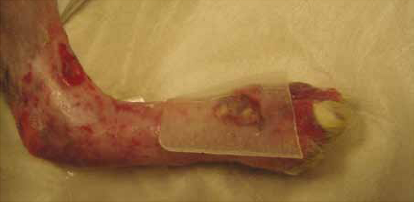
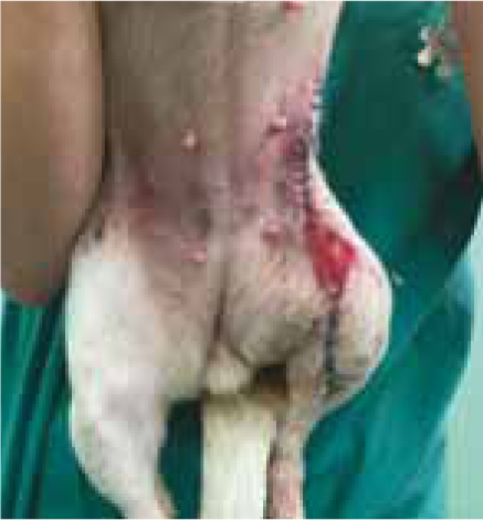
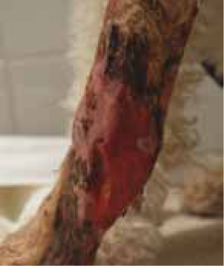
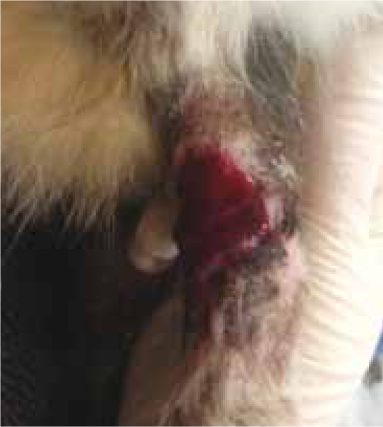
The dog presented to the practice with an avulsion injury and a large ‘pocket’ in her left hind limb following a road traffic accident. Image 1.
Presentation date
29th December 2014.
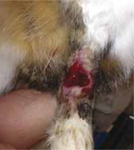
Initial management
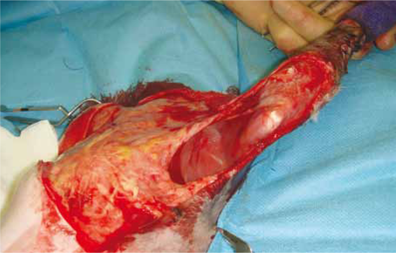
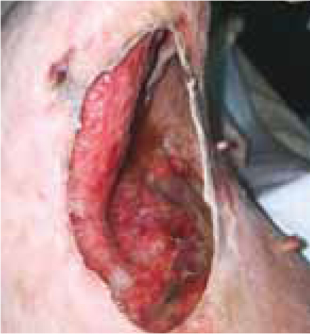
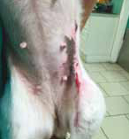
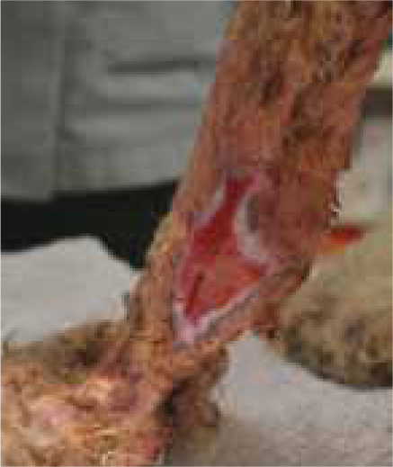
Initially the wound was cleaned and lavaged and some surgical debridement was performed around the skin margins. Debrisoft was then used to gently debride the sloughy material from the wound bed itself and the exposed muscle layers. Image 2.
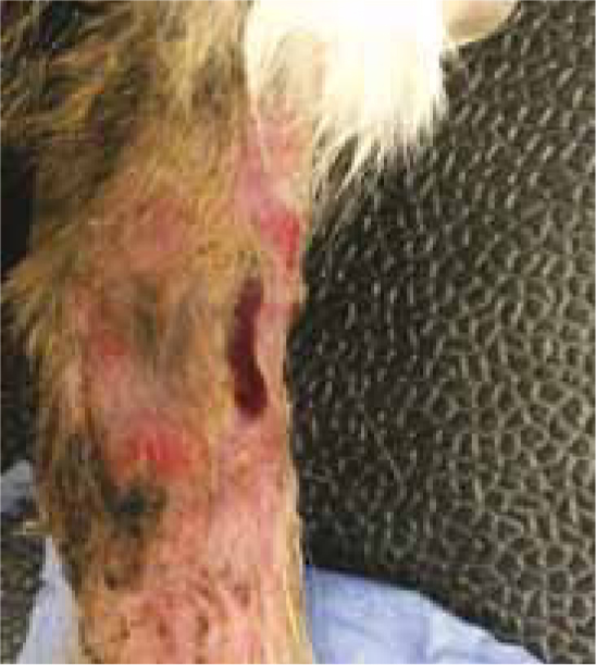
Ongoing management
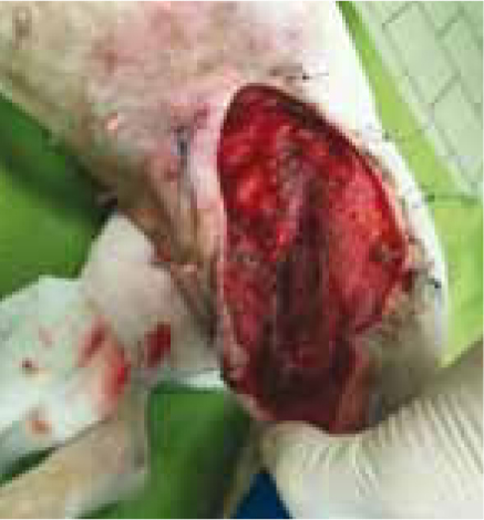
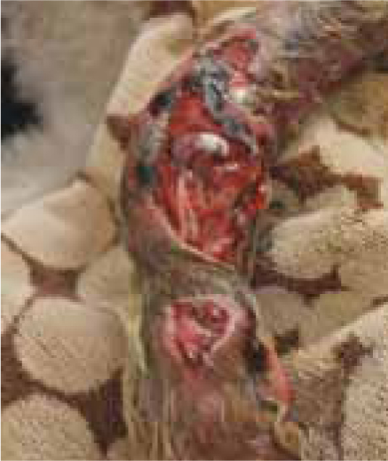
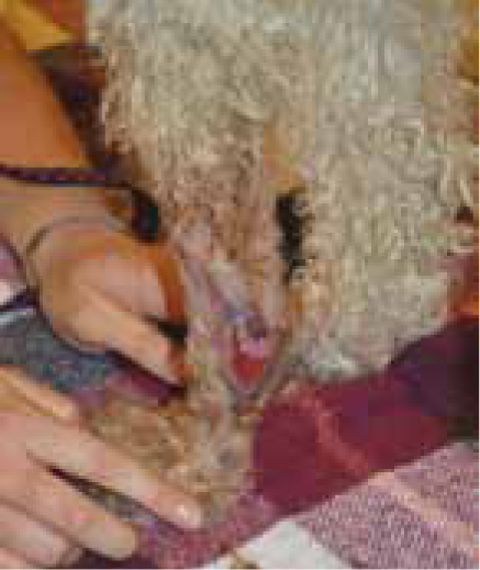
Stay sutures were placed in the healthy tissue surrounding the wound to allow for the use of a tie over (bolus) dressing. An antimicrobial tie over dressing was applied and debridement with Debrisoft took place daily at dressing change, for the following 3 days, allowing for gentle yet rapid debridement and removal of necrotic and sloughy material. Image 3.
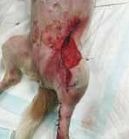
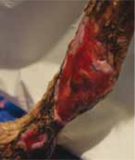
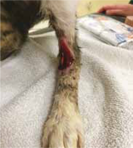
A staged approach to wound closure was taken in this case, with parts of the wound being sutured closed while the main pocket was left open to granulate and heal by second intention. Image 4.
Outcome
The wound went on to heal well without any complications and within 6 weeks from initial injury had almost healed. Image 5.
Product Summary
The Debrisoft monofilament debridement pad (Activa Healthcare supplied by Pioneer Vet Products) is made up of more than 18 million specially angled fibres that sweep and bind debris and slough into the pad, effectively yet gently debriding the wound to allow healthy granulation tissue formation. In this case Debrisoft allowed for the debridement of delicate tissue following the initial surgical debridement and offered an ongoing gentle debridement solution. Debrisoft offers veterinary professionals a simple to use, gentle and rapid debridement option where results can be seen in minutes.
Case courtesy of Louise O'Dwyer, MBA, BSc (Hons), VTS (ECC), DipAVN (Med & Surgery), RVN Clinical Support Manager, Vets Now Ltd
Case study 2
Patient
A female, medium-sized poodle cross.
Presentation
The dog was knocked down by a car and presented with bilateral abrasions.
Presentation date
19th March 2013.
Initial management
The dog initially presented to the veterinary practice with a heavily contaminated wound to her left fore, she had suffered a severe abrasion which revealed tendon involvement, but no fractures. There was not enough skin available to close the deficit immediately so open wound management was pursued to encourage a healthy granulation bed. Once granulating the decision could be made to either graft (although funds were short) or the wound could be allowed to close by second intention.
Ongoing management
Manuka Fill Honey dressings were used to dress the wound and to assist with debridement and dressings were changed initially every 2 days, and then every 3 days for the first 3 weeks.
Outcome
The wound went on to granulate and epithelise without any further complications and was practically healed 1 month after initial presentation.
Dressing summary
Manuka Fill honey dressings (Pioneer Vet Products) were used to assist with debridement — the osmotic action of the honey softens the necrotic tissue, assisting autolytic debridement. These dressings also provided antimicrobial support to what was a highly contaminated wound and helped to maintain a moist wound environment throughout the healing process.
Case Courtesy of The Veterinary Wound Library and Nora Janossy Dr MedVet
Case study 3
Patient
A male cat.
Presentation
The cat first presented to the practice following a suspected RTA and had severe trauma and degloving injuries to the medial aspects of both hind legs. X rays revealed no fractures and the decision was taken to allow the wounds to heal by second intention.
Presentation date
18th March 2014.
Initial management
A paraffin gauze was initially used as a primary dressing secured with a light bandage and changed every 3 days. The majority of the wounds healed well but one was problematic. A wound on the dorsal aspect of the left hind was struggling to heal and at this point exposed extensor tendon was still visible and the wound was failing to progress. On the 18th April the wound was debrided under sedation.
Ongoing management
At this point additional advice was sought via The Veterinary Wound Library and on the 12th May the wound was dressed with a Cyprofil collagen dressing. At the bandage change 1 week later ‘the improvement was amazing’ and although the tendon outline was still just about visible the wound was totally covered by granulation tissue and had contracted well. The Cyprofil collagen dressings were continued, with the bandages being changed every 3 days.
Outcome
Within a further 2 months the wound was practically healed and required only a couple of further dressing changes, although there was a little bit of surrounding soreness from some minor bandage rubs.
Product summary
The Cyprofil Collagen dressings (Pioneer Vet Products) were used to encourage the formation of a bed of health granulation tissue. The Cyprofil provides a scaffold and the protein fragments produced by degradation of the Cyprofil help to stimulate fibroblast proliferation, guide new collagen deposition and help to form extracellular matrix. This in turn allows for endothelial cell proliferation, wound contraction and ultimately healing. ‘At the second bandage change the improvement was amazing with the tendon outline still just visible but totally covered by granulation tissue and the wound contracting down well’.
Case Courtesy of Francesca Braid MRCVS & Catherine Darragh MRCVS, Companion Care Tunbridge Wells and Hove and The Veterinary Wound Library
Conclusion
In wound management, it is vital that veterinary staff have a good understanding of the processes involved in wound healing, along with good knowledge of the actions of wound dressings, so that an appropriate dressing can be matched to the specific stage of wound healing process, in order to optimise wound healing. Wound management is an ever-evolving field so it is essential that staff remain up to date on the novel technologies.
Key Points
- Decreasing the presence of these impairments to wound healing will enable the reduction of the level of contamination and the presence of necrotic tissue to a point where the closure technique chosen can be safely carried out.
- Debrosoft acts as an exciting new means of debridement, removing sloughy and necrotic tissue from the wound bed — its gentle action enables use in the conscious patient.
- Each of the wound healing phases overlap as they progress and hence influence the development and duration of the next phase.
- It is vital that veterinary staff have a good understanding of the processes in wound management, so that an appropriate dressing can be matched for the specific stage.
Conflict of interest: none.