

When collagen is an option
In veterinary medicine there is a growing trend to select ‘biological’ dressings that create a physiological interface between the wound surface and the environment. Collagen dressings, such as Cyprofil (Pioneer Vet Products) are therefore a real option in the management of hard to heal wounds in dogs and cats See Case study 3.
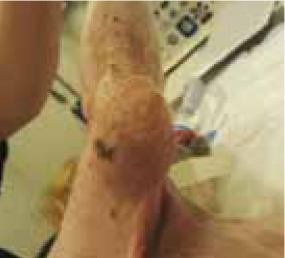
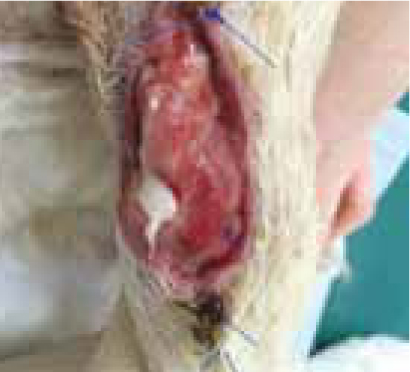
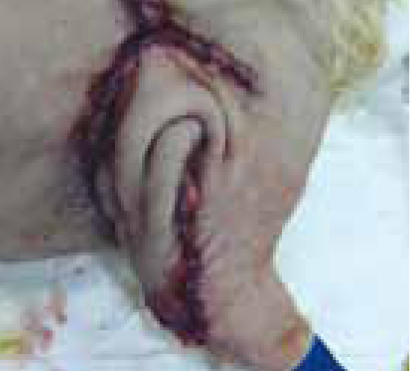
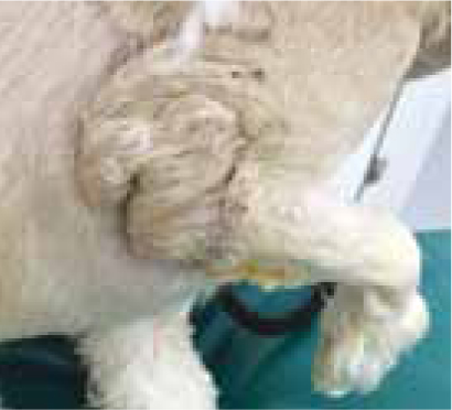
A 5-year-old male Lhasa Apso presented to our specialist veterinary hospital with a 4 cm diameter soft tissue sarcoma on his caudal elbow that required surgical excision. The surgery was initially successful however the wound dehisced and open wound management was started using wet-to-dry dressings, changed on a twicedaily basis. Cultures of the wound revealed a profuse growth of Streptococcus canis, sensitive to amoxicillin clavulanate, so open wound management was continued in combination with oral antibiotics. The wound, located over the olecranon, was in a difficult area to allow primary closure using the adjacent skin due to tension. It was therefore important that a healthy and clean granulation bed of tissue was obtained to enable a thoracodorsal flap placement. A Cyprofil collagen dressing was selected following control of the infection because of the known effects on inflammatory mediators and the proliferation phase of wound healing in addition to the structural support that it provides to the wound bed. The dressing was cut to the size of the wound, facilitating multiple uses of one dressing and applied directly onto the wound bed. The dressing was then secured with a secondary and tertially dressing layer. The collagen dressing was changed every 3 days. The wound was not inspected between these changes to allow integration of the dressing with the wound surface.
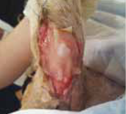
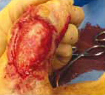
Eight days later the wound bed had a healthy bed of granulation tissue that was free of infection. Surgery was performed at this point and a thoracodorsal axial pattern flap was elevated to close the wound. This is the first report of the use of a collagen dressing to support a wound prior to the transfer of an axial pattern flap in the dog. The flap healed successfully and at the last re-examination, a year following surgery, the patient was clinically well with no evidence of tumour recurrence or wound problems.
Collagen was selected in this wound once the infection had resolved, as the function of collagen in infected wounds is unknown. However once infection had been controlled the benefits of the collagen in this particular case were: the integration with the wound surface, the non toxic and non-irritant properties and the promotion of a viable and healthy bed of granulation tissue that was suitable for flap transfer. The low frequency of bandage changes was also beneficial as it allowed the maintenance of a moist wound environment while reducing the risk of exposure to further infection. In this case the dressing selected was collagen type 1 of fish origin (Cyprofil, Pioneer Vet Products), a licensed product in dogs.
The role of collagen
Collagen is a natural product constituting more than 70% of the structure of skin. It would therefore seem logical that any wound dressing that is similar to the environment into which it is placed would have potential beneficial effects. These beneficial effects have been well described in the human and veterinary literature. The effects can be divided into two areas: collagen as an inactive scaffold and collagen as an active biological stimulator of wound healing.
As an inactive component of wound healing collagen acts as a extracellular matrix scaffold support for cells, growth factors and receptors that are essential to wound healing, mimicking the natural collagen of wounds in those wounds that are not healing as quickly as they should. Collagen is also thought to act as a sacrificial substrate, providing the wound with an alternative source of collagen for elevated levels of matrix metalloproteinases (MMPs) to degrade, thereby leaving the endogenous native collagen to continue normal wound healing (Brett, 2008). Perhaps, more importantly however, is the active function of collagen dressings in wounds. It has been shown that collagen significantly accelerates the neutrophil and macrophage recruitment to the wound site and increases the amount of endothelial cells present in wounds indicating an accelerated inflammatory phase and an improved vascularisation (Elgharably et al, 2013). Their positive role in the repair phase of wound healing has also been reported, with the rapid ingrowths of fibroblasts and collagen deposition using Type I collagen in animal wounds (Singh et al, 2009).
In conclusion, the successful use of collagen as a biological wound dressing has been described in this clinical case report and benefits of its use in hard to heal wounds lie in its ability to provide both structural support to the wound bed and promote the inflammatory and repair phase of wound healing. Collagen dressings should therefore be considered when selecting the appropriate wound dressing for such wounds.