Cardiopulmonary arrest (CPA) is the cessation of spontaneous ventilation and systemic perfusion leading to inadequate tissue oxygen delivery and death. Cardiopulmonary cerebral resuscitation (CPCR) is a technique utilized to reverse CPA. Effective chest compressions can restore circulation, maintaining cerebral and myocardial perfusion, and ventilation aims to maintain adequate oxygenation until return of spontaneous circulation (ROSC). Nurses play an important role in CPCR as they are often the first on the scene. CPCR can be divided into three stages: basic life support; advanced life support; and post-resuscitation care. This article provides a review of how to perform effective basic life support. Recent veterinary and human literature is examined to provide some suggested guidelines for CPCR in small animal patients. Advanced life support will be discussed in a future article.
Survival rates following CPCR
Studies have examined the survival rates of cats and dogs after CPCR and the survival to discharge rates are very low. Wingfield and Van Pelt (1992) report survival rates of 4.1% for dogs and 9.6% for cats after CPCR, which is similar to those reported by Kass and Haskins (1992) following full CPA. It is important to note that the reason for and the type of arrest profoundly affects survival. Wingfield and Van Pelt (1992) demonstrated that in patients with respiratory arrest alone, the success rate of resuscitation was much higher (28% of dogs and 58% of cats survived until discharge). All animals that survived had CPA due to drug overdose or general anaesthesia (Kass and Haskins, 1992). This demonstrates the importance of early intervention in respiratory arrest before full CPA occurs, and that the decision of whether to attempt resuscitation should take into account the reason for CPA as well as the owner's wishes. Cole et al (2002) suggest that if CPA is due to a reversible underlying disease, is vagally mediated or due to a drug overdose, then CPCR may lead to long-term survival and aggressive resuscitation attempts are warranted.
Recognition of CPA
As stated by Cole et al (2002), in addition to the underlying disease process, a successful outcome depends on the rapid recognition of CPA and prompt initiation of CPCR. A witnessed arrest provides the best chance of ROSC, therefore close monitoring is crucial for all patients at risk. Predisposing factors for CPA include: trauma, metabolic or electrolyte abnormalities, vagal stimulation, sepsis, anaesthesia, cardiopulmonary disease, anaemia, hypothermia, hypoxia, or drug overdose. Nurses should monitor at-risk patients for signs of impending CPA. Signs include: changes in respiratory rate, effort or pattern, altered mentation, obtundation, bradycardia or tachycardia, poor/lack of peripheral pulses, arrhythmias and hypotension. These progress to loss of consciousness and absence of spontaneous ventilation, heart sounds and central pulses. In order to optimize success, essential supplies required to perform CPCR should be available. Plunkett and McMichael (2008) and Cole et al (2002) recommend for every hospitalized patient that the veterinary surgeon should discuss with the owner the type of resuscitation to be attempted if required. These orders must be communicated to all staff members and will depend on the owner's wishes and the animal's disease process. Instructions include ‘do not resuscitate’ (DNR), ‘external CPCR’ or ‘all attempts to resuscitate including open chest’. This is crucial for every critical patient or those undergoing major surgery. However, Boller et al (2010) discovered that only 15.9% of the general practitioners surveyed obtained resuscitation orders for patients deemed ‘at risk’ of CPA.
Basic life support
The steps of basic life support are referred to as ‘airway, breathing, circulation’ (ABC) (Table 1) and they are the most important part of CPCR.
Airway
The airway should be secured promptly by orotracheal intubation using a well-fitting and cuffed endotracheal tube (ETT). The tube must be properly secured to prevent inadvertent extubation. Cleaning of the oropharynx or an ETT stylet may aid intubation. Correct placement should be confirmed by visualization using a laryngoscope and appropriate chest wall movement during intermittent positive pressure ventilation (IPPV). Unlike under anaesthesia, partial pressure of end expired carbon dioxide (PECO2) and capnography may not be a reliable way to check ETT placement initially during CPA, as readings will be zero or near to zero initially due to lack of pulmonary perfusion.
Breathing
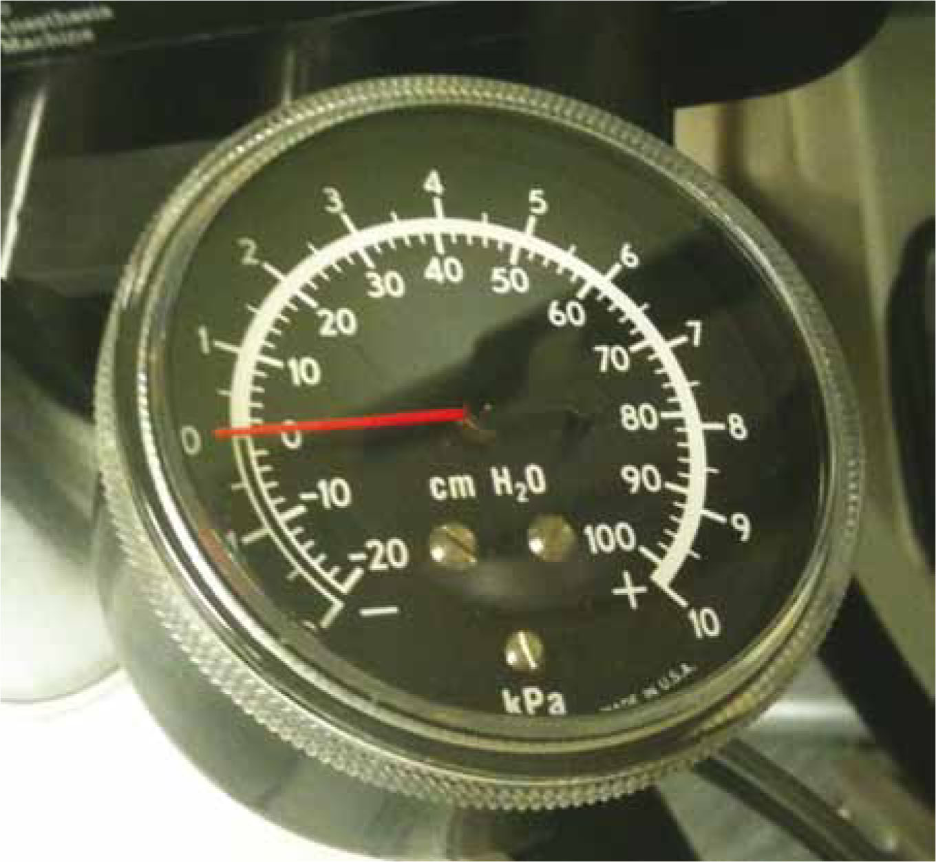
In order to maintain adequate tissue oxygenation, IPPV using 100% oxygen should be instigated. Varying ventilation rates have been suggested recently, however the emphasis is now on lower ventilation rates than previously suggested and a rate of 10–12 breaths per minute is generally recommended (higher for smaller dogs or cats). The ECC committee, subcommittees and task force of the American heart associations (2005) suggest administering two breaths 1–2 seconds apart. Signs of spontaneous ventilation returning can then be evaluated. If spontaneous ventilation resumes then full CPCR may be avoided. Breaths should last 1 second and be of sufficient volume to cause chest movement without overinflation, judged by visual assessment of the chest wall, and normal chest relaxation should occur after each breath. Over ventilation commonly occurs during CPCR. Care must be taken to ensure this does not occur, as Aufderheide and Lurie (2004) and Aufderheide et al (2004) demonstrate, excessive ventilation rates decrease the success rate of CPCR, as well as decreasing myocardial perfusion. Excessive ventilation pressures (>20 cmH2O) can also cause barotrauma and a manometer can be attached to the breathing system to ensure excessive pressures are not used (Figure 1). IPPV is not so crucial in early CPA, when the arterial concentration of oxygen is still high and oxygen delivery is limited by reduced cardiac output rather than hypoxaemia. This was demonstrated in a canine experimental study by Chandra et al (1994), which showed that adequate gaseous exchange was maintained for 4 minutes by chest compressions alone without intubation or ventilation. However after 4 minutes artificial ventilation is likely to be beneficial.
Table 1. Key points of basic life support
| Action | Comment | |
|---|---|---|
| Airway | Tracheal intubation with a cuffed endotracheal tube | Ensure tube adequately secured |
| Breathing | IPPV at 10–12 breaths/minute with 100% oxygen | Take care not to over ventilate |
| Circulation | Chest compressions at 100–120 compressions/minute | Frequent changing of staff to ensure adequate force and rate |
IPPV, intermittent positive pressure ventilation
Circulation
Chest compressions should begin if heart sounds and central pulses are absent. The goal of chest compressions is to pump blood from the chest to vital organs during the compression phase and to enhance venous return to the chest during relaxation of the chest wall. ECC committee, subcommittees and task force of The American Heart Association (2005) for human CPCR emphasize the importance of continued, uninterrupted chest compressions. They state that the number of interruptions should be kept to a minimum, as these lead to a decrease in coronary perfusion pressure and reduced likelihood of ROSC. Chest compressions should not be interrupted to allow intravenous (IV) catheterization, this can occur fairly easily during chest compressions if necessary, however any patient at risk of CPA should have an IV catheter placed prior to CPA. Advanced vascular access techniques will be discussed in a future article. Compressions may need to be briefly discontinued to allow electrocardiogram (ECG) interpretation, however the pause should be less than 10 seconds. Feneley et al (1988) demonstrated in an experimental canine study, that compression rates of 120 compressions/minute produced higher coronary perfusion pressure, as well as improved ROSC and 24 hour survival compared with lower rates of 60 compressions/minute. In light of this, Cole et al (2002) suggest chest compressions should be provided to small animals at a rate of around 100–120 compressions per minute, with a ratio of 1:1 compression to relaxation. In order to maintain force and rate to sustain adequate perfusion, the person performing compressions should be changed frequently — every 2 minutes is recommended in the human literature (ECC committee, 2005). Capnography can be a useful tool in assessing adequacy of chest compressions, using a decrease in PECO2 as a guide to changing personnel.

External chest compressions
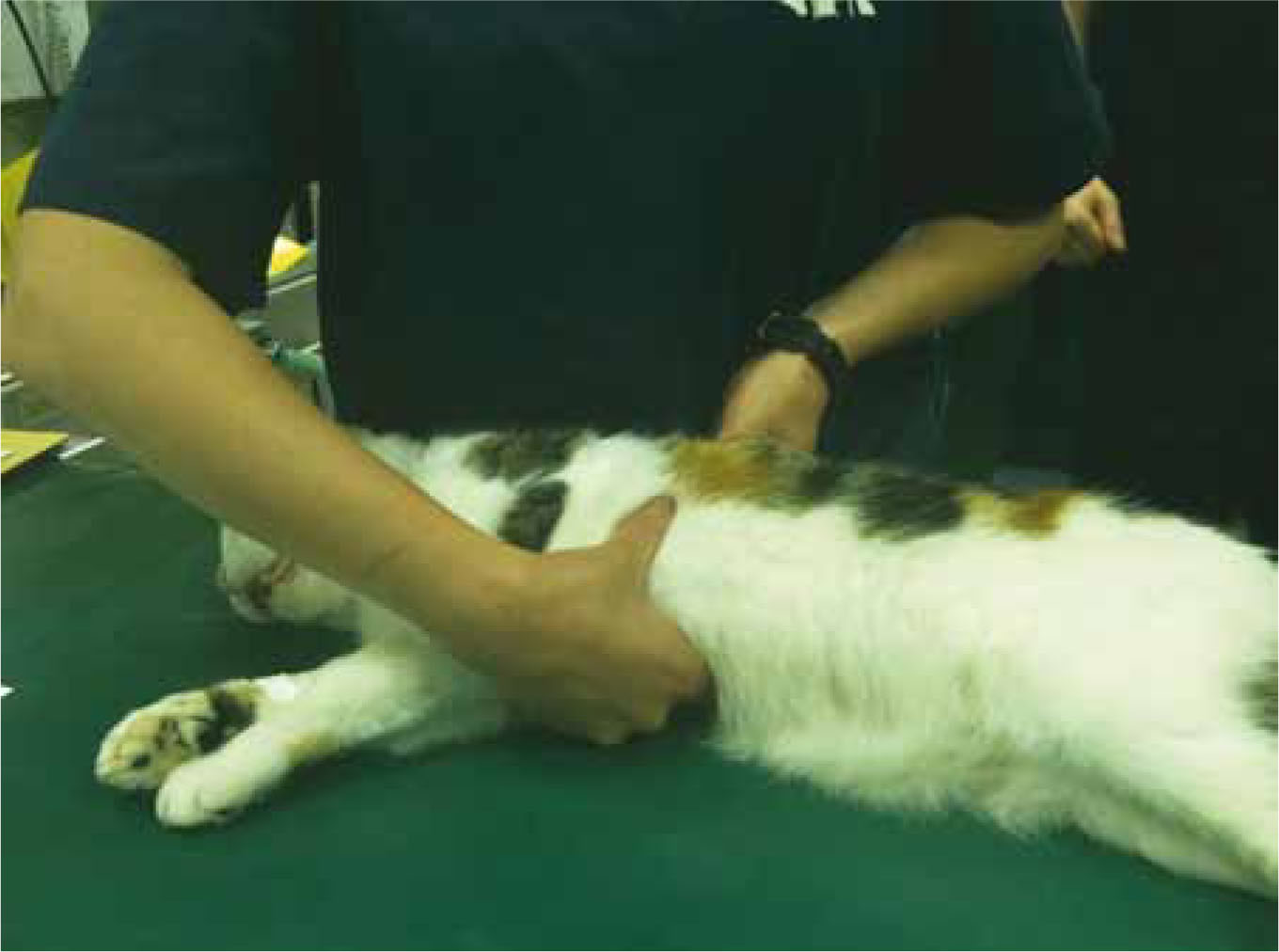
The patient should be placed on a firm surface in right lateral recumbency. The compressor should be over the chest with his/her front against the animal's spine for stability. In dogs and cats less than 15 kg the cardiac pump theory is thought to be the method responsible for blood flow. This is when arterial blood flow is caused by direct compression of the ventricles. The compressor should place their hands directly over the apex of the heart. Suggested landmarks are the 4th–6th intercostal space, at or slightly above the costochondral junction (Plunkett and McMichael, 2008). For cats and very small dogs this can be performed using one hand, however fingertips should not be used as they may cause myocardial damage (Figure 2). For medium and large dogs greater than 15 kg the mechanism responsible for forward blood flow is thought to be that stated in the well documented thoracic pump theory. Rhythmic pressure is applied to the chest wall creating forward blood flow by increasing intrathoracic pressure. Hands should be placed over the widest part of the chest and the chest compressed by about 30% (Figure 3). It has been suggested that dogs larger than 15 kg should be placed in dorsal recumbency to provide maximal changes in intrathoracic pressure, providing the patient can be positioned and stabilized effectively (Henik, 1992). Cole et al (2002) state that closed chest compressions produce less than 20% of normal cardiac output, so additional interventions to increase blood flow and cardiac output have been investigated. These include active compression–decompression, interposed abdominal compression and the use of impedance threshold devices. These techniques may increase ROSC, however there is not enough evidence suggesting increased survival to routinely recommend these techniques.
Internal chest compressions
Internal compressions use direct cardiac massage to generate blood flow and in animal models have been associated with increased ROSC (Kern et al, 1987; DeBehnke et al, 1991). In order to achieve better outcome in people, internal cardiac massage should be initiated early (ECC committee, 2005). Plunkett and McMichael (2008) suggest converting to open chest CPCR when external chest compressions have failed to achieve circulation within a few minutes of external CPCR, especially in dogs greater than 20 kg or when CPA is due to thoracic trauma or disease where thoracotomy would allow treatment and diagnosis. Lack of resources, staff expertise and intensive aftercare mean internal compressions are rarely performed in veterinary environments. Staff must be trained and have equipment prepared. In the author's experience, internal cardiac massage has been performed successfully during intra-operative CPA, particularly during thoracic or abdominal surgery where access to the heart can be easily achieved by incising the diaphragm.
Effcacy of CPCR
Kruse-Elliot (2001) suggests that CPCR success is indicated by palpable peripheral pulses, the detection of peripheral arterial flow via an ultrasonic flow detector (Doppler) and PECO2 exceeding 10 mmHg, also suggesting that patients with a PECO2 of less than 10 mmHg have a significantly reduced chance of survival. Capnography is a reliable tool in assessing efficacy of chest compressions during CPCR as it is an indirect indicator of cardiac output. As pulmonary perfusion increases during effective CPCR, PECO2 should increase. A Doppler probe placed on the cornea can be useful to detect the presence of blood flow to the head. An ECG is useful to evaluate heart rate and rhythm, enabling arrhythmias to be identified and treated. A human study (Abella et al, 2005) demonstrated that during in-hospital arrests, despite staff being trained in CPCR and attempting to follow CPCR guidelines, compression rates were often below the recommended 100 per minute and ventilation rates were often above the recommended rates. This concurs with studies demonstrating how common hyperventilation is during CPCR (Aufderheide and Lurie, 2004) and indicates the need for developing means of monitoring and supervizing efficacy of CPCR during resuscitation attempts. This could be achieved with a designated trained person leading the team, observing signs of effective/ineffective CPCR and counting compression and ventilation rates during resuscitation attempts.
Staff training
Although difficult to prove, level of staff training and degree of preparation is likely to have an impact on the success of CPCR. Continued education in CPCR, in-house training drills and staff debriefiings after CPCR are all important ways of improving resuscitation team efficiency. Boller et al (2010) reported that only 23.4% of general practitioners ran regular training drills for staff likely to be involved in CPCR. This demonstrates significant room for improvement in CPCR training in veterinary practices.
Discussion
Basic life support is performed following the ‘ABC’ mnemonic, however in light of studies demonstrating that adequate oxygenation can be maintained for at least 4 minutes by chest compressions alone (Chandra et al, 1994), recent discussion has arisen about whether ‘CAB’ would be more appropriate, emphasizing early instigation of chest compressions. This seems to be of particular relevance in humans, as a common reason for CPA is cardiac arrhythmias secondary to myocardial infarctions, when chest compressions are required until defibrillation can be performed. In veterinary medicine, the reasons for CPA are different, with most arrests not being primarily cardiac in origin. Primary respiratory arrest may be more common in cats and dogs and often responds to IPPV alone. Unlike human out of hospital arrests, veterinary practices have equipment for endotracheal intubation and provision of oxygen. In the author's experience, placement and adequate securing of an ETT is difficult once chest compressions have commenced and the detrimental effects of interruptions during chest compressions have been documented. For these reasons the ‘ABC’ approach remains more suitable in our veterinary patients. This also prevents aspiration of regurgitated stomach contents and allows 100% oxygen administration. In instances where a lone person has to provide CPCR, it may be appropriate to only administer chest compressions until help arrives, although a call for help should be made before instigating compressions.
Recommendations for veterinary practice
The ABC of basic life support should be executed efficiently in the event of CPA. IPPV should be provided at a rate of 10–12 breaths per minute and chest compressions at a rate of around 100–120 per minute. Nurses must be well informed with up to date literature and trained in CPCR in order to optimize success. Senior nurses should ensure their staff are up to date with current recommendations and run practical training drills to rehearse emergency situations. An experienced person should ‘lead’ the situation, taking responsibility for monitoring efficacy of compressions and ensuring guideline adherence. Emergency equipment should be well stocked, clean and easily accessible.
Conclusion
Good communication between all staff members is vital both prior to and during CPA. CPCR cannot be performed effectively by one person. Well executed, effective, continuous chest compressions are the single most important thing nurses can do in the event of a CPA in order to maintain sufficient myocardial and cerebral perfusion vital for ROSC. These should be instigated quickly once the trachea is intubated. Care should be taken not to over ventilate the patient with the emphasis on lower ventilation rates and more frequent chest compressions than previously recommended. Nurses should ensure that once started, chest compressions are not discontinued for any reason until ROSC or the decision is made to terminate CPCR. In order to improve survival rates, future research should concentrate on veterinary CPCR in clinical settings rather than human medicine and animal models with particular attention to monitoring efficacy of CPCR, adherence to guidelines and staff training. More research on additional interventions to increase cardiac output during CPCR such as impedance threshold devices and interposed abdominal compressions in veterinary patients would also be of interest.
Management of CPA does not end at ROSC. Many patients will require drug therapy and intensive nursing. A future article will discuss advanced life support.
Key Points
- Cardiopulmonary cerebral resuscitation (CPCR) is a technique utilized to reverse cardiopulmonary arrest (CPA).
- Rapid recognition of CPA and prompt initiation of CPCR is crucial for a successful outcome.
- The basic steps of basic life support should be remembered with the ‘airway, breathing, circulation’ (ABC) mnemonic and are the most important part of CPCR.
- Nurses should ensure essential emergency equipment is accessible, clean and well stocked at all times.
- Senior nurses should ensure their staff are up to date with current recommendations and run practical training drills to rehearse emergency situations.