Chronic gingivostomatitis (CGS), or recurrent oral ulceration (ROU), is identified in veterinary patients frequently, and histopathological examination of often severely inflamed tissues characterises the reactive cells that are within the oral mucosa (Lyon, 2005); this is obvious on conscious oral examination in these patients where proliferative ulceration and marker hyperaemia is evident. It has also been referred to as lymphoplasmacytic gingivitis stomatitis (LPGS) (Johnston 1998), lympho-plasmacytic stomatitis (LPS), plasmacytic stomatitis (PS), chronic ulcerative paradental stomatitis (CUPS) (Lyon, 2005) and chronic mucositis (CM) (Gengler, 2013). Gorrel (2011) highlighted that CGS can occur in canine patients, but is most frequently seen in feline patients, hence the label feline chronic gingivostomatitis (FCGS). A study by Healey et al (2007) reported a prevalence of 0.7% in a population of feline patients that visited first-opinion small animal veterinary practices, and Mihaljevic (2008) discussed that juvenile and adult clinical forms of this disease are often seen in purebred cats, domestic short hairs, Maine Coons and Siamese breeds. Due to this increased prevalence in felines this article will focus on FCGS.
Presentation
The owners of a FCGS cat may bring their cat to a veterinary nurse (VN) dental clinic initially for some advice as they may have a few concerns, but they may not be fully aware of the extent of the problem or the implications associated with treating the condition. The clinical signs associated with FCGS can include one or all of the following:
Aetiology and pathogenesis
FCGS has been extensively researched, but unfortunately the aetiology is still largely unknown (Southerden, 2010; Johnston, 2012; Lommer, 2013). From the research there appears to be a higher incidence of the condition in cats with calicivirus, but any causal relationships have not been definitively established.
Although research suggests that FCGS is a multifactorial condition, in general it is accepted that plaque bacteria is a major contributing factor to the development of FCGS, as gingival and oral mucosal tissues overreact to the presence of plaque bacteria (Gorrel, 2011; Lommer, 2013). The chronic nature of the condition is thought to be partially attributable to an underlying immune abnormality of the host (Mihaljevic, 2008; Southerden, 2010; Gorrel, 2011). These ideas were further developed, and sufferers are therefore usually grouped in two main ways:
To which group a patient belongs often remains unknown; it depends on whether the owners want to do further viral testing to rule those in or out as the cause, or if they want any other forms of investigative procedures to try and diagnose an underlying disease process. Investigations to identify whether the inflammation is secondary to another underlying condition would involve more than simple haematological and biochemical analyses, which can obviously start to get quite costly for the owners before even considering the treatment possibilities and their associated costs.
Inflammation
Inflammation is a normal response to an insult or injury, initiated by the body; this process however can cause problems itself. Recurrent, chronic, generalised or localised inflammation of the oral mucosa and gingiva may be a common finding associated with a wide spectrum of oral cavity disorders (Lommer, 2013). Oral inflammatory lesions occur as a result of an ulcerative condition, a vesiculobullous disease, or they are proliferative lesions. Potential underlying causes associated with the initiation of the inflammatory response include: dental disease; idiopathic causes; infectious conditions; mucosal and cutaneous immune-mediated disorders; reactive lesions; and neoplastic changes (Gorrel, 2008; Southerden, 2010; Gorrel, 2011; Johnston, 2012; Lommer, 2013). Inflammation restricted to the gingival and alveolar mucosa is therefore generally associated with periodontal or endodontic diseases, whereas acute ulceration of the oral mucosa, potentially including the tongue, could be the result of calicivirus, herpesvirus, panleukopenia, leukaemia (FeLV), immunodeficiency virus (FIV) or parvovirus.
Gingivitis is the term that refers to inflammation of the gingival tissue at the gingival margin; FCGS is described when there a little more tissue involvement than just the gingival margin tissue alone. FCGS also tends to affect large amounts of attached gingiva, right up to the mucogingival junction; this is a key difference between the two conditions.
The ‘stomatitis’ component refers to inflammation associated with any other soft tissues of the oral cavity, which can be anywhere. The usual sites for stomatitis development include the caudal oral cavity, the palatoglossal folds, the buccal mucosa towards the back of the mouth, the ventral tongue, the soft-palate tissues and the oropharyngeal areas. Lommer (2013) surmised that because tooth resporption and periodontitis can be associated with inflammation in the oral cavity, the presence of inflammation in these more caudal areas is one way of distinguishing FCGS from other types of inflammation.
Clinical examination
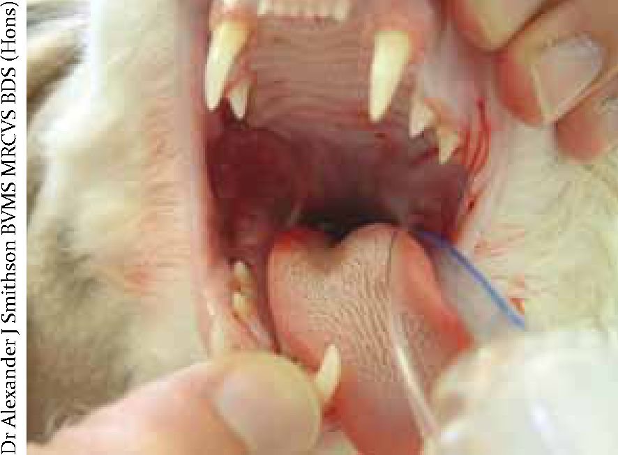
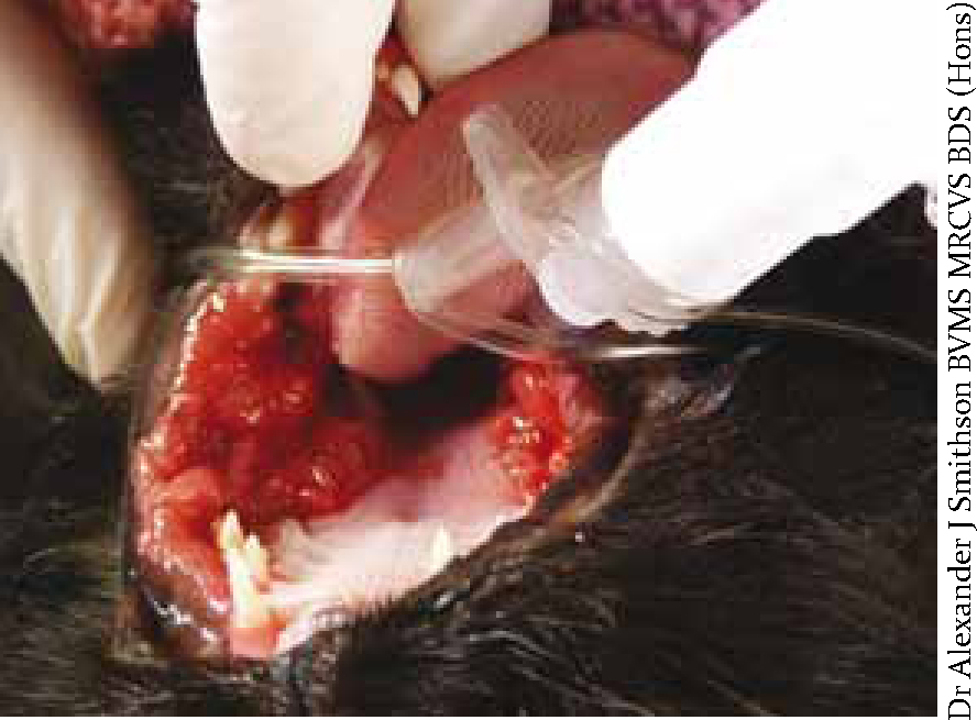
Lewis (2013) outlined that a patient's history coupled with a thorough physical examination are necessary so the veterinary surgeon (VS) can make a correct diagnosis of FCGS, but it is also important to assess the risk versus benefit ratio associated with any proposed dental procedures and interventions. A thorough assessment of the patient should be undertaken to determine the severity of their disease, and steps must be taken to rule in or out any underlying disease processes which could be affecting the patient's immune status and functioning. As well as noting the extent of the problem, the VN also needs to pay close attention to the actual physical appearance and characteristics of the inflammation (Figure 1) and lesions visible on oral examination (Gorrel, 2008); recording this qualitative information adds to the overall clinical picture and also provides a point of reference for future examinations, to decide whether the inflammation is getting better or indeed if the condition is deteriorating. The inflammation that is FCGS could just be the typical red, widespread inflammation that immediately comes to mind when thinking about gingivitis, or the inflammation could appear ulcerative and/or hyperplastic to differing degrees (Figure 2).
The assessment of an FCGS patient can be difficult; it must be remembered that these patients will be uncomfortable orally to differing degrees, so very careful and gentle handling during examination is warranted. A summary of a typical way to work through such a case with the VS is as follows:
(Lyon, 2005; Gorrel, 2008; Southerden, 2010; Johnston, 2012).
Treatment options
Treatment of FCGS can be quite involved and technically difficult as cat teeth can be difficult to extract successfully, and protracted. Lyon (2005) emphasised that all patients should be treated as individuals, as they will all have their own unique set of underlying contributing factors, and Lewis (2013) discussed that evidence-based practice is desirable to justify all therapeutic decisions. Treatments the VS may use for treating FCGS include: analgesics, antibiotics and extractions.
Analgesics
Analgesics are indicated in cases of FCGS as they are usually in considerable discomfort at the very least, and displaying overt signs of pain at worst. Cats tend to hide their feelings quite well when it comes to pain, so careful questioning of the owner about the patient, its usual routine and any subtle or insidious changes is indicated here.
Antibiotics
Extractions
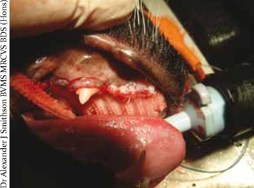
Extractions are also indicated as part of the treatment regimen for patients with FCGS, and are generally classed as the current recommended gold-standard treatment (Lommer, 2013) (Figure 3); this should follow full radiographic evaluation of all of the dentition to check for retained root remnants, and it is good practice to take pre and post-extraction radiographs (Lemmons, 2013). Each VS will have their own preferred approach to treatment, and this will often be based on their previous experiences with these kinds of cases. There are generally three phases when using extractions as a part of the treatment protocol for gingivostomatitis cases.
Phase one treatment
Phase one treatment involves extraction of the carnassials and upper first molar teeth, gingivectomy and gingivoplasty (Table 1). Post-operative care for phase one treatment would include continuing with analgesics as there will be inflammation post surgery. Continued antibiosis is often indicated, sometimes for up to 6 to 8 weeks. Chlorhexidine rinse should be used twice a day (BID) until the tissues are more amenable to tooth brushing; again the degree of inflammation needs to reduce. Tooth brushing should be performed BID ideally and used in conjunction with chlorhexidine 0.18–0.2% rinse, as impeccable plaque control is essential to the success of these cases. FCGS patients that have had extractions should return to the surgery for a 3 day post-operative check up with the VS, and then return weekly so healing, plaque control and tooth brushing efficacy can be monitored, gradually extending the intervals between checks.
| Treatment | Comments |
|---|---|
| Extraction of the carnassials (108, 208, 309, 409) and the upper first molar teeth (109, 209) | These teeth are the ones that cannot be brushed as well/effectively |
| Gingivectomy and gingivoplasty | Removal of all hyperplastic tissue evident and recontour the gingival margin |
| Extraction of root remnants evident | If root remnants are left and have communication with the oral cavity there will not be a resolution of the problem. If radiographs show the roots are resorbing they should be fine left in situ |
| Perform a complete and thorough ‘dental’ prophylaxis — probe and chart, scale and polish, other extractions as required | Any other diseased teeth or those likely to cause/promote plaque accumulation should also be extracted |
| Rinse the oral cavity with chlorhexidine solution | Provides about 12 hours of protection against plaque build up |
If the VS deems it necessary, a feeding tube may have been placed while the animal was under general anaesthetic to ensure its daily nutritional requirements are met, as nutrition is essential to healing and recovery from any illness. Some cases will be managed well with a naso-oesophageal (NO) feeding tube, but if the VS wants to bypass the head area completely they may choose an oesophagostomy (O) feeding tube; administering foodstuffs down O tubes is much easier than NO tubes as they have a wider diameter. To encourage oral food intake, using the cat's favourite food and warming it up to enhance the smell can be beneficial, and the patient will be able to eat in this manner despite having a feeding tube in situ.
After having phase one treatment many of the cases will significantly improve to the point where they return to their normal demeanour; it has been found that roughly 60 to 80% of cases (Gorrel 2011; Lommer 2013) will significantly improve following the extraction of the premolars and molars, as large plaque-retentive surfaces have been eliminated. The appearance of the oral cavity in patients with FCGS will rarely ever return to what would be classed as ‘normal’, but so long as they are comfortable and pain free the VS and owner will be satisfied.
When the cat has finished its course of antibiotics it is likely to have recurrences of the problem. So long as the owner is aware of this and knows that a flare-up and its severity is likely to be proportionate to the level of plaque accumulation on the cat's teeth they can monitor progress and hopefully remain motivated with the oral homecare regimen recommended.
When a cat is presented with a flare-up the VS will decide what the most appropriate next stage of treatment should be in order to regain control of the situation. If the owner's efforts have ceased, or are not as effective as they were originally, the oral homecare can be instigated again. It may be necessary to completely re-educate the owners about homecare regimens at this stage, and it is sensible to ensure they return for dental checks with the VN on a regular basis to ensure they remain motivated for the benefit of their pet.
Phase two treatment
Flare-ups, unfortunately, can occur on a regular basis in some cats after phase one treatment, which means the VS may recommend re-investigating the condition and moving on to phase two treatment (Table 2) in which all teeth, except the four canines, are removed. It is thought that most FCGS cases will reach this point eventually, despite best efforts to prevent it.
| Treatment | Comments |
|---|---|
| Extraction of all teeth except the four canines | Removing all other teeth removes the plaque retentive surfaces. Leaving the canines is ideal as it maintains jaw integrity, maxillofacial structure and tongue position etc |
| Scale and polish the canines | Probing and charting the teeth would always be sensible as a record of their health at that point in time |
| Intra-oral radiography | It may be wise to take some more radiographs at this point to check again for root remnants — consider whether radiographs were taken during phase one and whether something may have been missed. It is easy to misinterpret radiographic findings with such small pieces of tissue being involved |
Post operatively the treatment, follow up, aftercare and homecare regimens would be much the same as for phase one. The application of chlorhexidine should be even easier in these circumstances as the canines are much more accessible than the more caudal dentition.
Usually, after phase two treatment there will be an even more significant improvement in the cat's condition when everything is healed and the degree of inflammation has reduced. There are considerably-fewer teeth for plaque to accumulate on, thus reducing the chance for recurrence even more; any flare-ups can again be managed with antibiotics when necessary.
Phase three treatment
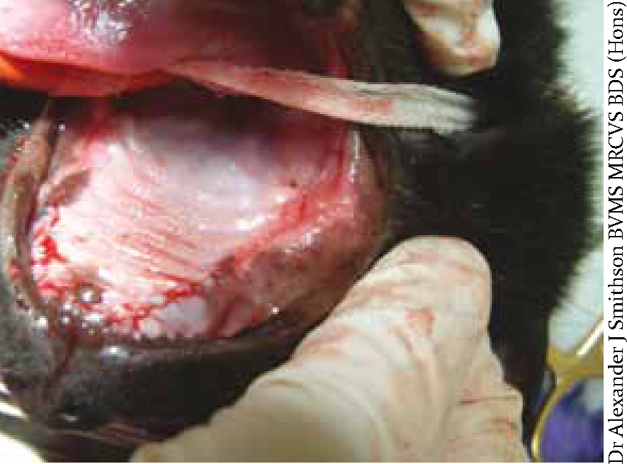
Sometimes phase two treatment still does not keep the problem under control, and if the patient is still suffering then phase three must be considered and discussed with the owners. This involves extraction of the remaining teeth (Table 3) (Figure 4). Post operatively there is not much oral homecare required when there are no teeth left! The patient does however need to come back at 3 and 10 days post operatively so the VN can check healing of the extraction sites. The patient should still be able to eat a kibbled diet as most cat foods are formulated from small kibbles that can be swallowed whole.
| Treatment | Comments |
|---|---|
| Extraction of the remaining canine teeth | Both upper canines can be removed |
| It is not advisable to remove both lower canines during the same surgery as a cat mandible is very fragile and does not comprise a significant amount of bone which could lead to pathological fractures, or even iatrogenic fractures unfortunately | |
| The second lower canine should be left for at least 6 weeks after the first lower canine removal to allow the alveolar bone to remodel and become as stable as possible before disturbing the contralateral tissues. You could be called phase 3b! |
Further treatment and management advice
There are a few other noteworthy points regarding treatment and management of FCGS cases, including:
Other therapies that have been considered for use in combination with the treatments mentioned thus far include the following:
Conclusion
It is apparent that FCGS is a very difficult condition to manage and treat successfully in the first instance, and can be distressing for both the patient and the owner. The owners should be made aware from the outset that it could be a quite invasive and protracted treatment, which is likely to be quite costly, and which due to the nature of the disease may require a concerted effort to medicate their pet and provide effective oral homecare if any of the earlier phase treatments have a chance of being successful.