

The investigation and management of otitis externa is similar to any skin problem, and involves the collection of a history, a physical and dermatological/otic examination before undertaking diagnostic tests (Griffin, 2006; Bloom, 2009). An appreciation of the aetiology helps in that process. The aetiology and pathogenesis of otitis externa can be divided into four parts represented by the initialism PPSP. The first P represents primary causes (Table 1) that are present in every case of otitis externa. Primary causes, which are almost always skin problems, are responsible for the initiation of the inflammatory process in the ear that in turn leads to disease.
Table 1. The most common primary causes of otitis externa
| Primary factors | |
|---|---|
| Allergy | Atopic dermatitis, cutaneous adverse food reaction, contact allergy |
| Endocrine | Hypothyroidism, hyperadrenocorticism |
| Autoimmune/immune-mediated | Pemphigus foliaceus, bullous pemphigoid, discoid lupus erythematosus, erythema multiforme, vasculitis, drug eruption |
| Keratinisation disorders | Sebaceous adenitis, primary idiopathic seborrhoea |
| Ectoparasites | Otodectes cynotis, Demodex spp. |
| Foreign bodies | Grass awns, sand, powder |
The second P in the initialism represents predisposing factors, which contribute to ear disease but do not cause inflammation. There is some overlap between some of these factors and primary causes, e.g. obstructive disease caused by neoplasia can be a perpetuating factor and a primary cause. Important predisposing factors are listed in Table 2.
Table 2. The most common predisposing factors in otitis externa
| Predisposing factors | |
|---|---|
| Conformation | Stenotic canal, pendulous pinnae, hairy ear canals |
| Water | Dogs that swim, water-based medication, bathing |
| Obstructive disease | Polyps, neoplasia, canal hyperplasia |
The third P is for the perpetuating factors that drive the disease process once it has been established and are most important in long-standing disease. Where they are not resolved they will prevent effective therapy, and the otitis will inevitably relapse. Important perpetuating factors are found in Table 3.
Table 3. The most common perpetuating factors in otitis externa
| Perpetuating factors | |
|---|---|
| Chronic change | Hyperplastic changes in the canals, ceruminous gland hyperplasia |
| Otitis media | Infection, chronic change within the tympanic bulla |
Infection in canine otitis externa is never a primary problem hence the S in the initialism stands for secondary infection. Infection, which can be with either bacteria or yeast, develops because of inflammation produced by primary causes usually in combination with perpetuating and predisposing factors. Effective therapy of infection is essential but where this is treated without making any attempt to identify and treat other factors, then recurrence will inevitably occur. Table 4 details the most common bacterial and yeast infections identified from ears of dogs.
Table 4. The most common causes of secondary infection in otitis externa
| Secondary infection | |
|---|---|
| Bacteria | Acute disease — Gram-positive bacteria Staphylococcus spp.,Streptococcus spp.Chronic disease — Gram-negative bacteria Pseudomonas spp,Proteus spp. |
| Yeast | Malassezia spp. |
The veterinary nurse can play an important role in every part of the disease process in cases of otitis externa. They are well placed to work with their veterinary surgeon to help identify primary causes; to undertake basic investigations of the ears to provide the veterinary surgeon with diagnostics information; and to use their communication skills to advise owners on appropriate ear cleaning, the administration of medication and the need for follow up and ongoing therapy.
The nurse's role in helping to identify primary causes
As in any dermatological disease the investigation of otitis should involve a general, dermatological and otic history. Experienced nurses are capable of taking a history from clients. This can be undertaken using dermatology history sheets to help guide questions and preventing the omission of important facts. Thorough history taking provides the opportunity to try to establish the primary, perpetuating and predisposing factors. In the author's experience, owners often find it easier to chat to nurses than veterinary surgeons about their pet's problem and are often more open about the challenges they face with giving therapy. A full physical examination is useful to identify any signs of disease that may give an indication of an underlying primary cause, e.g. the presence of a bradycardia may give a clue to hypothyroidism. A dermatological inspection may find evidence of skin disease such as pedal saliva staining suggestive of allergy, or footpad hyperkeratosis in pemphigus foliaceus. A report of the clinical finding to the attending veterinary surgeon can then lead to recommendations for further investigations such as endocrine function tests, the institution of a food trial or allergy testing.
The nurse's role in helping to manage secondary infection
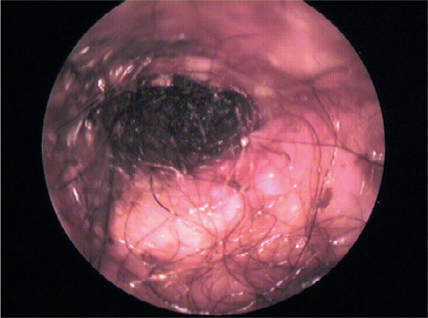
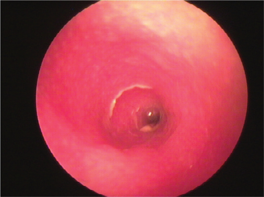
After a general assessment of the dog, the ears should be examined. This can help establish the presence of infection both by the gross appearance of the ear, the discharge and the cytological findings. The ear should be examined with a hand-held otoscope and cytology should be performed to look for bacteria, yeast and parasites (Angus, 2004). The appearance of the ear will vary enormously with the different types of infection (Cole, 2004). Where yeast is present the discharge is thick and ceruminous, the canal tends to be swollen and pruritic but is rarely ulcerated (Figure 1). Where bacterial infection is present the canal is more inflamed, may be ulcerated and the discharge tends to be more purulent. Where Gram-positive bacterial infection such as Staphylococcus spp. is present, the purulent discharge is usually yellow/brown and cerumino-purulent. Where Gram-negative bacterial infection is present, especially with Pseudomonas spp., changes within the ear canal are more severe. The canal is usually swollen, painful and ulcerated and the discharge is greenish/yellow and muco-purulent, often with accompanying haemorrhage (Paterson, 2012) (Figure 2). Samples of discharge can be collected from the ear using a cotton tipped applicator which can then be rolled along a glass slide (Griffin, 2006). It is important to assess all samples for parasites especially Demodex spp., which can influence the choice of topical therapy as well as infection. The author would therefore generally take two samples, one to check for parasites and the second for pathogens. The second sample can be heat fixed using a hand drier or hair drier and then stained using a Romanowsky stain such as Diff Quik. If cytology reveals infection the next step is to establish if it is yeast or bacteria and what type of bacteria. If yeast or cocci bacteria are identified then therapy can usually be selected on an empirical basis, as it is likely the yeast will be Malassezia spp. and the cocci will be Staphylococcus spp. If a Diff Quik stain is used and rods are found then precise identification of the bacteria can be more difficult. Organisms in these cases may be indicative of Gram-negative infection with Pseudomonas spp. or Proteus spp. The author would therefore always recommend cultures when rods are found on cytology.
The nurse's role in managing predisposing factors
Where predisposing factors are identified these need to be managed to maximise the response to therapy and minimise the risk of relapse. The nurse is well placed to advise on these, as many of these factors, which relate to breed conformation or lifestyle, can be managed with appropriate ear cleaning.
Ear cleaners can be divided into cleaners that remove wax and those that remove purulent exudate and create an environment within the ear that is hostile to the over growth of pathogens (Nuttall and Cole, 2004).
Wax removing ear cleaners
The two main groups of products with wax removing capabilities are the ceruminolytic and the ceruminosolvent cleaners. Both of these types of cleaners are useful in animals with conformational characteristics that leads to a reduction in ventilation in the ear, allowing wax build up, that may predispose them to the development of disease, e.g. dogs with pendulous pinnae such as the Basset Hound or Spaniels (Figure 3), or dogs with stenotic ear canals such as the Sharpei (see Table 5). Ceruminolytic ear cleaners are generally easy to use and do not leave a greasy discharge in the ear which the oilier lubricant cleaners can. However, they can be irritant in sensitive or ulcerated ears and are reported as being ototoxic if the ear drum is damaged. Examples of ceruminolytics include the surfactant ceruminolytic, dioctyl Na or Ca sulphosuccinate (DOSS) and the humectants ceruminolytic, carbamide peroxide. Lubricant ceruminosolvents soften and loosen wax rather than disrupt it. They tend to be gentler in an inflamed ear than a ceruminolytic. Examples of lubricant cleaners are squalene, triethanolamine polypeptide oleate condensate, propylene glycol and mineral and vegetable oils. Wax removing ear cleaners are important as part of any routine grooming in any breed with strong predisposing factors, but are also important where yeast infection is present. Removal of wax where secondary infection with Malassezia spp. is present allows penetration of the topical antimycotic drug to maximise the response to therapy.
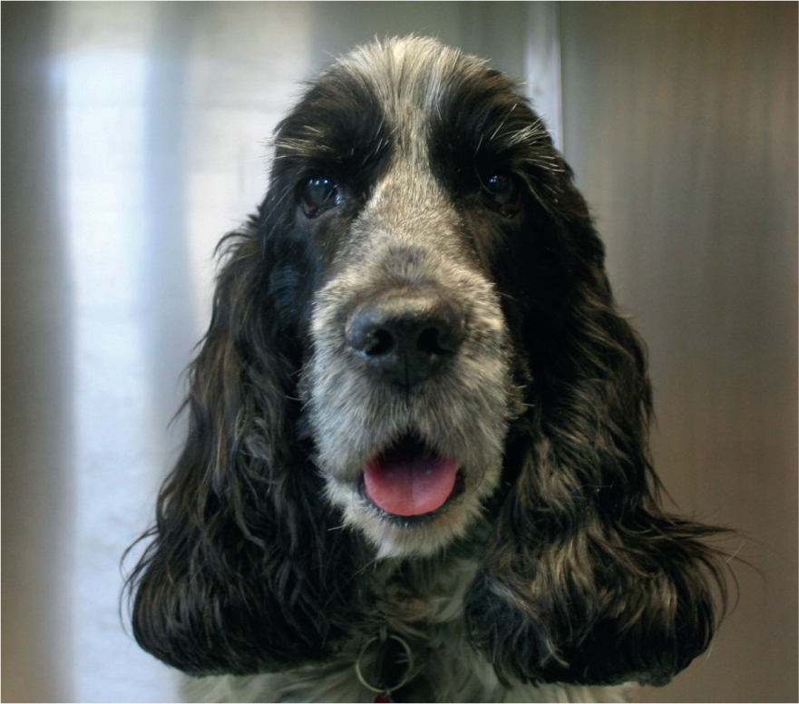
Table 5. Predisposing factors in the development of otitis externa
| Predisposing factor | Breed predisposition |
|---|---|
| Pendulous ear pinnae | Basset, Blood hound, Spaniels |
| Hairy ear canals | Poodle, Cocker spaniel, Cockerpoo, Labradoodle |
| Stenotic ear canals | Sharpei |
| Swimming | Labrador, Newfoundland, Spinoni |
Anti-bacterial ear cleaners
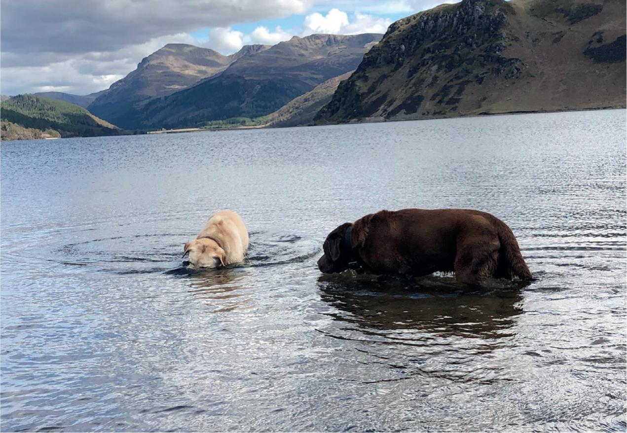
Conformation can predispose to the development of otic infection, but lifestyle factors, particularly dogs that swim and those that put their heads underwater, can also predispose (Figure 4). In these cases, drying antiseptic ear cleaners can be used after dogs have swum to prevent maceration of the canal and create an environment within the ear to reduce the risk of infection. Antibacterial ear cleaners are also important as part of any treatment protocol. Where infection is present, water-based antiseptic products help break up the muco-purulent discharge. Acidic solutions, such as lactic acid and acetic acid, are known to have antibacterial effects. Chlorhexidine is also recognised as having broad spectrum antibacterial properties. Its activity is enhanced when combined with EDTA tris (Swinney et al, 2008).
The nurse's role in managing perpetuating factors
Once perpetuating factors develop, they can be among some of the most challenging factors to manage in cases of otitis externa, often requiring referral to a specialist centre for appropriate treatment or surgery. However perpetuating factors develop as a result of recurrent disease, which can be as a result of a range of factors. This may be because of a failure to manage the underlying primary cause or because of poor owner compliance as a result of their inability to use medication or a misunderstanding on the part of the owner about the need for reassessment and ongoing maintenance therapy. Nurses can play an invaluable role therefore in acute disease to help advise and support owners with therapy so they understand the need to complete a course and return for recheck appointments. Follow-up appointments are important even in apparently simple cases. Treatment should only be discontinued when follow-up cytology reveals no evidence of microorganisms or an inflammatory infiltrate. Once this end point has been achieved, the dog should be switched onto maintenance therapy. This will usually be an appropriate ear cleaner to reduce the risk of disease recurrence, as well as measures to monitor or treat the primary factors.
Conclusion
Ear disease is commonly encountered in primary care practice. The skills of the veterinary nurse should be employed at all stages of the management of otitis. As part of the veterinary surgeon-led team, they can contribute to the investigation of primary factors by history taking, dermatological inspection and sample collection. By advising owners on the need for appropriate cleaning, the correct use of topical medication and the requirement for reassessment, they can help deal with secondary infection and reduce the risk of predisposing factors. Finally, by advising on maintenance therapy to help recurrence of disease they can reduce the risk of chronic disease and the development of perpetuating factors.
KEY POINTS
- Nurses play an important role in all aspects of the management of otitis externa.
- Visual examination and cytology of the otic discharge by the nurse can help the veterinary surgeon in selection of therapy.
- Ear cleaning should be an integral part of the management of otitis, experienced nurses are well placed to advise clients on this.
- Nurses are excellent communicators and can help advise owners around the appropriate use of medication in both the treatment and maintenance of ear disease.