
Rabbits have become more frequent patients in small animal practice and are now the most popular small mammal exotic pet in the UK (Pet Food Manufacturers Association, 2013). With this increase in domestic rabbits seen in practice there will also be an increase in demand for high quality veterinary care. However this is a relatively new speciality area within veterinary medicine (Huynh and Pignon, 2013).
Rabbits typically suffer from unique conditions that might seem daunting for common small animal practices which mostly treat dogs and cats. These patients also have to be approached from a different angle appreciating their different anatomy and physiology. Rabbits are hind-gut fermenters, have an unusual calcium metabolism, high metabolic rates and are catecholamine driven prey animals that stress easily (Fisher, 2010). Their calcium metabolism is considered unusual as they absorb all the calcium they can from their diet, and excrete the excess through their urine (Girling, 2013). Catecholamines, such as norepinephrine, epinephrine and dopamine, are hormones responsible for the flight-or-flight reflex (Colville and Bassert, 2008). Most diagnoses in these animals are gastrointestinal, possibly due to their sensitive gastrointestinal system and specific dietary requirements, such as a high fibre diet. The most common gastrointestinal condition diagnosed is gut stasis, which is one of the most challenging problems in practice when dealing with rabbits due to the non-specific clinical presentation associated with the condition and the fact that there are several factors that can trigger it: environmental factors such as stress and an inappropriate diet, but also other underlying conditions that cause pain and stress. This creates challenges for the veterinary team when trying to find and treat the primary cause of the condition (Fisher, 2010).
To understand how to treat and monitor the gut stasis patient, knowledge and an understanding of possible causes is needed to reverse the condition. The origin of the condition needs to be identified to aid in treatment as well as reducing the chance of encountering the problem again.
Even though gut stasis is one of the more common conditions that are encountered in veterinary practice, it is rarely a primary disorder. There are many factors that can trigger it and each and every one needs to be investigated to identify the correct cause (Krempels et al, 2000). Any underlying conditions causing pain and distress in the rabbit will result in depression and anorexia. Dental disease is very common in rabbits, as a result of their open rooted teeth. Insufficient dental care will result in malocclusion and eventually pain when the rabbit tries to eat (Fisher, 2010). Incorrect diet is another common cause, where a rabbit has been fed a diet that is high in carbohydrate and protein, instead of high in fibre. The rabbit is also a fragile creature, very susceptible to environmental stress. A new environment or a new handler could be enough to initiate anorexia (Fisher, 2010). Dehydration will also reduce gut movement and aid in the formation of trichobezoars (a mass found trapped in the gastrointestinal system), which can cause intestinal obstruction if they cannot pass through the gut (Johnston, 2005).
Nursing the gut stasis patient can be challenging but also very rewarding. Intensive nursing care is necessary to support the rabbit while the underlying cause of the condition is identified. This article will review the key aspects of care where the veterinary nurse will be the primary advocate of the rabbit while in practice.
Measuring blood glucose
When a rabbit is brought into practice with gastrointestinal symptoms it is important to rapidly establish whether there is an obstruction requiring immediate emergency surgery or if gut stasis is the diagnosis which requires immediate medical treatment. Harcourt-Brown and Harcourt-Brown (2012) suggest that blood glucose measurement in anorexic rabbits can be a useful diagnostic aid if used alongside other examinations. Measuring blood glucose with a portable glucometer is not only inexpensive and simple but can easily be repeated if the results are inconclusive.
Normal glucose intervals in rabbits fall between 4.2 and 8.2 mmol/l although there are slight differences in published ranges. Hartcourt-Brown and Hartcourt-Brown's (2012) study showed that rabbits with confirmed gut stasis had a mean glucose concentration of 8.5 mmol/l while rabbits with a confirmed obstruction had a mean value of 24.7 mmol/l. This suggests that blood glucose measurement can be used as a measurable parameter to assess the severity of a rabbit's condition.
It is important to remember that stress from handling could raise the value of glucose above the reference range but any values above 20 mmol/l suggests a life-threatening condition such as hepatic lipidosis, enterotoxaemia, intestinal obstruction, bladder stones or mucoid enteropathy. Measuring blood glucose can help with monitoring the progression of conditions such as gut stasis and obstruction (Harcourt-Brown and Harcourt-Brown, 2012).
Recognising pain in rabbits
Gut stasis is a very painful condition and it is important that any symptoms of pain are recognised so they can be adequately managed before the condition gets worse. Although it can be difficult, it is important to be able to distinguish between anxiety and pain. Anxiety can easily be dealt with by altering the environment and enriching the rabbit's kennel, while pain indicates disease and will require analgesics. The difficulty in differentiating between the two comes from the fact that they are often combined and are presented with similar changes in behaviour. A stressed rabbit might be more jumpy and restless with eyes wide open to assess the environment, while a rabbit in pain is more likely to be inactive and have squinting eyes. A list of signs indicating stress and pain can be found in Table 1, where it can be seen that the signs are similar, but the two can still be differentiated.
| Signs of stress and frustration | Signs of pain |
|---|---|
|
|
|
| Hansen and Berthelsen, 2000 | Johnston, 2005; Leach et al, 2009; Wenger, 2012 |
As a prey species, rabbits are naturally quiet, reserved and quite anxious in an unfamiliar environment and they are inclined to hide any painful condition to try to maintain a normal appearance. Any presence of a potential predator, or even an observer, will most likely result in the rabbit freezing for long periods of time. This is a normal response to pain and stress in rabbits. Pain assessment has, for these reasons, been a challenge for the veterinary team and an understanding of normal behaviour is essential for accurate assessment (Wenger, 2012).
Each rabbit has to be assessed as an individual where tolerance and pain threshold will vary and accurate assessment relies on experience and professional judgement. Some rabbits will sit motionless in a corner but others will have periods of uncontrolled movement and struggle when being handled. A rabbit's normal heart rate can be very high making an increased heart rate difficult to detect and an unreliable clinical sign; however their respiratory rate will often change when they are in pain. Compared with a healthy rabbit's short and shallow breath a rabbit experiencing pain may exhibit a deeper breathing pattern with a pronounced nasal flare (Johnston, 2005).
Stress management and kennel enrichment
The rabbit is a catecholamine driven prey animal that stresses easily and requires specialised hospitalisation to reduce stress and aid in a more rapid recovery. Rabbits can potentially be stressed by multiple factors such as pain, being away from their normal handler and environment, transport, disruption of normal routine and the smell and sound of any potential predators such as dogs, cats and ferrets. In most cases, gut stasis is triggered by unalleviated pain or stress and so reducing stress in the hospital environment will not only promote a speedy recovery but also improve animal welfare while in practice (Fisher, 2010).
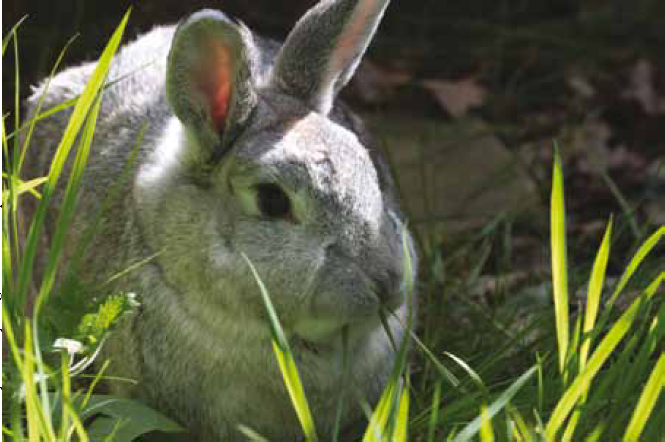
Accommodating rabbits in practice kennels will in most cases not satisfy their behavioural needs and environmental limitations could result in increased stress, restlessness and altered behaviours such as bar gnawing, excessive grooming and aggression. Although it could be difficult to accommodate for all of the rabbit's needs, a few alterations in how rabbits are accommodated could decrease stress and improve welfare. A simple wooden box should be placed in every rabbit's cage as not only does it provide shelter but it has been shown that rabbits like to use the top of the box as a resting place and look-out point (Hansen and Berthelsen, 2000). Also, allowing the rabbit the opportunity to move around and exercise on a daily basis will not only lower stress levels, but it will also help with gut movement (Figure 1). Having a bigger kennel or a separate run for the rabbits would be ideal (Hansen and Berthelsen, 2000).
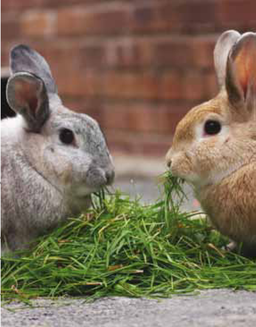
Where possible the rabbit should be accommodated away from dogs and cats and with another rabbit for company to satisfy their social needs (Figure 2). However if this is not an option a mirror could be placed in the rabbit's kennel to create the illusion of company if the rabbit is to be in the practice for a long period of time (Zotte et al, 2009).
Even after centuries of domestication rabbits still have the primal instinct to chew everything and dig everywhere. A litter tray filled with straw or a towel will satisfy their needs for digging and creating a nest, and a block of wood will prevent them from chewing at the kennel bars. These are simple ways to prevent unwanted behaviour (Crowell-Davis, 2006). A list of simple methods of enriching a rabbit's kennel in practice can be found in Table 2.
| Kennel enrichment |
|---|
|
|
| Hansen and Berthelsen, 2000;Crowell-Davis, 2006;Zotte et al, 2009 |
Abdominal massage
Abdominal massage is known for its therapeutic effects in increasing gut motility in both humans and rats (Sinclair, 2011; Chapelle and Bove, 2013) and is slowly being introduced into veterinary practice. It is a simple and inexpensive way to improve gut motility in rabbits and can easily be taught to the owner too. To perform the massage the rabbit should be placed on a table or on a lap depending on where the rabbit feels more secure. The abdomen should be gently massaged with the fingertips and palm of the hands as deeply as the rabbit will allow but taking care not to damage or bruise the rabbit's delicate organs. If the rabbit expresses pain, massage should be stopped immediately and retried at a later point and any further massage should be more gentle. Gently lifting the rabbit's hindquarters a few inches (the rabbit's head safely tucked into the handler's elbow and spine supported) could help gas to pass more easily and rabbits tend to find this comfortable after getting over the initial surprise at being held this way (Krempels et al, 2000; Fisher, 2010).
Nutrition
Maintaining high fibre intake is a considerable challenge for the veterinary team as a high fibre diet consisting of minimum 20–25% fibre is required for normal peristalsis and a healthy bacterial flora in the gut (Fisher, 2010). A health assessment should be conducted first prior to creating a nutritional care plan for critically ill rabbits to establish the severity of the situation. Any rabbits with anorexia for more than 1 day are already in danger of hepatic lipidosis which can lead to enterotoxaemia, sepsis and death (Krempels et al, 2000). Initiating early enteral feeding will not only decrease the chances of hepatic lipidosis but will also reduce pain as it helps gastric motility (Lichtenberger, 2007). A new feeding schedule should be created daily based on the animal's weight and the food given with careful consideration to the fibre content to accommodate the rabbit's requirements. Assisted feeding should continue until the rabbit is able to consume a normal amount of food and is producing normal faeces. When doing a nutritional assessment it is important to consider that rabbits have a high feed intake of 65–80 g/kg of bodyweight (Orosz, 2013).
Whenever hospitalised the rabbit should always be offered water and ad libitum good quality grass hay such as timothy, oat or meadow. High fibre and low protein pellets should also be offered at approximately 28 g/kg alongside fresh leafy greens. Due to the rabbit's sensitive gastrointestinal system, fruits and certain vegetables high in starch and sugar should be limited (Fisher, 2010).
Syringe feeding
Syringe feeding has long been the standard protocol for feeding anorexic rabbits as it is an easy way to force food into the patient and also makes it easier to introduce a diet high in insoluble fibre. However it could be argued that due to the rabbit's short gastrointestinal transition time they will require frequent feeding approximately every 2 to 4 hours which can create a very stressful environment for the rabbit (Rosen, 2011).
Syringe feeding also requires slow and careful administration in order to eliminate the risk of aspiration and should be avoided if the patient displays any signs of dental disease or oropharyngeal disease (Orosz, 2013).
Nasogastric (NG) tube feeding
Many critically ill rabbits are commonly too weak or nauseous to eat from a syringe and a nasogastric (NG) tube can be much less stressful than force feeding. It also allows for continuous nutritional support and easy administration of fluids and medication. Feeding rabbits through a nasogastric tube was previously not considered ideal due to the small diameter of the tube. This is because it precluded the administration of insoluble fibre which is necessary for normal gut motility (Lichtenberger, 2007). Since then however new products have been introduced to the market and some products such as Oxbow Critical Care Fine Grind are designed to fit through tubes as small as 3.5-French making NG tube feeding an achievable task (Oxbow Animal Health, 2014). Another critical care diet that is suitable and high enough in fibre is the Supreme RecoveryPlus.
The rabbit's anatomy and physiology needs to be considered when discussing placement of NG tubes. Rabbits are obligate nasal breathers and the nasal tube may obstruct their airway causing hypoxia. For this reason the size of the tube will have to be decided based on the size of the rabbit to allow for normal breathing and easy passage of food. It can also be irritating to the nasal mucosa so if the rabbit is showing any signs of either lower or upper respiratory disease prior to placement the tube should not be used and an alternative feeding protocol should be implemented. A rabbit with a NG tube needs to be closely monitored for any complications. If the rabbit shows any signs of nasal discharge or respiratory distress after placement of the tube it must be removed immediately and in most cases this will resolve the problem. If the rabbit is not a suitable candidate for a NG tube (e.g. respiratory distress, too small nasal passages, does not tolerate the NG tube), an oesophagostomy tube should be considered, with careful consideration taken to the general anaesthetic needed (Rosen, 2011).
Placement of NG tube
Equipment needed for this procedure can be found in Table 3. The length of the NG tube should be measured from the nares to the last rib and the NG tube marked using a permanent marker. Local anaesthetic should be applied into the nares 5 to 10 minutes prior to placement and the NG tube should be lubricated with either KY jelly or lidocaine jelly. The rabbit should be placed in ventral recumbency and restrained appropriately (a towel is a useful way to support the entire rabbit). The rabbit's head should initially be tilted up to place the tube into the ventral nasal meatus and should then be aimed ventrally and medially. When the end of the tube is in, the rabbit's neck should be moved back to a normal, flexed position to ensure the tube enters the oesophagus and not the trachea. Conscious rabbits should swallow the tube once it enters the pharynx. The tube should be advanced into the stomach to the level of the pre-measured mark on the tube and secured in place to the top of the head between the ears with butterfly tape and suture. A properly placed tube should not encounter much resistance and if the rabbit objects or struggles at any point the procedure should be stopped and more local anaesthetic administered into the nostril or possible sedation should be discussed. Placement should be confirmed with radiographs (however, this might require sedation), capnography and aspiration of stomach contents (Rosen, 2011).
| Equipment needed for placement of nasogastric (NG) tube |
|---|
|
|
| Lichtenberger, 2007; Rosen, 2011 |
Medical protocol
Pharmacological agents used in treatment of gut stasis include appetite stimulants, analgesics, prokinetic agents and anti-bloating agents. Vitamin B complex is often used as an appetite stimulant alongside these (Fisher, 2010). Cisapride, ranitidine and metoclopramide are possible prokinetic agents (Girling, 2013) that should be included in the medical protocol alongside anti-bloating agents, such as simethicone (Fisher, 2010). Robinson (2005) also recognises the need to address the colonisation of pathogenic bacteria that occurs during gut stasis with antibiotics such as ‘enrofloxacin’ to prevent further bacterial colonisation. Fluid therapy may also be necessary to restore hydration in rabbits that have also stopped drinking. Fluid therapy is also important to help restore gut motility and to help any trichobezoars pass, especially if they are the cause of the gut stasis. Normal maintenance value for rabbits is 80–100 ml/kg/day (Girling, 2013).
Owner education
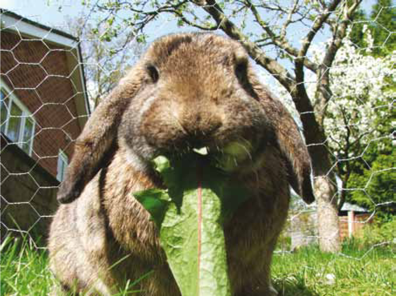
Prevention rather than cure! It is believed that gut stasis could be prevented with appropriate diet and housing at home (Fisher, 2010) (Figure 3) and the veterinary team is the best source of up-to-date information about housing and diet. A study by Edgar and Mullan (2011) highlights that many owners have limited knowledge of the needs of the rabbit, especially regarding dietary and social needs. This demonstrates the need for rabbit clinics and owner education (Edgar and Mullan, 2011). Many practices now offer ‘puppy parties’ where the owners learn about behaviour, training and nutrition but little is done to offer rabbit owners advice outside of consults. In order to promote and educate owners about responsible rabbit ownership the author suggests ‘rabbit parties’ could be implemented to educate owners about the importance of proper diet and environmental needs.
Conclusion
Gut stasis would be less of a challenge for the veterinary team if considerations were made for the special nature of rabbits; understanding how they present symptoms, their nutrition requirements and sensitive gastrointestinal tract and best practice with regard to their treatment and management as inpatients.
An appropriate protocol should be created taking into account all these things. Special attention should be given to appropriate accommodation and pain assessment as it is difficult to distinguish between anxiety and pain in rabbits. Having a nursing team that has knowledge of common rabbit health issues and basic critical care needs and methods will greatly improve patient care and case success as well as improve animal welfare (Fisher, 2010).
The International Society of Feline Medicine (2013) stresses the importance of having a cat advocate — a member of staff that ensures a high level of cat welfare. This should prompt a discussion about advocates in practice for exotic pets. The rabbit has special needs that need to be accommodated for and it could be argued that it is time to introduce the exotic advocate to the veterinary practice to improve exotic animal care.