

Thoracic trauma is commonly seen in small animal practice. It can be in the form of penetrating bite wounds, falling from heights or being hit by cars. The pattern of injury is dependent on the cause, and different causes will require different management techniques; each case should, therefore, be assessed individually in order to achieve the best outcome for the patient.
It is believed that road traffic accidents (RTAs) are the leading cause of blunt force trauma for small animals in the UK, with young male cats being the most likely to be hit (Conroy at al, 2018). Blunt force trauma typically results in diaphragmatic rupture and/or damage to the intrathoracic organs (Barnes, 2019). Despite this, damage to the thoracic wall itself is uncommon because of its high compliance. Rib fractures are not uncommon, and the RVN should be aware of the increased risk of intrathoracic organ damage as a result (Barnes, 2019).
Triage
Triage is the process of categorising and prioritising patients based on their severity of condition. Nurses should be confident in their abilities to triage patients efficiently and categorise correctly. Triaging a patient can be broken down into the ‘ABC’ (see Box 1).
Box 1.The ABC of triageA:Does the patient have a patent airway?B:Is the patient breathing? Is the breathing appropriate? Is intubation required?C:Does the patient have a heartbeat? What is the heart rate? Can you palpate pulses? Are the pulses synchronous with the heartbeat?
The initial assessment should take no longer than 60–90 seconds, including obtaining a brief history from the owner. If any level of dysfunction is noted in any of the major body systems (respiratory, cardiovascular, neurological, renal), then immediate intervention is required (Davis, 2001). Physical examination in patients with thoracic trauma may be brief as they frequently present dyspnoeic and/or tachypnoeic. Oxygen therapy should be provided immediately for these patients.
Initial stabilisation
The early steps in stabilising the patient allows the veterinary surgeon to decide on the most suitable diagnostics and therapeutic approach (Sharp, 2015).
The first step is to increase oxygen delivery with or without sedation, depending on the status of the patient. Flow by oxygen is readily available in most veterinary practices, however it is not always well tolerated, and it does not provide a high level of inspired oxygen (FiO2). If available, the patient should be placed in an oxygen rich environment such as an oxygen cage where a higher percentage of oxygen can be delivered to the patient with a minimal amount of stress (Sharp, 2015). In extreme cases, the animal may require intubation and intermittent positive pressure ventilation (IPPV).
Auscultation of the thorax allows identification of changes in lung sounds. Quiet lung sounds are typical in those patients with pleural space disease. Gas within the pleural space rises, therefore in the case of a pneumothorax, the lung sounds will be quieter dorsally. If the lung fields are quieter ventrally, it is suggestive of an effusion. It should be noted that these clinical signs will not improve without the performance of a thoracocentesis (Sharp, 2015).
Box 2.Emergency database
- Packed cell volume/total protein (PCV/TP)
- Blood glucose
- Blood urea nitrogen (BUN)
- Blood smear
- Electrolytes
- Blood gases
Crackles are caused by a gas/fluid interface, such as pulmonary contusions, pulmonary haemorrhage, pulmonary oedema. Borborygmi (gut sounds) are often heard with a diaphragmatic rupture; the gastrointestinal tract has migrated into the thoracic cavity. This diagnosis will require confirmation via imaging before being surgically repaired (Callan, 2002).
Placement of an intravenous catheter should be performed as soon as possible, patient condition permitting. This procedure can be quite stressful for patients and a staged approach may be more beneficial. This may include clipping the insertion site and placing a numbing cream, e.g. EMLA, onto the skin and then returning the patient to their kennel to allow the cream to work prior to placement. A cephalic vessel may not always be appropriate depending on patient temperament. Saphenous vessels can also be used; however it is important not to use excessive restraint, and allow the patient to maintain a comfortable position to maximise air flow and avoid further respiratory distress.
The largest bore catheter should be selected and placed aseptically. Bloods for an emergency database can be taken at this time (Box 2). Intravenous access allows for the administration of analgesia and fluid resuscitation. Blood gas analysis can also be performed at this point.
Arterial blood gas analysis is the gold standard for assessing a patient's lung function. The PaO2 (amount of oxygen in arterial blood) and the PaCO2 (amount of carbon dioxide in arterial blood) are used as indicators of lung function. Both parameters are influenced by oxygen supplementation and this should be taken into consideration when the sample is taken. An arterial blood sample is obtained through direct puncture of an artery and this is more painful than taking a venous sample. It is slightly more challenging than a venepuncture but is a procedure that RVNs can perform. Care should be taken to ensure than a mixed venous/arterial sample is not obtained by accident. Arterial spasm or haematoma formation can prevent further collection of samples from the same artery. It should be noted that this technique should not be performed in those patients that are suspected to be suffering from a coagulopathy, or from a fractured limb. The desired vessel is located by feel, not sight as for a venous sample. The dorsal metatarsal arteries are normally the vessels of choice; however, the femoral and lingual arteries can also be used. The area should be clipped and cleaned, then the arterial pulse should be re-palpated. Once the person taking the sample has confidently located the arterial pulse, the arterial syringe should be inserted through the skin firmly at a 45° angle. Arterial syringes are pre-heparinised and should be drawn back prior to use. The arterial flow will fill the syringe without the need to draw back. Once the appropriate volume has been collected, the needle can be removed, and a pressure bandage should be applied immediately for up to 5 minutes. The sample should be analysed immediately to prevent contamination with room air (Snyder, 2009).
There are challenges to obtaining arterial blood samples, mainly through technique. The procedure is painful, and this may cause the patient to react negatively, potentially pushing the patient into respiratory arrest. Obtaining the sample can be slow. If oxygen therapy is being withheld during this time, then the patient may suffer. If serial arterial samples are required, then it may be in the patient's best interest to be sedated once stabilised and an arterial catheter placed (placement of this is beyond the scope of this article).
The patient should also be checked for the presence of any wounds. Open wounds should be covered to prevent the development of nosocomial infections and further managed once the patient has been stabilised. Patients with open wounds along the thorax are at greater risk of developing a tension pneumothorax (Maritato et al, 2009). This condition arises when a one-way valve is created allowing air to enter the pleural space during inspiration but is then unable to leave during expiration. The build up of air within the thorax results in progressive dyspnoea and if left untreated will result in death. The only treatment option is an emergency thoracocentesis.
Oxygen delivery
As mentioned previously, flow-by oxygen is the easiest method of supplemental oxygen provision, however it is not the most effective and may not be appropriate. This technique provides an inspired oxygen level of between 25–40% oxygen, only twice that of room air. Not all patients will tolerate this method of delivery, and if the patient is panting the volume of oxygen inhaled will be decreased. The use of oxygen masks can help increase oxygen delivery, but these, too, are not always tolerated and can increase patient hypercapnia.
The gold standard technique is to place the patient in an oxygen kennel. This is a controlled stress-free oxygen environment and allows the patient to receive higher levels of oxygen, up to 100% (Waddell, 2016). Oxygen kennels are temperature and humidity controlled; however, they take time to reach the desired oxygen percentage and oxygen therapy is interrupted when the door is opened to administer treatments. If the oxygen kennel is to be used, the use of an extension line attached to the intravenous catheter allows for drug administration to be provided without opening the kennel door.
Oxygen prongs and nasal catheters are other techniques that can be employed for oxygen delivery. Both prongs and catheters can be placed by RVNs but are case dependent. It is not uncommon for those animals that have been involved in a road traffic accident to suffer some form of facial trauma. This may mean that it is inappropriate for facial instrumentation in order to deliver oxygen. A summary of oxygen supplementation can be found in Table 1.
Table 1. Methods of oxygen provision
| Technique | Flow rate | Approximate percentage of oxygen in inspired air | Advantages | Disadvantage |
|---|---|---|---|---|
| Oxygen cage | 10–12 litres/minute | Up to 80 |
|
|
| Face mask | 2–5 litres/minute | 40–50 |
|
|
| ‘Crow collar’ | 2–5 litres/minute | 30–40 |
|
|
| Nasal prongs | 50–100 ml/kg/minute | 40–50 |
|
|
| Nasal catheter | 50–100 ml/kg/minute | Unilateral: 40–50Bilateral: 60–70 |
|
|
| Flow-by | 2–10 litres/minute | 30–40 |
|
|
The dyspnoeic patient is difficult to physically assess as they can decompensate rapidly, and stress is a significant environmental factor to consider. Anxiety as a result of dyspnoea can be exacerbated by a stressful hospital environment. Increased levels of stress increase the respiratory rate and oxygen consumption, preventing the patient from being able to meet their required oxygen demands, and this may result in respiratory arrest. To minimise stress and prevent decompensation, the ‘hands off’ technique should be used, allowing the patient to become calmer in an oxygen rich environment and stabilise. Anxiolytic drugs such as butorphanol, acepromazine and dexmedetomidine can be given intramuscularly to help calm the patient (see Table 2) (Sharp, 2015). These can also be combined with an opioid such as methadone to provide analgesia and make the patient more comfortable. Sedatives are also beneficial for performing diagnostics and procedures.
Table 2. Sedative drugs
| Sedative drug | Dose range |
|---|---|
| Butorphanol | 0.1–0.4 mg/kg IV/IMEvery 1–4 hours |
| Acepromazine | 0.005–0.05 mg/kg IV/IMEvery 4–8 hours |
| Dexmedetomidine | 0.01–0.1 IV/IM0.01–0.2 µg/kg/min as a continuous rate infusion |
IV = intravenously, IM = intramuscularly
The choice of drug is the decision of the veterinary surgeon and is case dependent. Butorphanol has a shorter duration of action, 1 to 2 hours, but cannot be easily reversed. Acepromazine has a longer duration of action, approximately 4 to 6 hours, but may have undesirable effects on the cardiovascular system and is unable to be reversed. Dexmedetomidine also causes cardiovascular depression, but is reversible and can be titrated to effect (Sharp, 2015). These drugs should always be given at the lowest dose possible to begin with and increased to obtain the desired effects. The RVN should be aware of the drugs given and the route by which they are administered.
Diagnosis
Point of care ultrasound (POCUS) is a highly sensitive and specific diagnostic tool, which can be used to identify the presence of fluid/gas in the pleural cavity. It does not require chemical restraint and can be repeated at the patient bedside allowing for serial monitoring of the patient (Powell, 2011).
Therapeutic thoracocentesis may be indicated in an emergency if the build-up of fluid and/or air within the thorax is causing severe respiratory compromise, and respiratory arrest is imminent. It is often done conscious and the patient may be unaware of the procedure because of the distracting injury. This can also be repeated easily by a competent veterinary surgeon. Nurses are routinely involved in this technique, supporting the patient, and assisting the veterinary surgeon. In some cases, the placement of chest drains may be required; management of these is beyond the scope of this article.
Radiographs are not typically indicated in the early assessment of thoracic trauma patients, but should be obtained later on for diagnosis of conditions such as rib fractures and diaphragmatic hernia. The radiograph should be taken on the peak of inspiration to maximise air flow in the lungs and create the most contrast in order to obtain the best diagnostic image (Mauragis and Berry, 2011). If the lungs are under inflated, they will appear to have a higher density (whiter in appearance) increasing difficulty in diagnosis of the condition (Fox, 2005). Both lateral and dorso-ventral views should be obtained for the most complete interpretation; lateral views may compromise patient respiration if performed conscious (Fox, 2005). General anaesthetic or sedation may not be appropriate for the patient immediately, hence there may be a delay in obtaining radiographs until the patient has stabilised.
How to perform a thoracocentesis
To perform a thoracocentesis, an appropriate section of the thorax should be clipped and cleaned. Unless stated otherwise, it should be assumed that the condition is bilateral. The height of the needle insertion site will vary; if fluid is suspected, the designated site should be between the eighth and ninth intercostal space at the costo-chondral junction ventrally. If air is anticipated, then the site should be at the same intercostal point, but more dorsal (Maritato et al, 2009). Care should be taken to avoid the edge of the rib as this is where the neurovascular bundle is located, and this will cause unnecessary pain.
The nursing role during this procedure varies: although this technique involves entering a body cavity and therefore can only be carried out by veterinary surgeon, RVNs should be fully aware of the procedure. The RVN should be confident in setting up the equipment required and supporting the veterinary surgeon. There should be suitable oxygen provision for the patient and continuous monitoring should be performed. Nurses are often required as an ‘extra set of hands’ drawing back on the syringe as the veterinary surgeon focuses on placement of the needle; this can be more difficult if the procedure is ultrasound guided as the veterinary surgeon's attention will be on the screen and not on the patient (Looney, 2001).
Patient care
The RVN is a crucial member of the team for both stabilising and assisting with the diagnostic procedures, but also for the recovery period during patient hospitalisation.
Nursing these patients can be very demanding, and routinely involves serial monitoring of respiration rate and effort, allowing the identification of trends and early detection of patient deterioration. These observations can be performed as frequently as necessary, and where possible, thoracic auscultation should be performed.
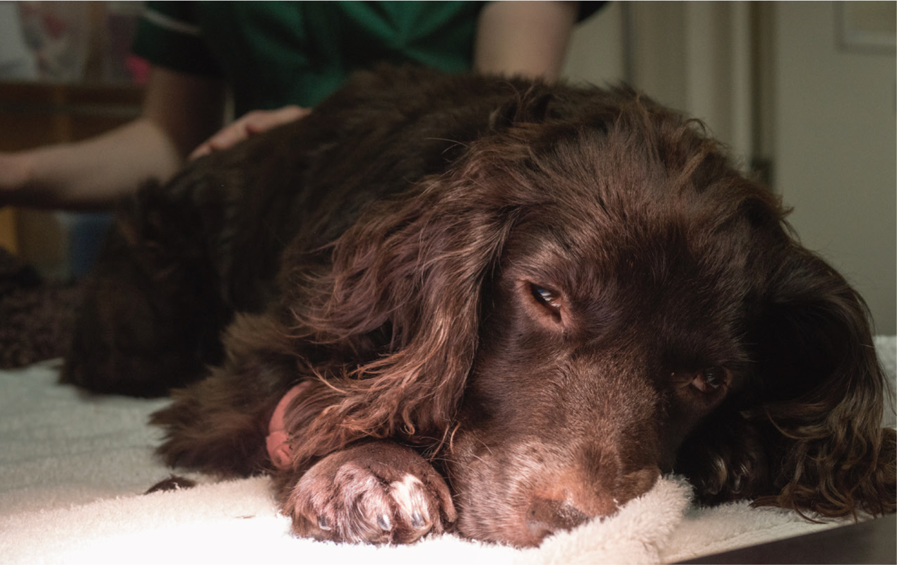
Patients should be kept in sternal recumbency where possible in order to maximise lung capacity (Figure 1). If this is not possible, then the patient should be turned regularly to prevent atelectasis. Patient comfort is paramount. Remaining in a fixed position for prolonged periods of time encourages the development of decubitus ulcers and muscle soreness. If the patient is elderly then passive range of motion can help alleviate stiffness; this is dependent on the condition of the patient, and fractured limbs should not be moved. Bedding should also be checked regularly to ensure that it is not soiled to prevent patient scalding.
Regular pain scores should be instigated in order to make sure that the level of analgesia delivered to the patient is appropriate. Local anaesthesia can also be used for those patients with thoracostomy tubes.
Nebulisation can be carried out on these patients to help loosen secretions and deliver certain drugs like acetylcysteine (Ackerman, 2015), patient permitting. This can be done with a hand-held nebuliser, or some oxygen cages provide this function.
Conclusion
Respiratory distress as a result of trauma is commonly seen as an emergency in small animal practice and patients in respiratory distress should be prioritised. Prompt identification of the cause of the respiratory issue is crucial for a positive outcome. The RVN plays a key role in the welfare of these patient from triage to stabilisation and in-patient care.
KEY POINTS
- Thoracic trauma is frequently seen In small animal practice, and has various presenting pathologies ranging from superficial wounds to penetrating trauma.
- Complications with pathologies will result in rapid decompensation of the patient.
- Initial assessment and basic interventions are paramount for patient survival to discharge.
- Oxygen can be provided through various methods in order to suit each individual patient.
- Registered veterinary nurses provide multiple levels of care in order to nurse these appropriately.