

Most of the domestic species have a dense haircoat that protects them from excessive exposure to sunlight. However, in areas of the body not naturally covered in hair, or where hair is lost for other reasons, the exposed skin becomes susceptible to damage from sunlight (Miller et al, 2013). In the dog, cat and horse phototoxicity and photosensitivity are the two forms of sunlight induced disease that are of primary concerned to the veterinary clinician (Miller et al, 2013). Both forms of damage are caused by light in the ultraviolet (UV) spectrum. UV light can be divided into UVA, UVB and UVC light.
Phototoxicity is the classic sunburn reaction and is dose dependent. Both UVC and UVB are phototoxic. Fortunately the earth's ozone layer protects us from the most damaging UVC (<290 nm). However, UVB (290–320 nm) does reach the earth's surface and is the wavelength most commonly associated with phototoxic reactions. UVB is often referred to as the sunburn or erythema spectrum, and is regarded as being 1000 times more erythemogenic than UVA (Walker et al, 2003).
Photosensitivity occurs when the skin has an increased susceptibility to non-burning doses of UV light (Miller et al, 2013). UVA (320–400 nm) penetrates further into the skin than other forms of UV and is the wavelength associated with photosensitivity reactions. Unlike phototoxicity, photosensitivity can occur on haired skin, but the hair is generally unpigmented. The increased susceptibility of the skin to sunlight in photosensitivity is due to the production of (e.g. congenital or acquired porphyria), ingestion of (e.g. drugs), or contact with, photodynamic agents (Miller et al, 2013). Photosensitivity has been recognised in domestic species for almost 50 years (Berry and Merriam, 1970; Hudson and Florax, 1991; Dolowy, 1996; Ivens, 2011; Puschner et al, 2016); it is most commonly seen in horses.
Solar exposure can also induce or exacerbate the lesions of some autoimmune skin diseases, notably discoid lupus erythematosus (Figure 1), systemic lupus erythematosus, pemphigus (especially pemphigus erythematosus) and pemphigoid (Miller et al, 2013). These diseases will not be discussed in this article, but where a history of exacerbation of disease occurs in relation to sunlight exposure, the same principles of sun protection should be applied as for other UV-exposure associated diseases.
Most people are aware of the health benefits of sensible levels of sunlight exposure. Most are also aware of the need for protection against the damaging phototoxic effects of UVB, for themselves or family members, but few are aware of the need to protect their pets from the potentially damaging effects of the sun.
Phototoxicity (sunburn)
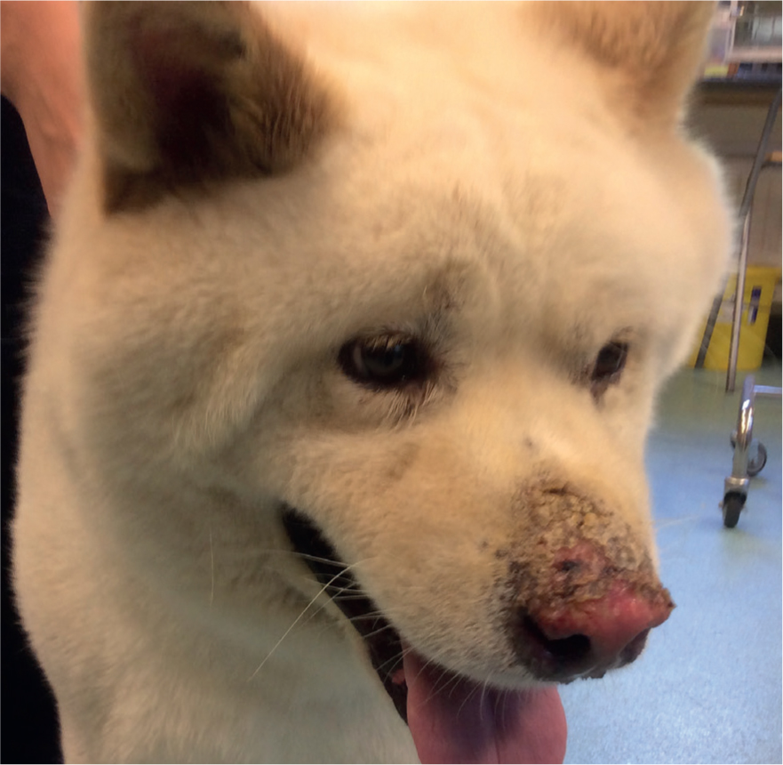
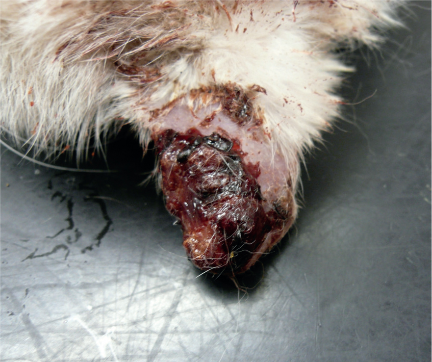
Phototoxicity, often referred to as solar dermatitis, is purely a reaction to over-exposure to the sun's rays. Although the condition develops most commonly when white or non-pigmented skin is exposed to direct or reflected sunshine, especially where there is little or no hair covering (Figure 2), it can occur in dogs with dark hair and a full hair coat. One case report in the literature describes full thickness necrosis of the skin on the dorsum of a dachshund after excessive sun exposure (Sumner et al, 2016). The speed of onset of the condition and the severity of clinical signs are related to a range of factors, which include duration of sun exposure and the intensity of the sunlight (Miller et al, 2013). The sun's rays are most intensive during the summer months, particularly during the middle of the day from 9.00 am to 3.00 pm, but especially from 11.00 am to 2.00 pm. A simple way to assess the intensity of sun exposure was described by Downham (1998) and has since been developed by other authors (Downham, 1998; Lorentzen, 2002). As a general rule of thumb, when shadows are shorter than objects throwing them, avoid direct sunlight, when shadow and object are of equal length restrict sun exposure to half an hour, and when shadows are twice the length of objects 1 hour in the sun is permissible (Table 1).
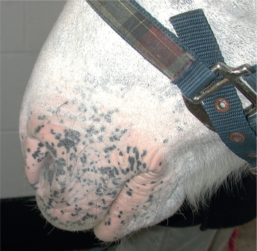
Table 1. Simple guide to sunlight exposure based on shadow length (adapted from Lorentzen 2002)
| Length of shadow of object | Risk of sun burn | Advice |
|---|---|---|
| Shadows shorter than object throwing them | High | Avoid direct sunlight or if sun exposure unavoidable wear high factor sun protection |
| Shadows equal length to object throwing them | Moderate to high | Limit unprotected sun exposure to 30 mins. After this time wear high factor sun protection |
| Shadows twice the length of object throwing them | Low | Sun exposure is permissible for up to 1 hour before avoidance or sun protection is required |
Another important factor that influences sunlight intensity is altitude. The sun's intensity increases by 4% for every 300 m (1000 ft) (Nogier, 1950; Miller et al, 2013) altitude rise. Animals out at pasture at altitude, or dogs out hill walking with their owners, are at higher risk than those at lower levels. The pathogenesis of phototoxicity is incompletely understood, but damage is known to affect the epidermis, leading to the formation of vacuolated keratinocytes (so called sunburn cells), and the blood vessels of the superficial and deep dermis. Solar dermatitis can affect the dog, cat and horse. The most common forms of phototoxicity are canine nasal solar dermatitis, canine solar dermatitis of the trunk and extremities, feline solar dermatitis and equine solar dermatitis. Details of the different diseases together with breed and/or phenotypic predisposition are found in Tables 2 and 3.
Table 2. Solar dermatitis in the dog, cat and horse
| Species | Condition | Clinical signs |
|---|---|---|
| Canine | Nasal solar dermatitis | Initially erythema and scale of sparsely haired, non-pigmented nasal skin. Prolonged exposure causes exudation, crusting and ulceration. Chronic lesions heal by scarring |
| Solar dermatitis of trunk and extremities | Initial signs erythema and scale of exposed skin. With excessive exposure the skin becomes tender and necrotic. With repeated episodic exposure actinic folliculitis, actinic follicular cysts with dermal fibrosis are seen. Chronic exposure leads to erosions, ulceration and comedones | |
| Feline | Solar dermatitis | Initially erythema and scale of susceptible areas, especially pinnal margins. Early lesions are not painful. Chronic sun exposure leads to marked erythema, skin peeling and crusting often with curling of pinnal tip with associated pain. Similar signs can be seen on lower eyelids and nose |
| Equine | Solar dermatitis | Initial signs of erythema and scale of sparsely haired non-pigmented skin. Prolonged exposure leads to exudation, crusting, peeling of the skin and ulceration |
Table 3. Breed predispositions for solar dermatitis
| Condition | Breed and/or phenotypic predisposition |
|---|---|
| Canine nasal solar dermatitis | Dogs without pigment in their nasal skin, especially those born without pigment or breeds that have undergone spontaneous non-inflammatory depigmentation such as vitiligo (Rottweiler, Belgian Tervuren, Doberman, Old English Sheepdog) (Mahaffey et al, 1978, Wisselink, 1993)Dogs with active or resolved traumatic or inflammatory conditions of the nasal skin that causes hair loss or depigmentation |
| Canine solar dermatitis of trunk and extremities | Breeds predisposed to truncal solar dermatitis include the Dalmatian, American Staffordshire Terrier, German Short-haired Pointer, white Boxers and white bull terriers. Any dog that sunbathes will develop signs on the areas with the highest sun exposure. This may be the flank or the ventrum depending on how the dog lies. They may also develop signs of extremities (Miller et al, 2013) |
| Feline solar dermatitis | White cats or coloured cats with white areas on the face (eyelids and nose) and ears. Blue-eyed white cats most susceptible (Almeida et al, 2008) |
| Equine solar dermatitis | Horses out at permanent pasture in the summer months commonly affected on areas of non-pigmented skin on noses, around eyes and heels. Can affect almost any breed, because nearly all breeds produce horses with white facial and leg markings |
Photosensitisation
Photosensitivity is an abnormal reaction of the skin to light, caused by photodynamic agents in or on the skin. It is most commonly seen in the horse (Fadok, 1995; Ivens, 2011; Puschner et al, 2016) and has been recorded on rare occasions in the dog (Hudson and Florax, 1991; Dolowy, 1996). Unlike solar dermatitis, lesions of photosensitivity can occur on well-haired areas of the body. However, melanin in pigmented hairs has protective properties, so that lesions are exclusively seen in the white regions of black and white dogs and cats, or on the white areas on horses. In animals where the darkly pigmented haired areas remained unaffected and only the white haired areas develop signs of skin disease, photosensitisation is very likely. Photosensitivity can be either systemic or contact. The different types of photosensitisation with potential triggers are detailed in Table 4.
Table 4. Types of photosensitisation with potential causes
| Type of photosensitisation | Mechanisms | |
|---|---|---|
| Systemic | Congenital | Porphyria: abnormal accumulation of photodynamic porphyrins in blood and body tissue due to a congenital defect. Porphyrins absorb UV light leading to free radical formation and tissue damage |
| Acquired | Primary disease caused by ingestion of preformed photodynamic agents, e.g. drugs such as antibiotics, or plants. Plants may have pre-formed photodynamic agents, e.g. St John's wort, perennial rye grass, or may produce photodynamic agents when pasture is lush, e.g. clover, alfalfa, lucerne, vetch and oats. | |
| Hepatogenous photosensitisation occurs when liver damage inhibits elimination of phylloerythrin produced in the gastro-intestinal tract from plant chlorophyll. As phylloerythrin excretion is decreased, it builds up in the circulation where it accumulates in the skin to cause photoactivated skin disease in about 25% of affected animals. | ||
| Contact | Acquired | Primary disease is caused by direct contact with the skin of photoactive plants. Plants most commonly implicated include alfalfa, clover and almost any grass, especially when rapid growth occurs after rain. Environmental sprays and topical medication, including sun blocks, may also be contact photosensitisers |
Recognising phototoxic skin reactions
A carefully taken clinical history is one of the first factors that helps identify sunlight induced disease. The appearance of lesions on poorly haired areas, especially extremities, after prolonged exposure to strong sunlight is most typical. This may be a horse that has been out at pasture without a shady field shelter on a warm summers day, or a dog that has fallen asleep in an unprotected area of the garden in full sun, or a cat sun bathing all afternoon on the roof of the garage in the summer. The initial signs will be similar in all cases, consisting of varying degrees of erythema of the skin with associated scaling. Where exposure has been prolonged in high intensity sunlight, the skin may be painful, exudative, blistered and ulcerated. Only the hairless, exposed areas will be affected, which of course may depend on the position of the animal in relation to the sun.
Recognising photosensitive skin reactions
The history for photosensitisation reactions may be similar to that for phototoxic reactions. However, for lesions to develop, both a photodynamic agent and a period of prolonged sunlight exposure must be present. Identification of the photodynamic agent should be undertaken to try to prevent further damage to the skin, as well as sunlight avoidance and symptomatic therapy of the lesions. In all cases the lesions will affect the white-haired areas of the body. All of the white-haired areas of the body are affected in systemic disease, while in contact photosensitisation only the areas in contact with the photodynamic agent develop lesions. In this latter case this will often be the unpigmented lower legs and face. Initial signs consist of erythema of the skin in the white-haired areas, leading with prolonged exposure to exudation, ulceration, hair loss and peeling of the skin.
Prevention of sunlight induced disease
Once phototoxic or photosensitive lesions develop, therapy can be challenging as lesions are erythematous, often ulcerated and painful, meaning that many animals resent therapy. Animals need to be taken out of the sun to prevent further damage. The sun is at its most powerful between 11.00 am and 2.00 pm, so sun exposure during this time should be avoided. Further exposure to sunlight will inevitably lead to a recurrence of cutaneous lesions, and where skin is damaged on repeated occasions actinic damage can develop into neoplastic disease such as squamous cell carcinoma (Figure 3) (Hargis et al, 1977; Walker et al, 2003). Where animals have had previous sunlight-induced disease, sun avoidance should be advised, and where sun light exposure cannot be avoided sun protection is essential. Owner education is also important during warm weather, to encourage the preventative use of sunblock before lesions develop.
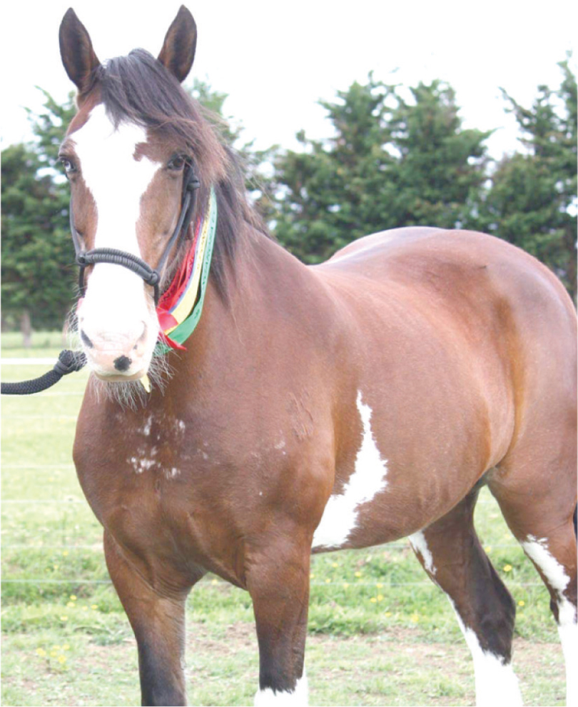
Case Study 1: Muriel
Muriel is a Clydesdale Cross, with very sensitive skin on the whole of her white face. Reacting in this area in particular, as it has been sun-damaged in the past — her delicate skin is affected by the sun, grass seeds, insect bites and mud.
In the past she has had an adverse reaction to many creams, so therefore requires something that doesn't cause her to have an allergic response — redness, inflammation, crusting and peeling of the top skin layers — similar to sunburn.
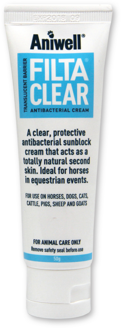
FiltaClear is tolerated well and keeps her sparsely-haired nose pink and soft all year round. Because FiltaClear rubs in to near clear it doesn't stand out when eventing or riding out. Aniwell produce a range of topical skincare products for all animals, specifically for sun, wind, rain and wound protection. Their product FiltaClear is a total sun-blocking, reflective, pale white cream with antibacterial qualities within the cream that rubs in to near clear. Filtaclear has an SPF of 25+ and rated superior protection for UVA/UVB.
FiltaClear can be applied daily to all areas requiring protection, with thorough washing of the applied area using warm water or a non-soapy cleanser every third day to prevent residue build up on the skin.
None of the ingredients in FiltaClear appear on the FEI Prohibited substances list 2018. www.aniwell-uk.com
Sun blocks
Topical sun screens may act physically or chemically. Chemical screens usually contain aminobenzoic acid or benzophenone derivatives. These compounds work by being absorbed into the epidermis to absorb UV rays. Although these actives are commonly used in people because they are clear, cosmetically acceptable lotions or gels, they are of little benefit if the skin is ulcerated when the epidermis has sloughed. Physical sunscreens include ingredients such as zinc oxide and titanium dioxide, which act to scatter light by forming an opaque barrier. These products are not as pleasant to use as the chemical screen but are often more persistent in animals and offer better protection if the skin is ulcerated. Photodecomposition can be a problem with some products, as they lose their efficacy and need to be applied 3–4 times daily. However, both zinc oxide and titanium dioxide products are more persistent and so do not need to be applied as frequently (Miller et al, 2013). Topical sun blocks are normally rated by a sun protection factor (SPF). In animals, water-resistant sun blocks with a high SPF are preferable. The recent introduction of a new sun block into the veterinary market promises to be beneficial in the management of sunlight induced disease (FiltaClear, Aniwell). This product can be used as both a preventative and a treatment in phototoxic disease. It provides total sunblock with an SPF of 24–27. FiltaClear contains titanium dioxide to provide a physical barrier against sun exposure and is compounded as a microfine formulation to make application easier. It leaves a fine white film on the skin after application, which acts as a guide as to when re-application is needed. As the white colour starts to fade it should be reapplied. FiltaClear also contains Bitrex, a bittering agent which prevents it being groomed from the animal's skin. In animals with sunburn it provides a soothing protective antibacterial barrier to help heal damaged skin and prevent further damage.
Conclusion
Animals suffer from sunlight-induced diseases; both phototoxic (sunburn) and photosensitising reactions are recognised in dogs, cats and horses. In an ideal situation sunlight exposure should be limited to prevent disease occurring. However, in circumstances where excessive sun exposure is unavoidable or where an animal is at high risk of developing disease due to their coat colour or life style, or where an animal has a previous history of sunlight-induced disease, topical sun protection should be used. The best sun protection should provide high levels of physical sun block, in excess of 25 SPF, and optimally should be in a coloured base to guide the owner when it needs to be reapplied. FiltaClear is a new topical sunscreen that provides these benefits.
Where animals develop phototoxic or photosensitising reaction, further exposure to sun should be strictly limited to prevent further damage. Topical sun blocks in these cases can help prevent any further exposure and can help act as a soothing, antibacterial barrier to promote healing.
Key Points
- Phototoxicity is mediated by UVB radiation and affects white or non-pigmented skin especially where there is little hair covering.
- Sunlight intensity is at its greatest in the middle of the day, from 11.00 am to 2.00 pm.
- Photosensitisation is an abnormal reaction to sunlight caused by the accumulation of photodynamic agents in the skin.
- Photosensitisation may be systemic (congenital or acquired) or contact (acquired).
- Topical sun screens may provide physical or chemical protection.
- Chemical sun screens usually contain aminobenzoic acid or benzophenone derivatives and work by being absorbed into the epidermis to absorb UV rays.
- Physical sunscreens include ingredients such as zinc oxide and titanium dioxide, which act to scatter light by forming an opaque barrier.