Electromagnetic radiation is composed of a continuous spectrum of wavelength of which ultraviolet (uv) light is of particular importance in precipitating or potentiating a number of skin lesions. This article focuses on the two main subdivisions of photodermatitis, namely phototoxicity and photosensitivity which are of primary concern to the veterinary surgeon. Pastern (and cannon) leukocytoclastic vasculitis is a specific disease of the equine skin that often occurs in the summer and has been associated with strong sunlight exposure (Stannard, 2000).
Phototoxicity
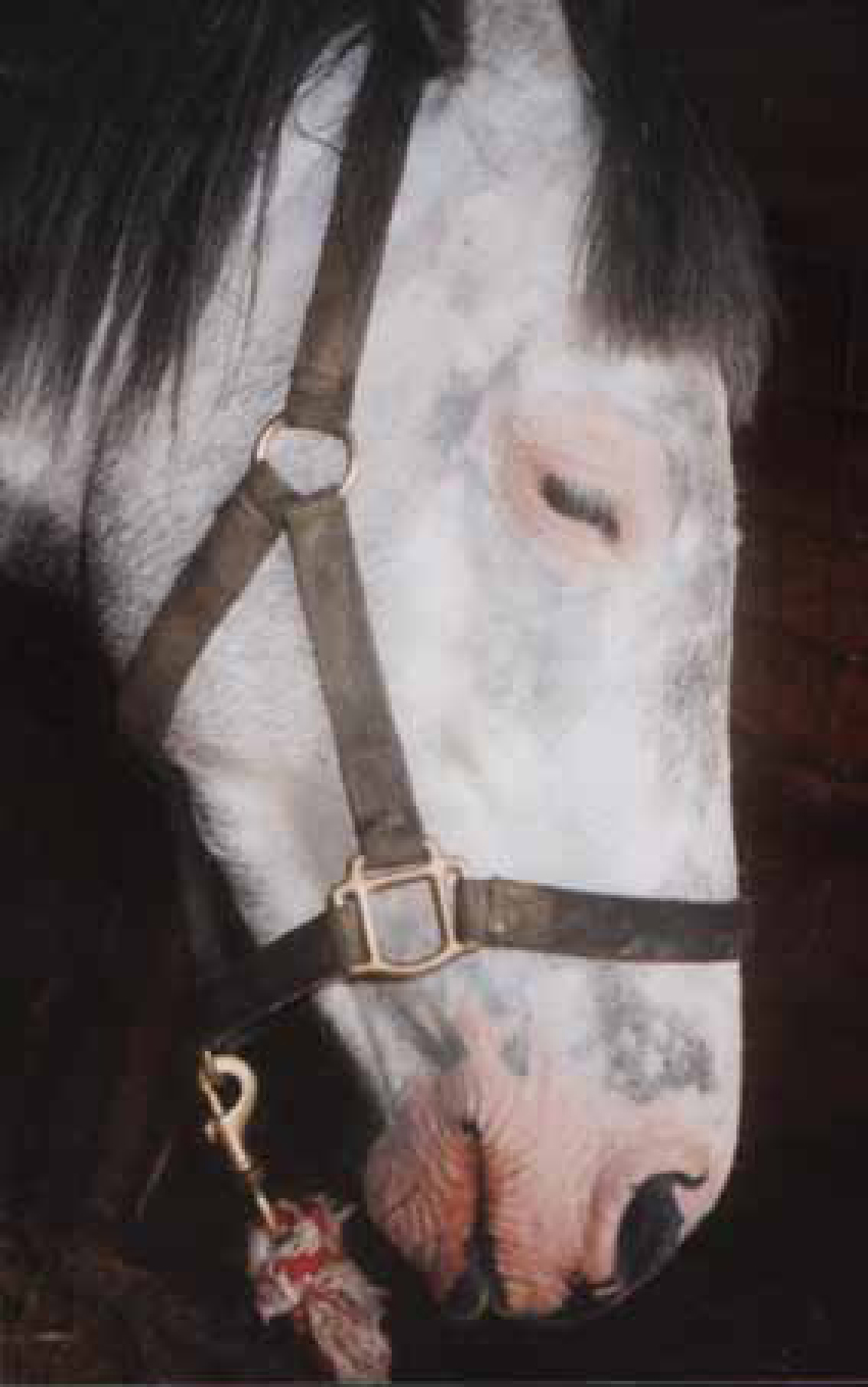
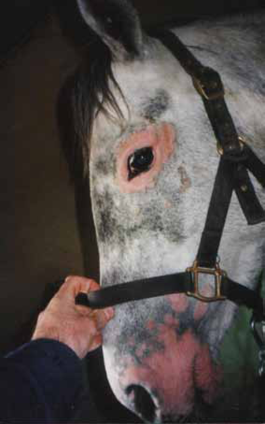
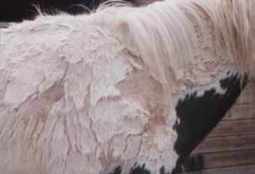
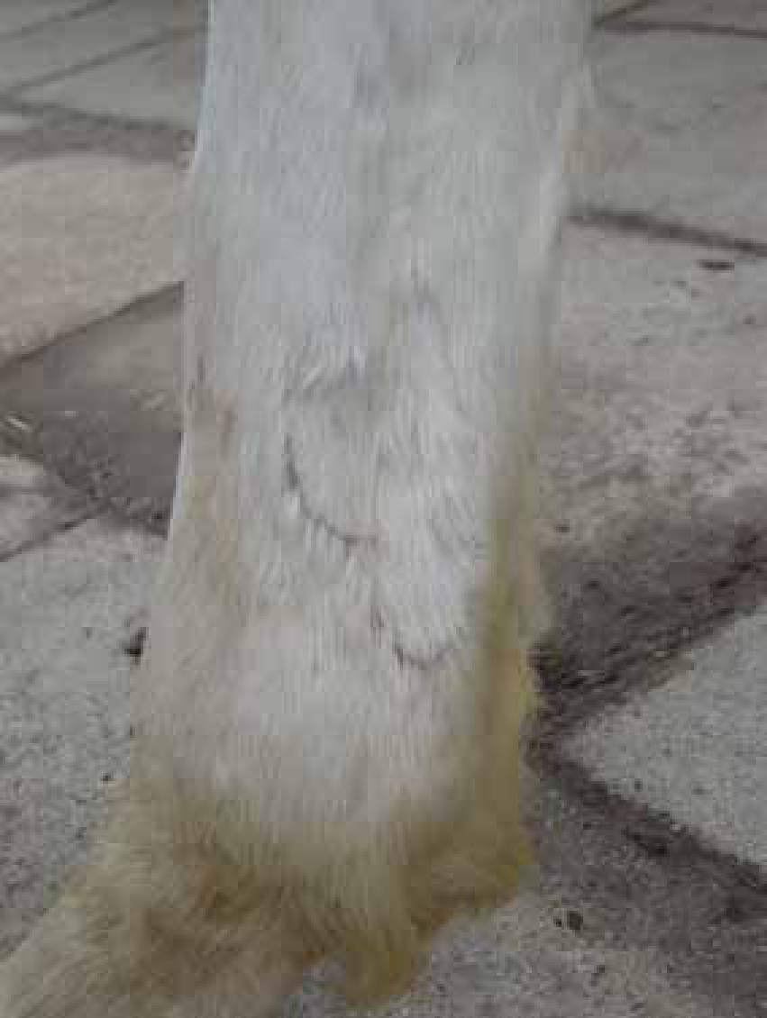
Phototoxicity is the classic sunburn reaction and is a dose-related response to light exposure. White skin, depigmented or scarred areas are most susceptible especially if there is poor hair covering (Figures 1 and 2). The pathogenesis is not fully understood but involves damage to the epidermis and blood vessels of the deep plexuses. In severe cases, secondary crusts may form and it may become very painful. Topical treatment with emollients and topical glucocorticoids is usually sufficient after removing from sunlight.
Management
There are a number of ways that phototoxicity can be managed:
Photosensitization
Photosensitivity is when the production, ingestion, injection or contact with a photodynamic agent leads to increased susceptibility to the damaging effects of uv light. Photosensitization can be classified according to the source of the photodynamic agent. Although rare, two types are mainly seen in practice.
Primary photosensitization
Photodynamic agents, such as hyperium contained in Hyperium perforatum (St Johns' wort), are ingested and reach the skin via the circulation. These agents absorb specific wavelengths of light, become activated and pass the extra energy to the surrounding cells which results in their damage (Stannard, 2000). The lesions can vary tremendously from urticarial reactions and areas of erythema to severe necrosis of the skin.
Hepatocutaneous photosensitization
In hepatocutaneous photosensitization phylloerythrin levels are elevated in the blood. The phylloerythrin is a breakdown product of chlorophyll that is formed in the hind gut of herbivores. Some phylloerythrin is absorbed by the hepatic portal system and excreted in the bile. When the liver function is damaged the phylloerythrin accumulates in the animal tissues. The pyrrolizidine alkaloids found in Senecio spp (ragwort) is the most commonly reported cause of liver damage. However other toxins and other causes of liver damage can result in the same mechanism (Scott and Miller, 2003).
Contact photosensitization
Most recognized cases of contact photosenstization have occurred in horses grazing paddocks rich in clover (Stannard, 2000).
Clinical signs
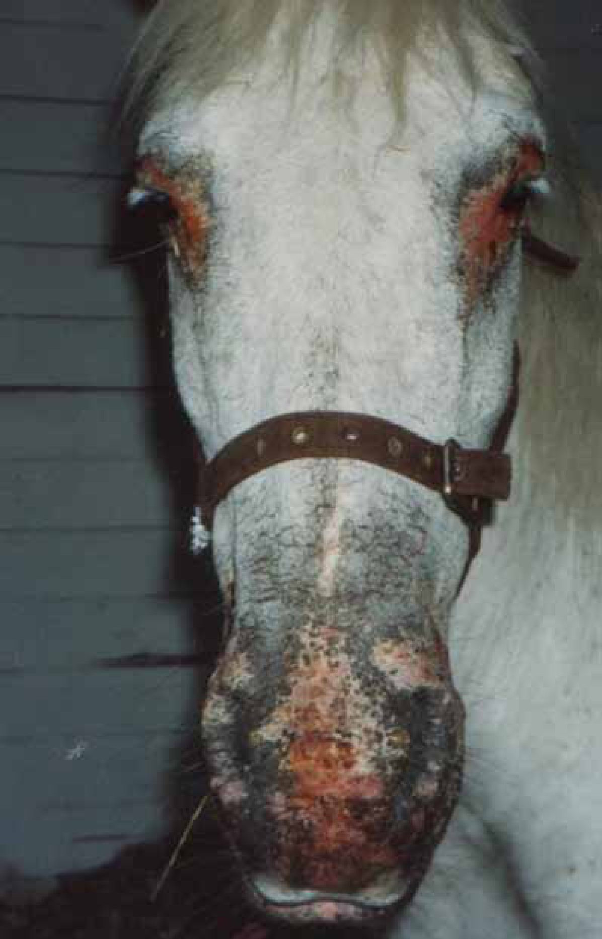
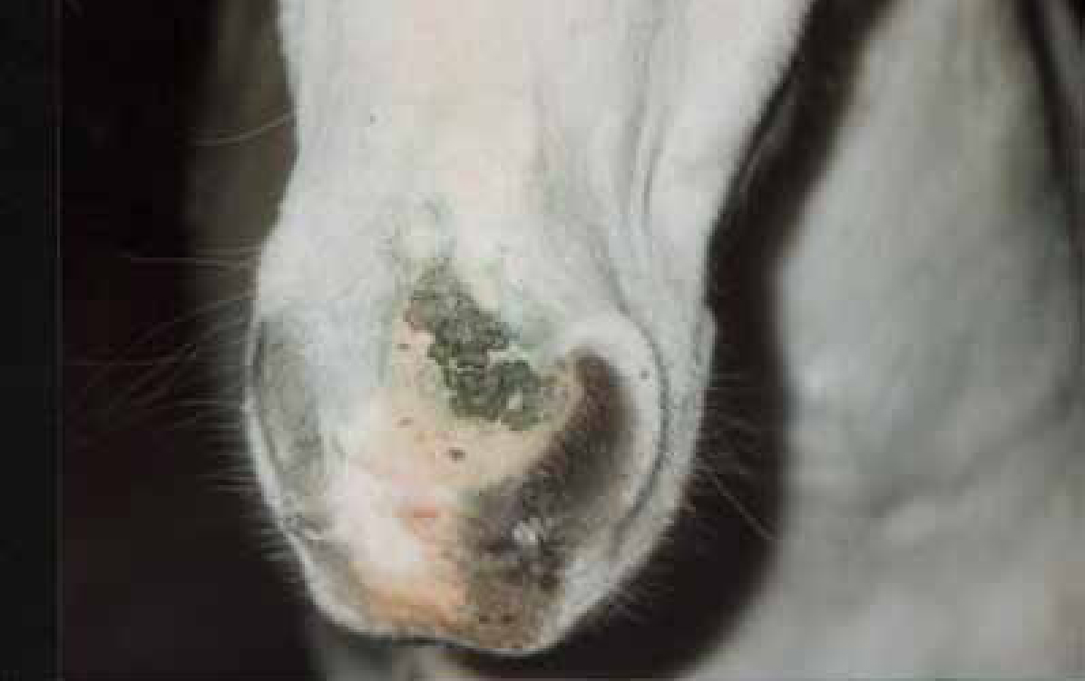
The most severe lesions resulting from photosensitization will involve non-pigmented skin that lacks hair, for example, around the eyes and muzzle. In severe cases, especially in hepatogenous photosensitization, lesions can be severe with erythema, oedema, ulcers, crusts and scaling. Secondary bacterial infections (Figures 3 and 4) are frequent and in chronic cases necrosis and sloughing may occur (Figure 5).
Diagnosis
Diagnosis is based on history including determining the number of animals at risk compared with the number of animals affected. If more than one is affected then the photodermatitis is photosensitive compared with being photoallergic when only one animal is affected (Scott and Miller, 2003). Clinically if the photodermatitis is confined to the distal limbs and/or muzzle then this is suggestive of a contact photosensitization (pasture plants (phytophotodermatitis), sprays, topical medications) or photoactivated vasculopathies. Laboratory findings should rule out the presence of liver disease. Histopathology on photosensitization cases is often unrewarding as they are rarely taken from the acute lesion (Scott and Miller, 2003).
Treatment
Treatment of horses with photosensitization involves:
Prognosis
The prognosis is good for primary photosensitization but poor when there is liver involvement.
Pastern leukocytoclastic vasculitis (photoactivated/photoaggravated vasculitis)
Photoactivated vasculitis is one of the causes of pastern dermatitis. It has no sex or breed predilection although those breeds with an increased proportion of non-pigmented extremities have increased frequency. The disease is more common in the summer with the exposure to abundant sunlight. Only one animal in a field may be affected and in contrast to photosensitization conditions, often only one limb is affected (Stannard, 2000).
Aetiology and pathogenesis
It is believed to be a vasculitis. However, the pathogenesis is not fully understood. The restriction of the lesions to the non-pigmented skin in most cases is suggestive of uv light involvement. However only one of the non-pigmented limbs may be affected and there is often no record of contact with a photosensitizing agent, which suggests that it is not a true photosensitizion. (Knottenbelt, 2009). Liver function is usually normal (Sloet van Oolruitenborgh-Oosterbaan et al, 2000).
Clinical signs
The medial and lateral aspects of the pasterns and fetlocks are the preferred sites. In the initial phases, erythema, oozing and secondary crust formation predominate. Superficial ulcerations and erosions may develop with extensive oedema in some cases. The lesions are painful and in the chronic phase the crusts are firmly attached often with a ‘leading edge’ which resists mechanical removal (Figure 6). Secondary infection with Staphylococcus spp, Dermatophilus congolensis, and Streptococcus spp is common.
Diagnosis
Diagnosis is based primarily on taking of an accurate history and the clinical appearance, usually on the lateral aspect of a non-pigmented pastern or cannon. Microscopic examination of the crusts should be undertaken to identify the presence of bacteria or dermatophilus, and swabs for culture may be necessary if the condition has been prolonged or is non responsive.
Biopsy in the early lesions shows a leukocytoclastic vasculitis with vessel wall necrosis and thrombosis. For differential diagnosis see Table 1.
| Primary dermatophilosis |
| Pastern folliculitis |
| Dermatophytosis |
| Photosensitization including hepatogenous photosensitization |
| Pemphigus foliaceus |
Clinical management
Management of photoactivated vasculitis involves reducing further exposure to sunlight by stabling in daylight hours or the use of leg wraps. Systemic glucocorticoids (prednisolone 1mg/kg q 12h with tapering doses) may also be useful (White, 1998), although the use of steroids at relatively high doses and the potential risk of laminitis must be evaluated (Bailey and Elliot, 2007). Topical treatment involves:
Prognosis
The long-term prognosis is good but recurrence is possible in the same or subsequent years (Knottenbelt, 2009).
Conclusion
Exposure to uv light is greatest in the summer months and is an important factor in potentiating or precipitating a number of skin diseases in the horse. Its pathogenic role in other skin conditions such as pastern and cannon leukocytoclastic vasculitis is uncertain at present but it is known that uv light has local and systemic immunologic consequences.