
An electrocardiograph (ECG) has two main purposes: to record the heart rate and the heart rhythm. An ECG can assist the person who is monitoring the patient in many situations, for example during anaesthesia, help assess emergencies, to monitor post-surgical cases, or if an arrhythmia is suspected. Furthermore, continuous monitoring systems such as telemetry, allow monitoring from a distance, so a patient can be observed without disturbance. Anecdotally, nurses want to use ECGs but are limited by a lack of understanding, frequently worried whether what they are looking at on screen is serious or not. This can distract nurses to the point where the ECG is ignored, or even not attached in the first place. This article aims to make interpretation simpler, help nurses become more confident in using ECGs, and provide a quick and simple guide to help the nurse know when to be concerned and seek advice.
The ECG trace
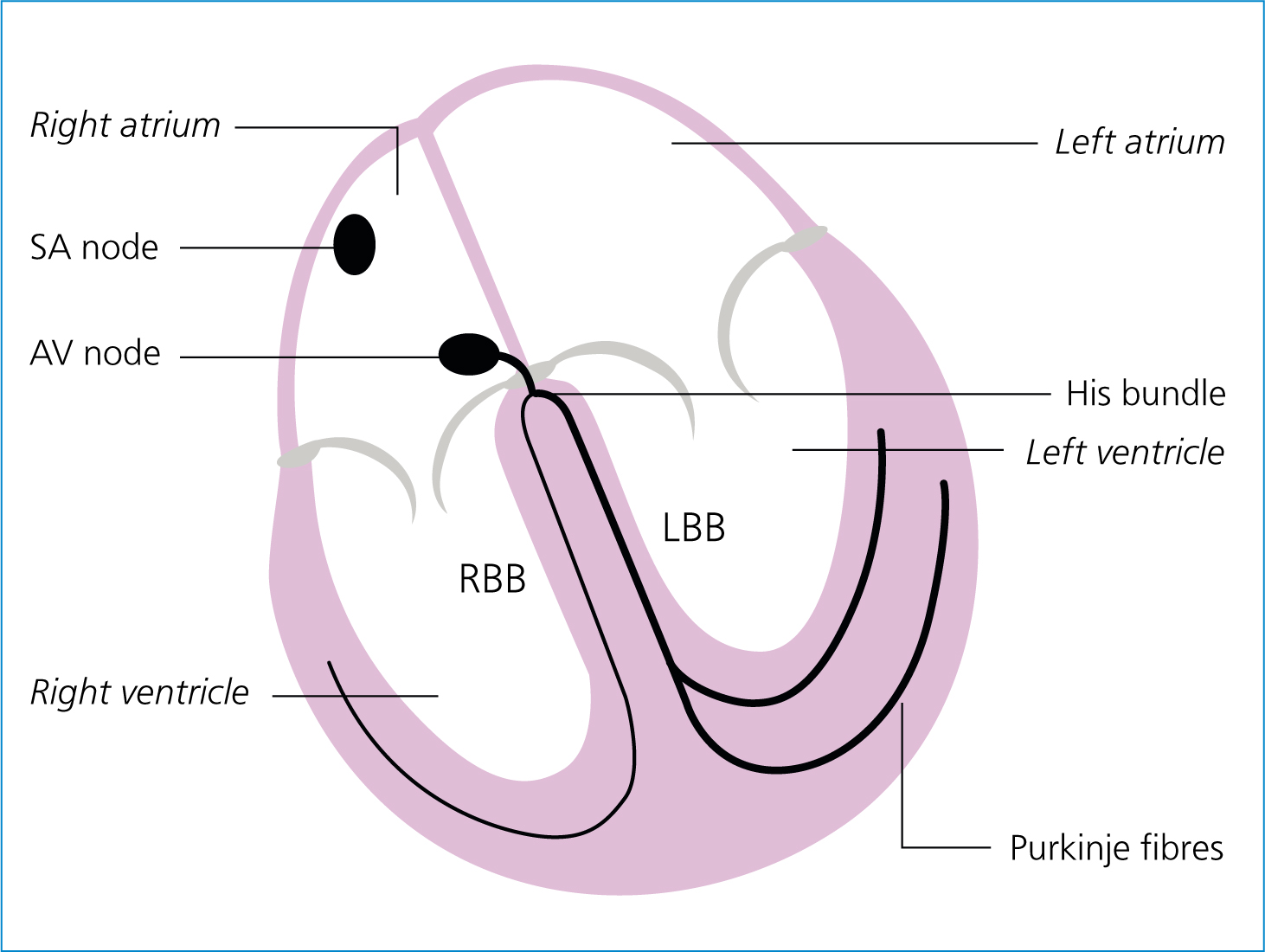
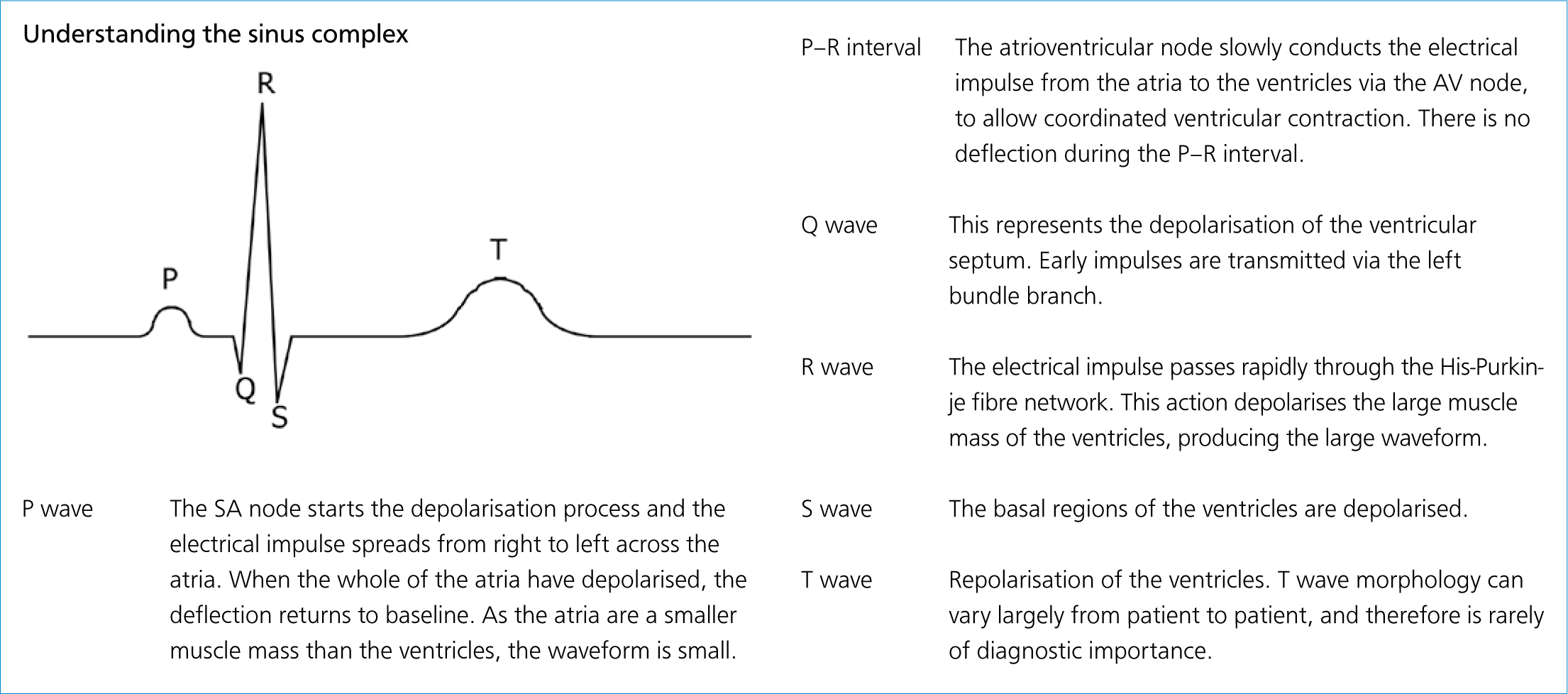
The ECG trace is based on a conduction system within the heart (shown in Figure 1). An electrical impulse is generated within the sinoatrial (SA) node, and this impulse travels across the atria depolarising the myocardium as it goes, and arrives at the atrioventricular (AV) node. The impulse then transmits via the right and left His bundle, and down through the Purkinje fibre network and depolarises the ventricular myocardium. This electrical activity can be recorded on an electrocardiogram. When the conduction system works correctly, it produces a normal or sinus complex, consisting of a P wave, a QRS complex and a T wave (Figure 2).
Recording a good quality ECG
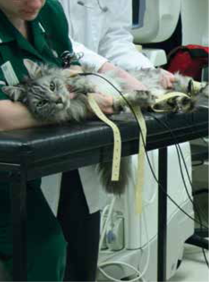
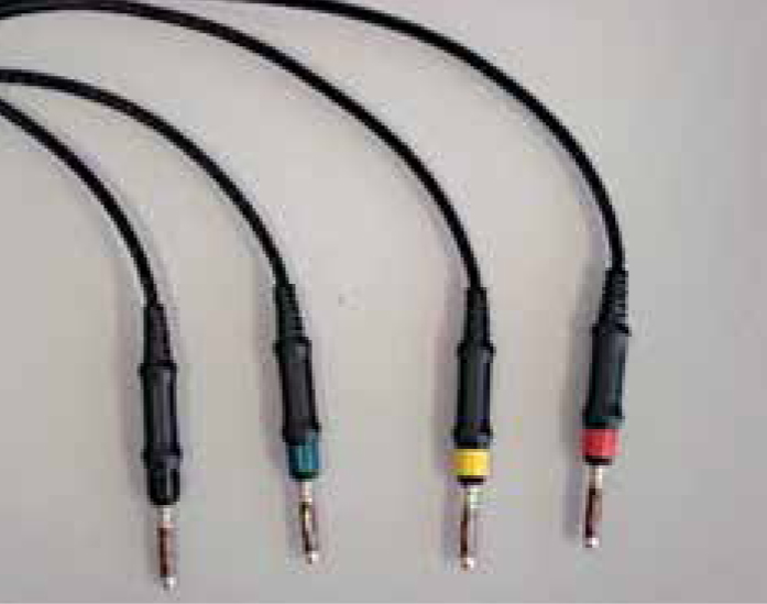
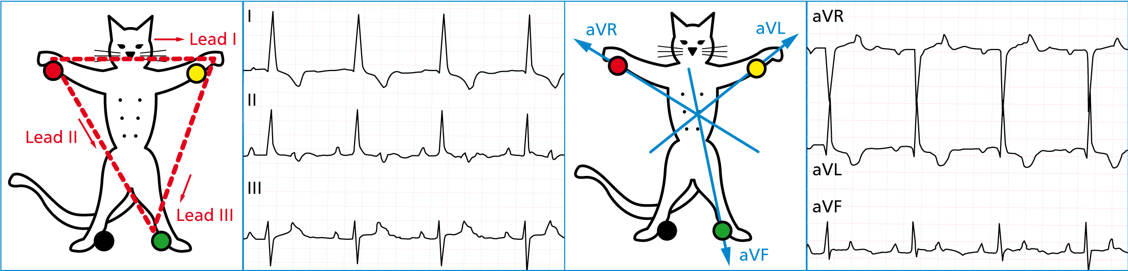
As far as possible, the patient should be calm and relaxed. Gold standard positioning for an ECG recording is right lateral recumbency with the patient lying on a vetbed or non-conductive mat (Figures 3 and 4). However, for cats it is thought that body positioning may be less important, and patient compliance to be improved with a more relaxed position (Willis, 2010). If a patient is dyspnoeic, an ECG should be taken in sternal recumbency and done with minimal stress. Standard electrode configuration on ECG machines consists of four cables and electrodes, which includes one earth electrode (Figure 5). These electrodes translate to a six lead ECG trace, with leads I–III, and aVR, aVF and aVL (Figure 6). Borgeat and Knowlson (2016: p163) describe the different leads as each having a ‘different point of view on the heart’. However, for ease of use, lead II is generally used for interpretation. The additional leads allow for further investigation, into things such as conduction abnormalities or cardiac chamber enlargement.

Electrode placement for British and American machines is shown in Table 1. Electrodes can be placed anywhere on the limbs, although it has been reported that when electrodes are attached to footpads, R wave amplitude (size) is reduced (Ferasin, 2006).
| Electrode | Colour for British machines | Colour for American machines |
|---|---|---|
| Right forelimb | Red | White |
| Left forelimb | Yellow | Black |
| Left hindlimb | Green | Red |
| Right hindlimb | Black | Green |
Adapted from Willis (2010: p69)
Movement artefact, such as panting or muscle tremor should be minimised by untangling cables and placing them so that they do not cross the thorax. Either conduction gel or surgical spirit can be applied to the electrodes, but remember surgical spirit is flammable if a defibrillator might be needed. Avoid electrical interference, such as clippers or fans, as these will cause artefact (Figure 7) and make interpretation difficult.

Machine settings
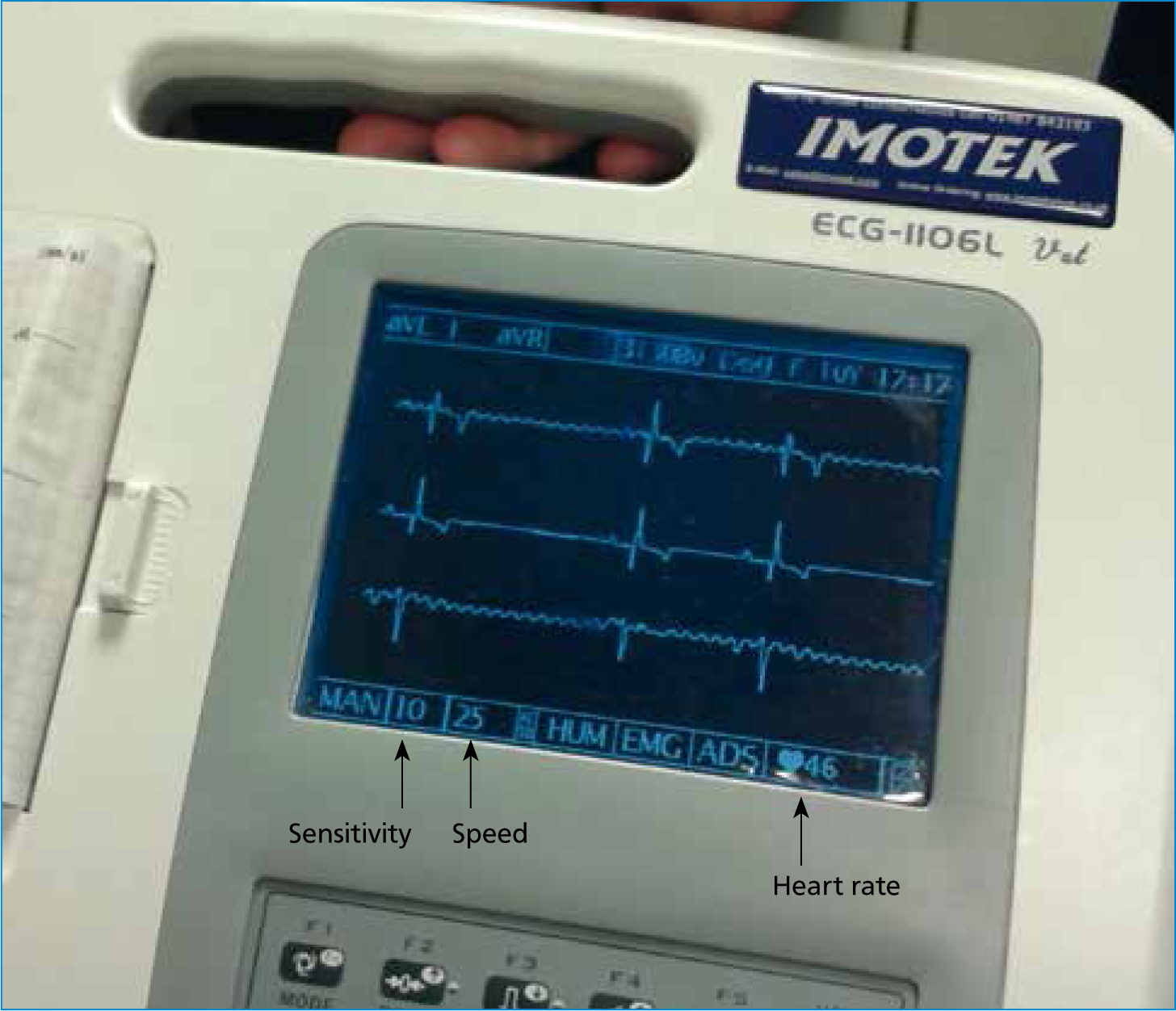
Recommended settings suggest that the filter is turned off, paper speed set at 25 mm/second, and the gain or sensitivity (which adjusts the size of the complexes), is set to optimise the recording, so that the complexes are big enough to be identified, but not so large as to overlap other leads (see Figure 8). If the heart rate is fast, the veterinary surgeon may ask for the paper speed to be increased to 50 mm/second, as this spreads the complexes wider, allowing for easier interpretation.
Interpretation
Once a good quality trace has been recorded, it needs to be understood. A series of five questions will help give a systematic approach to interpretation. For ease of interpretation lead II is always used.
Question 1: What is the heart rate?
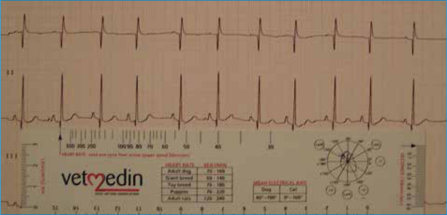
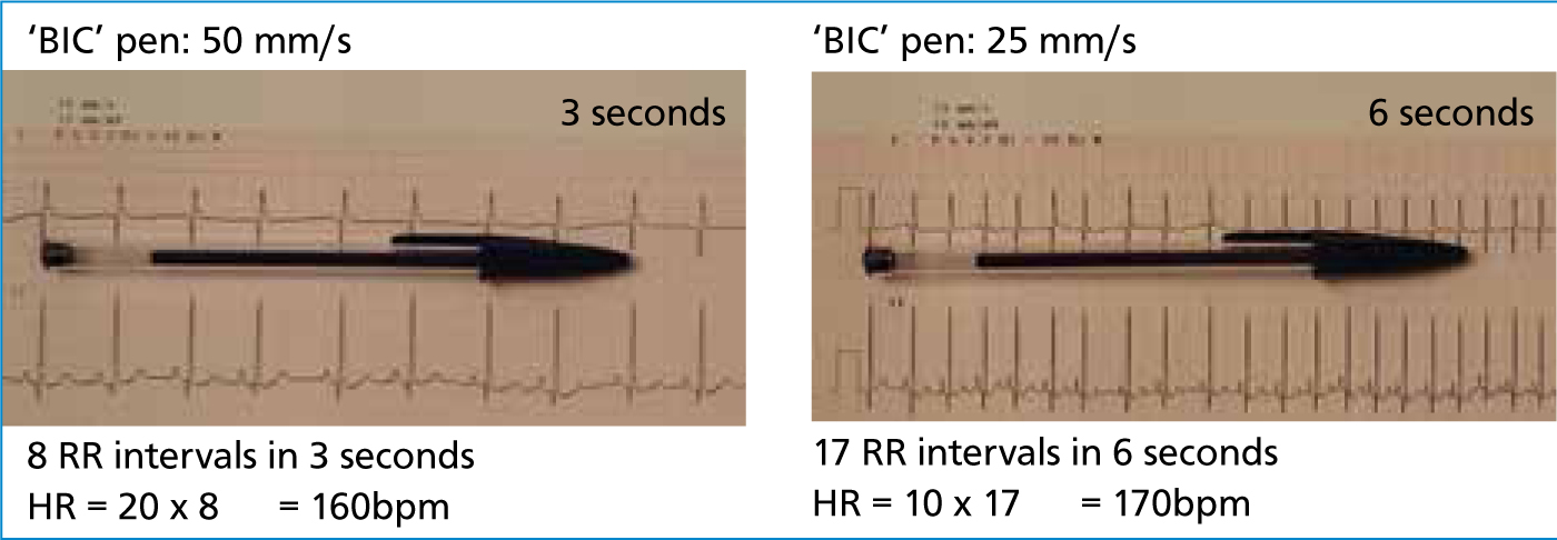
Determining whether the heart rate is fast, slow or normal is the best way to start working out whether there is a problem or not. Some machines will provide the heart rate on screen, but in some cases, this might need to be done manually, or need to be double-checked. This can be done with an ECG ruler or a BIC pen, with the lid on (Figures 9 and 10). Table 2 shows a quick glance guide to arrhythmias associated with different heart rates. It should be remembered that the heart needs time to fill, pump blood out through the ventricles, and then have enough time to refill, so it can repeat the process. If the heart rate is too fast or too slow to maintain sufficient blood pressure, cardiac output may be compromised.
| Tachycardia | Normal | Bradycardia |
|---|---|---|
| Sinus tachycardia | Normal sinus rhythm | Sinus bradycardia |
| Atrial fibrillation/flutter | Sinus arrhythmia | Atrioventricular (AV) block |
| Supraventricular tachycardia | Atrial fibrillation | Sinoatrial block/arrest |
| Ventricular tachycardia | Atrioventricular block | Escape rhythms |
| Premature complexes (atrial or ventricular) | Premature complexes (atrial or ventricular) | Atrial standstill |
Questions 2–4: The P-QRS relationship
Question 2: Is there a P wave for every QRS complex?
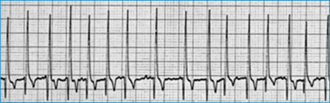
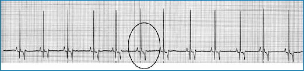
As previously discussed, each sinus complex should have a P wave and an associated QRS complex. Again, there are easy interpretation guides which can be used in conjunction with the heart rate table to narrow down possible answers. ECG traces 1 and 2 demonstrate arrhythmias with a lack of discernible P waves.
| Is there a P for every QRS? | |
| Yes | No |
| Are they consistently and reasonably related? |
Atrial fibrillation |
| Atrial standstill | |
| Ventricular tachycardia | |
| Premature complexes (atrial or ventricular) | |
| Escape complexes | |
Question 3: Is there a QRS complex for every P wave?
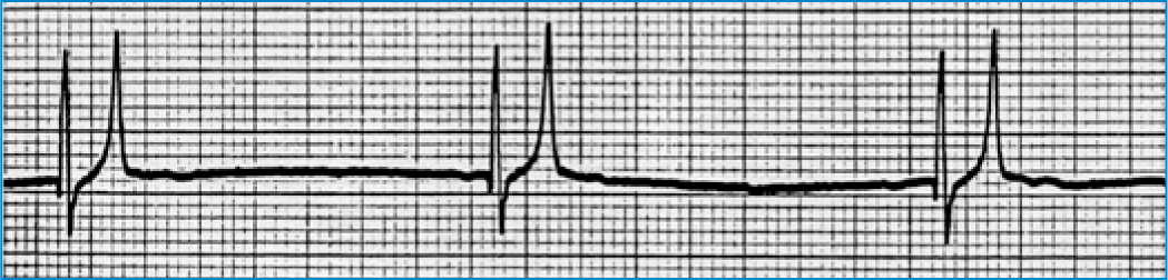
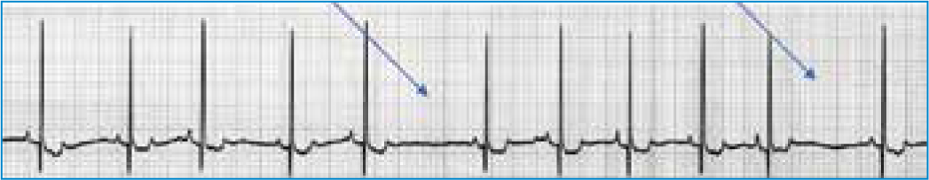
Equally, every P wave should be followed by a QRS complex. ECGs 3 and 4 illustrate when P waves are present, but there is no corresponding QRS wave.
| Is there a QRS for every P? | |
| Yes | No |
| Are they consistently and reasonably related? |
2nd degree AV block |
| 3rd degree AV block | |
| Atrial flutter | |
Question 4: Are they consistently and reasonably related?
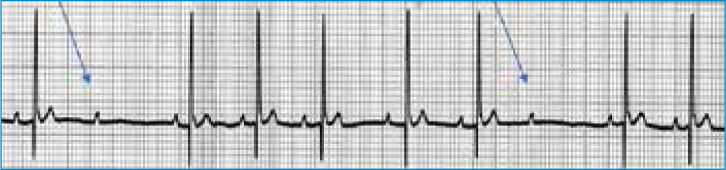
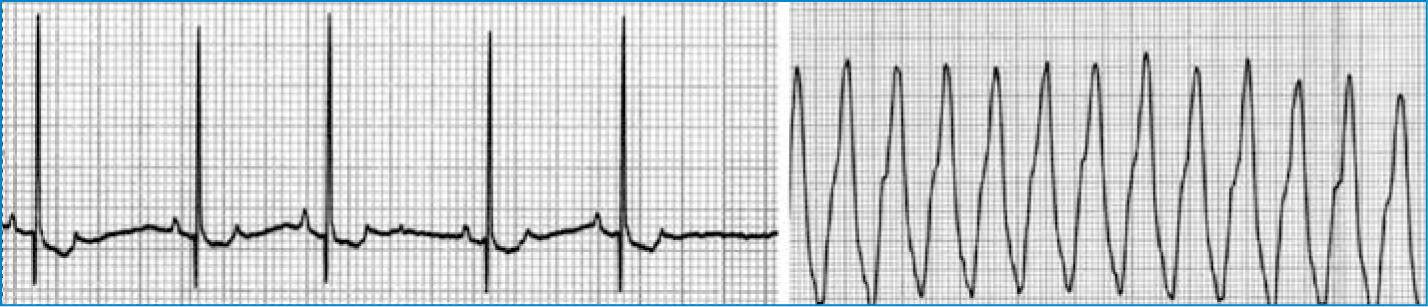
The only examples of where P-QRS complexes may be seen but not related are listed below. ECG 4 shows 3rd degree AV block, which has a characteristically slow heart rate. ECG 5 shows an example of ventricular tachycardia, which as the name suggests, is fast. Examples are also provided of a sinus rhythm (ECG 6) and sinus arrhythmia (ECG 7), where the P-QRS complexes are consistently and reasonably related.

| Are they consistently and reasonably related? | |
| Yes | No |
| Sinus rhythm | 3rd degree AV block |
| Sinus arrhythmia | Ventricular tachycardia |
| Sinus bradycardia | |
| Supraventricular tachycardia | |
| Atrial premature complexes | |
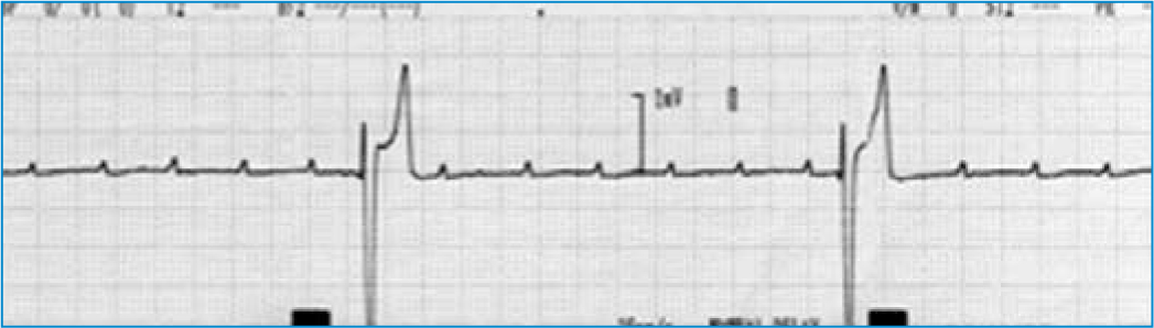
Question 5: What is the QRS morphology? Is it tall and upright or wide and bizarre?
If the electrical impulse has originated within the atria, the QRS is usually tall and narrow. If the impulse has originated in the ventricles, the QRS complex will generally be wide and bizarre. This is because the impulse has not travelled through the normal conduction system, but instead has had to depolarise the myocardium cell by cell, which takes longer, making the complex wider.
Putting theory into practice
Questions 1–4 provide a rule out process, but interpretation can take time until it becomes second nature. Furthermore, while trying to interpret the ECG, the nurse needs to be monitoring the patient, and this is where putting into practice ECG theory, can become challenging. However, if the patient is conscious, clinical signs can give clues. The primary role of the heart is to provide cardiac output. This cardiac output maintains blood pressure. Therefore if the heart rate is too fast or too slow, cardiac output is compromised. If this happens, the patient will display clinical signs such as collapse, weakness, poor or absent pulses, cold extremities, and pallor. If this happens, immediate veterinary attention is required. An ECG attached at this time will allow for a diagnosis by the veterinary surgeon, influence drug therapy and monitor response to treatment. Patients that are at high risk of life threatening arrhythmias are those in critical conditions, those with heart failure and those under anaesthesia. If the patient is anaesthised, an ECG will help the nurse monitor the patient. Things such as AV block or sinus arrhythmia can be seen, and adjustments made to anaesthesia depth as necessary. Patients that present an arrhythmia risk for anaesthesia are listed in Box 1. Below is a list of the some of the more common causes of arrhythmias seen in practice:
If these patients are attached to an ECG, either to the traditional machine or using a telemetry system, the nurse can recognise and identify problems quickly, seek assistance earlier, therefore enhancing patient care.
Conclusion
ECG monitoring can only benefit the patient if the veterinary nurse is comfortable using the ECG machine, understands its settings and is able to troubleshoot common problems. However, used repeatedly, it can become a valuable part of monitoring patients during surgery and after surgery, and help keep close attention to those who could be at risk of developing life threatening arrhythmias. Only until in those situations, will nurses know how to react appropriately. A lot of arrhythmias will arise from pain, stress and the underlying disease process, but only with practice will the nurse know when to alert the veterinary surgeon. One point that should always help the nurse is to consider what is happening to the patient at that moment, and consider whether cardiac output is being compromised, which requires immediate attention, or whether pain or stress are contributing factors, and nursing can be adapted to accommodate.