
Auscultation is a cheap and easy diagnostic tool available in every veterinary practice. Yet if we ask ourselves if it is used to its full potential, the answer is probably no. While it seems like an easy enough task to perform, it is frequently over complicated. Two things hinder auscultation technique. First, optimal listening conditions are rarely achieved; and second, cardiologists agree that recognising abnormal heart sounds, and gaining proficiency in the technique is difficult (Pedersen et al, 1999; Naylor et al, 2001; Ware, 2007).
Primarily, auscultation of the heart is used to record heart rate and rhythm, and to determine heart sounds. As with most things in life, practice is key, so that abnormal sounds can be readily distinguished from normal heart sounds. If abnormal sounds are heard, it is recommended to determine the timing of the additional sounds, the point of maximal intensity (PMI) — or where the sound is heard the loudest — and finally, the intensity or loudness of the sounds in relation to a grading system.
Equipment
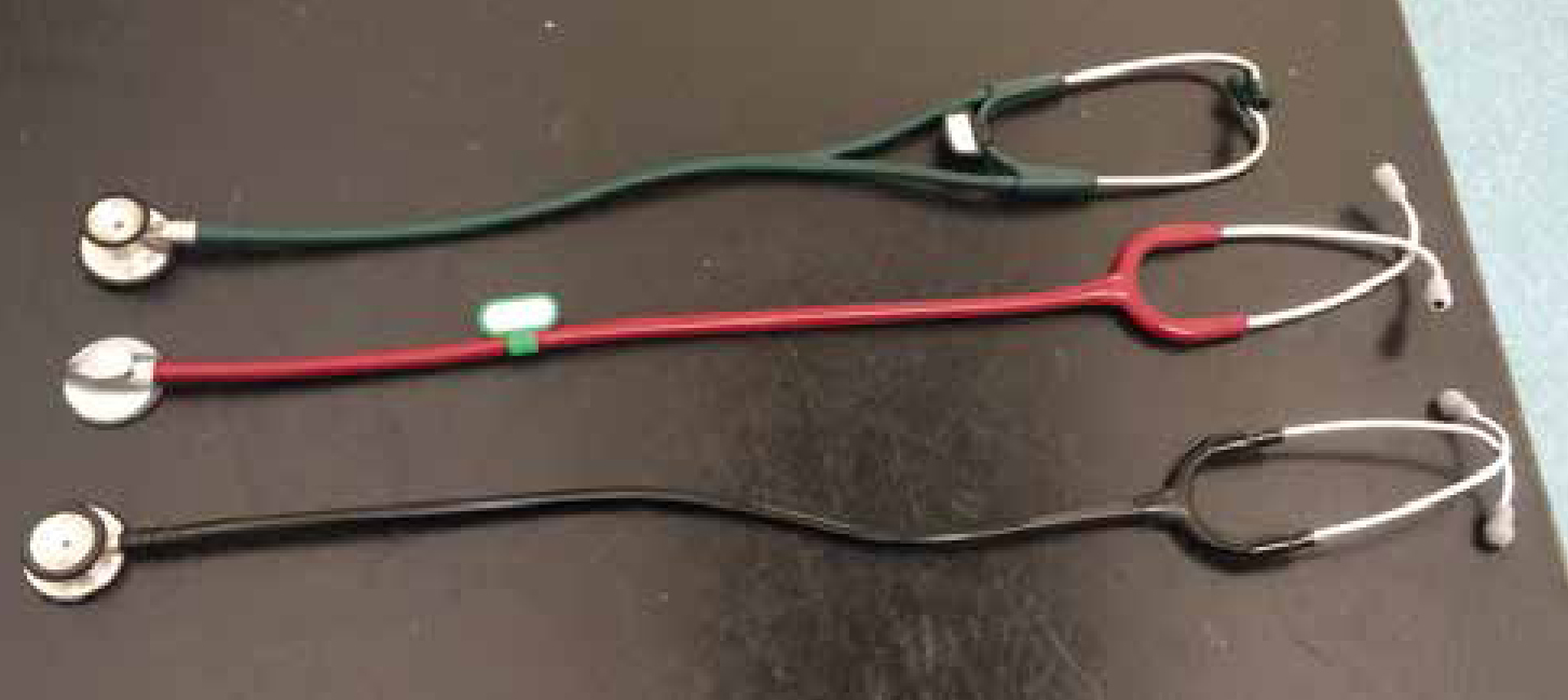
There are a few different types of stethoscopes available on the market (Figure 1). The better quality stethoscopes are adequate for veterinary use. Paediatric stethoscopes are not recommended for most auscultation because a larger bell is needed to maximise amplification of heart sounds. The main components are ear pieces, tubing, bell, and diaphragm (Figure 2).
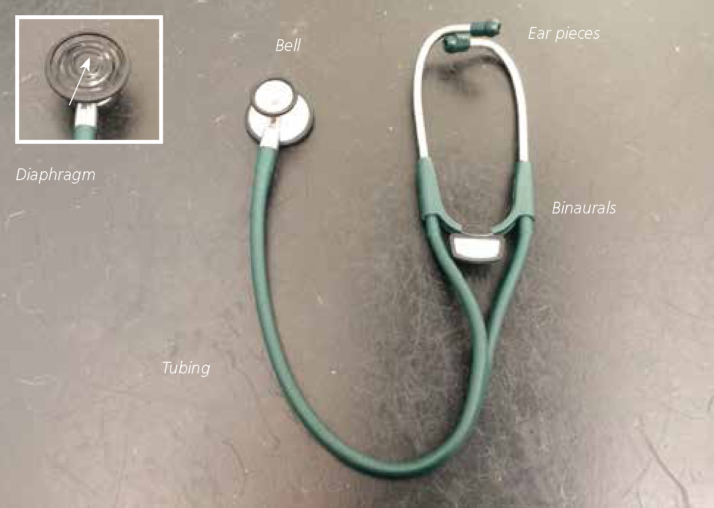
The binaurals should face forward (Figure 3) and be placed snugly into the ears. It may be necessary to move the tubing to better fit the ear canal and minimise sound leakage. Sound leakage and/or environmental sounds are a common problem when attempting to detect lower heart sounds or identify heart murmurs. The diaphragm is used to hear high-pitched sounds in the lungs and heart. It is best for heart sounds one and two (S1 and S2). The bell is used for lower-pitched heart sounds and extra heart sounds (S3 and S4). Some stethoscopes do not have a separate bell and diaphragm, and so fingertip pressure should be used to distinguish high and low pitched sounds.

Environment
Consider where the auscultation is to be performed. Ideally, a quiet room and gentle handling is recommended, so as not to excite or distress the patient. Panting dogs can be a problem; gentle closing of the mouth can allow a few seconds of undisturbed auscultation. Sometimes cats may purr, which can make auscultation impossible. Holding a finger across the nose may stop purring for a moment, or turning on a tap near to the cat may provide some distraction. If the owner happens to be present during auscultation, it may need to be explained that silence is required for optimal auscultation. Owners quite often like to talk during quiet periods.
Technique
First, it is important to know what is expected to be heard. There are two normal heart sounds in the dog and cat:
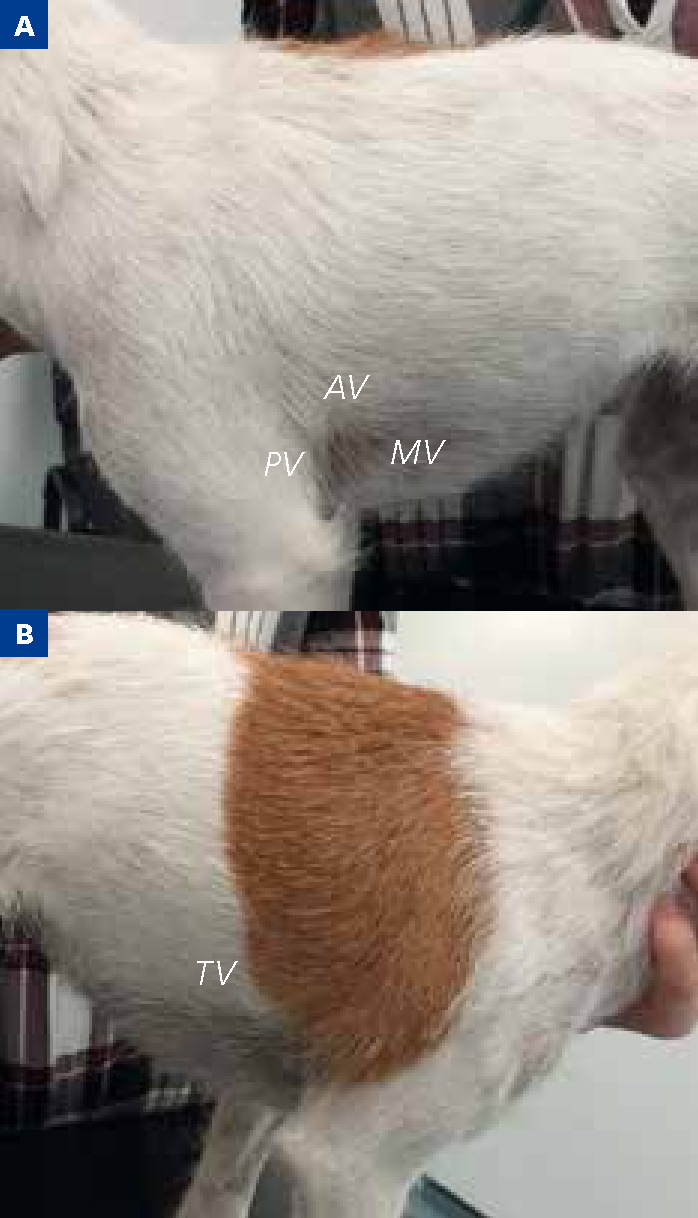
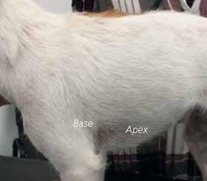
S1 is a louder, longer, and duller sound than S2. It is best heard in thin and young animals, or those with high sympathetic tone, and those with tachycardia, mitral regurgitation, systemic hypertension, or anaemia. S2 is a shorter higher pitched sound, which is loudest over the pulmonic and aortic areas. Any other sounds are described as additional heart sounds. These extra sounds are described by location (the PMI), timing in the cardiac cycle, and intensity of the sound (loudness). Table 1 outlines the PMI in dogs and cats.
| Structure | Location |
|---|---|
| Mitral valve (left apex) | Dog — left side, 5th intercostal space at costochondral junction. |
| Aortic valve (left base) | Dog — left side, 4th intercostal space, just above costochondral junction |
| Pulmonic valve (left base) | Dog — left side, 2nd–4th intercostal space, just above sternum. |
| Tricuspid valve (right apex) | Dog — right side, 3rd–5th intercostal space near costochondral junction |
Timing of additional heart sounds is important. Sounds heard between S1 and S2 occur during systole. This is the most common type of heart murmur recorded in small animals. Sounds heard between S2 and S1 are diastolic. Diastolic murmurs are rare in small-animal medicine. Murmurs throughout systole and diastole are called continuous murmurs. The most common cause of a continuous murmur is a patent ductus arteriosus. Distinguishing when additional heart sounds occur, will help the veterinary surgeon to focus their differential diagnosis. An approach, such as the one described below, should be followed to reliably perform auscultation:

| Intensity | Grade | Loudness |
|---|---|---|
| Low intensity | I | Low intensity murmur heard in a quiet environment only after careful auscultation over a localised cardiac area |
| II | Low intensity murmur heard immediately when the stethoscope is placed over the PMI | |
| Moderate intensity | III | Murmur of moderate intensity |
| IV | High intensity murmur that can be auscultated over several areas without any palpable precordial thrill | |
| High intensity | V | High intensity murmur with a palpable precordial thrill |
| VI | High intensity murmur with a palpable precordial thrill that may even be heard when the stethoscope is slightly lifted off the chest wall |
Grading
The loudness of a heart murmur is graded between I and VI (Table 2). Grading can be useful to assess disease progression and/or severity of certain cardiac conditions, such as aortic or pulmonic stenosis or mitral regurgitation. However, it is less useful to assess myocardial disease severity. Furthermore, ventricular septal defects will give a louder sound when the hole is smaller because of the higher pressure across the hole. Therefore, grade does not always indicate disease severity.
It is also important to remember that veterinary professionals will not always agree on the presence or loudness of a murmur. Pedersen et al (1999) conducted a study evaluating different people and their auscultation. The study used dogs diagnosed with mitral valve disease, and they found that interobserver agreement on presence and absence of mildly-diseased dogs varied between 63–88%. In a similar study, Höglund et al (2004) showed high interobserver variance in dogs with aortic stenosis. Another study looking at the comparison of cat cardiac auscultation, showed only moderate interobserver agreement (Wagner et al, 2010).
What is a murmur?
A murmur can be best described as an abnormal heart sound of prolonged duration (Ware, 2007). Blood flow normally passes through blood vessels and the heart with minimal turbulence, which does not cause a murmur. A murmur arises from a vibration of structures within the heart, created by high velocity, abnormal blood flow, turbulence or reduced blood viscosity. Heart murmurs can occur without the presence of heart disease. Innocent or physiological murmurs can be quite normal for puppies and kittens, and disappear by the age of 6 months. These murmurs are usually left sided and between grades I–III/VI. Innocent murmurs can still be present in adult cats and dogs, but if no disease is suspected, no treatment is required.
Also, in cases of severe anaemia a heart murmur can be caused by low viscosity of the blood. When the anaemia is corrected, the murmur can disappear. Heart murmurs caused by heart disease are seen when there is an obstruction to blood flow, seen with aortic stenosis, or valvular incompetence such as mitral valve disease. For a summary of heart murmur causes see Table 3.
| Heart disease | Flow murmurs | Other causes |
|---|---|---|
| Leaking valves (e.g. mitral valve disease) | Innocent murmurs | Anaemia |
| Stenotic valves (e.g. pulmonic stenosis) | Physiological murmurs | Hyperthyroidism |
| Holes in the heart (e.g. ventricular septal defects) |
Other heart sounds
Occasionally, it is possible to hear other heart sounds. These sounds, either an S3 or S4, are known as gallop sounds. They are lower pitched than the S2 sound and, when heard, can sound like a galloping horse. They are both heard in diastole; distinguishing one from the other is very difficult, and requires some experience and practice. An S3 sound occurs because of ventricular dilation and myocardial failure. It can sometimes be heard in dilated cardiomyopathy, or advanced valvular heart disease. An S4 sound is associated with atrial contraction in dogs and cats that have abnormal ventricular relaxation and stiffness. In cats, it can be heard with advanced hyperthyroidism or hypertrophic cardiomyopathy. Sometimes, it can be a transient finding in older, stressed, or anaemic cats.
When a heart murmur is detected in a cat or dog
As already discussed, a heart murmur does not always signify heart disease. For example, a 2010 study in healthy cats, showed that 34% of apparently healthy cats had heart murmurs (Wagner et al, 2010). The same study also showed that of those recorded as having a murmur, only half had heart disease on echocardiographic examination. Furthermore, of those that did not have a heart murmur, 16% did have heart disease when echocardiography was performed.
Payne et al (2010) even showed that a heart murmur was a good prognostic indicator in cats. However, in dogs with mitral valve disease, heart murmurs can be an indicator of disease severity; for example, the more degeneration of the mitral valve, the louder the murmur. However, a study published in 2015 showed that only 46% of dogs with mitral valve disease died as a result of their disease (Lopez-Alvarez et al, 2015). They also showed that a murmur of grade III/VI or above was associated with a higher risk of mortality.
Conclusion
As with many things in life, practice makes perfect in the case of carrying out auscultation. Using a quiet room with a standing, calm and complicit animal is recommended. Using a systematic approach is helpful, starting with heart rate and rhythm, then recognising normal heart sounds, moving from left to right side across the thorax, focusing auscultation over the PMI areas.
If a murmur or additional heart sounds are auscultated, location, timing and intensity should be noted, recorded on the patient file, and highlighted to the veterinary surgeon. It is to be expected that there will be interobserver differences, but taking a calm and systematic approach will help reliability. A heart murmur can be an indicator of heart disease, but does not necessarily mean a death sentence.