
This article reviews the use of v-gel® anaesthetics masks (Docsinnovent®). The v-gel® is a supraglottic airway device that offers an alternative to endotracheal intubation.
Endotracheal tubes were first used in 1880 by William Macewen (Brandt, 1986). Since then endotracheal tubes have been used routinely in the human and veterinary fields. These are generally viewed as the standard technique for administering oxygen, nitrous oxide and volatile anaesthetic agents to patients under general anaesthesia or sedation. Endotracheal tubes are available in a variety of materials, e.g. red rubber, siliconized rubber or various types of plastic, e.g. PVC (Dugdale, 2010).
The use of endotracheal tubes in feline patients is a subject of great debate. The trachea of the cat comprises a cartilaginous and membranous tube that functions as an airway between the larynx and the bronchi. The trachea is composed of a series of hyaline cartilage rings which are joined by connective tissue and the trachealis muscle that runs along the complete length of the trachea. These incomplete rings and the trachealis muscle are beneficial because they give the trachea a great deal of flexibility and elasticity (Adshead, 2011). However, this area may be damaged during excessive cuff inflation, resulting in a longitudinal tear down the join between the trachealis muscle and the tracheal rings (Kästner et al, 2004).
In rabbits endotracheal intubation can be equally if not more difficult. Rabbits have a narrow gape, long incisors and a fleshy tongue, all of which can make the visualisation of the larynx difficult. Rabbits also have a relatively small glottis and, as with cats, laryngospasm can occur during endotracheal intubation, which may influence the selection of induction agent, and whether endotracheal intubation is attempted or not. Endotracheal intubation should be attempted in a gentle manner in rabbits, and cats, to avoid excessive laryngeal trauma (Dugdale, 2010).
Alternatives to endotracheal tubes
Supraglottic airway devices (SGADs) were developed for use in human anaesthesia to offer a simple and effective alternative to endotracheal intubation. SGADs are defined as devices that ventilate patients by delivering anaesthetic gases/oxygen to a patient above the level of the vocal cords (Vaida, 2004).
SGADs provide an alternative to endotracheal tubes as they do not sit within the airway, instead they function by protecting and ‘isolating’ the glottis. SGADs provide an alternative to endotracheal intubation in patients where intubation may be deemed as high risk, or difficult to achieve successfully. In human anaesthesia there are various types of SGADs, for example the i-gel® (see below) and the laryngeal mask (LMA). LMAs have a soft inflatable cushion which sits on or around the larynx with the idea of securing the airway. One potential disadvantage of using standard ‘human’ LMAs in veterinary medicine is that the seal is not always very good without excessive inflation pressure, and therefore a patent airway, should laryngospasm occur, cannot be ensured. Human SGADs are obviously not designed for the upper airway anatomy of veterinary species. It is therefore not surprising that there are issues with correct placement and achieving an effective seal. Over inflation of LMA cuffs presents a risk of pharyngeal or laryngeal trauma and could increase post-operative morbidity. However, when compared with endotracheal intubation in some species, e.g. rabbits and cats, it takes very little skill to place SGADs, in particular the v-gel®(see below) which is the only SGAD designed for use in animals, as direct visualisation of the larynx is not required (Dugdale, 2010).
The final method of ‘gas’ administration would be via face mask which is associated with its own inherent concerns. For example, many face masks increase dead space and the risk of leakage of volatile anaesthetic agent is very high due to the inability to create a seal against the animal fur/hair.
Although masks should be tightly fitted to reduce leakage, slipping caused by prolonged holding of the mask can result in the edge of the mask rubbing on the eye, creating severe corneal trauma.
In a 2008 study, The Confidential Enquiry into Perioperative Small Animal Fatalities (CEPSAF), it was demonstrated that rabbit anaesthesia carries a far higher anaesthetic risk than the anaesthetic risk posed to dogs and cats (Brodbelt et al, 2008). It was suggested that one of the reasons for this may be that endotracheal intubation is more difficult to perform in rabbits, as a result of their narrow pharyngeal inlet, small larynx and minimal pharyngeal and laryngeal visibility during oral examination.
Because of these difficulties encountered, many general practitioners use face masks rather than attempt intubation, which may not preserve the airway, and makes controlled ventilation difficult or impossible. If intubation is attempted then repeated intubation attempts in rabbits might result in laryngeal trauma, which could then result in airway obstruction following extubation.
The 2008 study also emphasises the anaesthetic risk posed to cats from use of endotracheal tubes and highlights that dogs are also at significantly higher risk when compared with humans (Brodbelt et al, 2008).
SGADs
In a 2004 study suggestion was made that instead of intubation or the use of face masks, the use of a SGAD would make airway management easier for practitioners (Smith et al, 2004). Various studies have been carried out using LMAs for rabbit anaesthesia (Smith et al, 2004; Yuri et al, 2007) including one that involved the use of human paediatric devices (Kazakos et al. 2007). It was also suggested that species other than rabbits, e.g. cats, may also benefit from changes to airway management, and the 2008 study concluded that endotracheal intubation may be associated with increased risk of anaesthetic death (Brodbelt et al, 2008); cats have the additional complication that they are at high risk of laryngospasm, the risk of which can be reduced using the application of a topical local anaesthetic, e.g. lidocaine, to desensitise the larynx before intubation is attempted.
The i-gel® tube
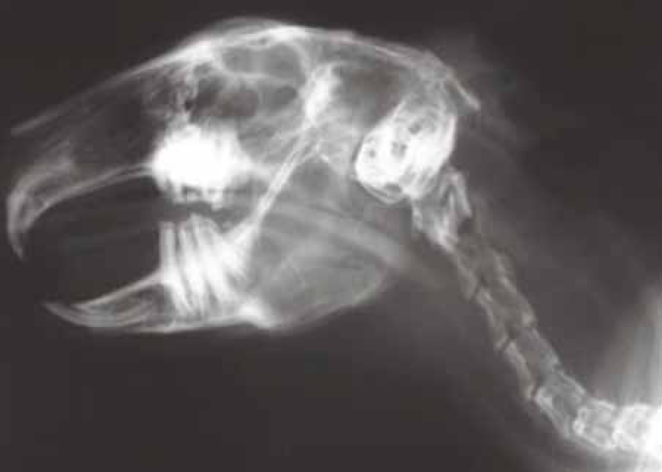
The i-gel® (Intersurgical, UK) is a novel human SGAD developed by Dr MA Nasir (Levitan and Kinkle, 2005). Work was carried out to see whether a non-inflatable SGAD could be produced that was appropriate for the upper airway anatomy of common companion animal species (Crotaz, 2010). This non-inflatable SGAD (v-gel®, as this new device was known) would be designed to consistently self position over the laryngeal inlet in small companion animal patients such as rabbits, cats and small dogs. Post-mortem studies were performed on various companion animal cadavers with all owners giving informed consent; neck dissections of seven cats, five rabbits and four dogs (all below 10 kg bodyweight) were performed.
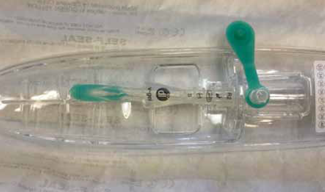
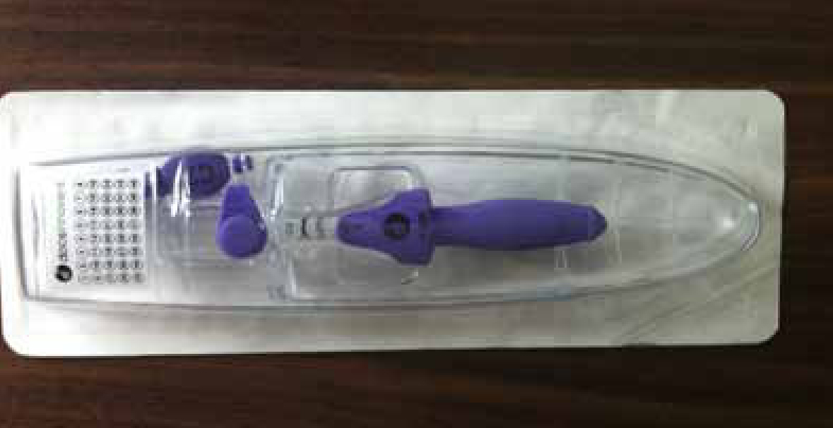
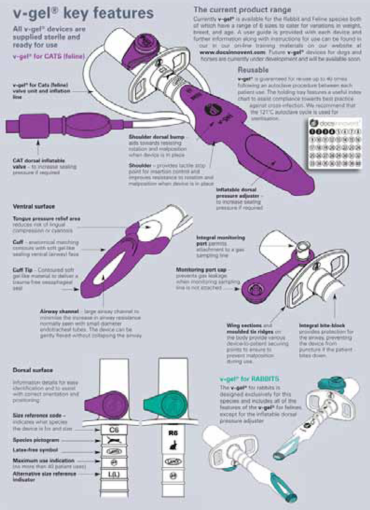
The study used the size 1 i-gel® (human SGAD) device as a starting point, each device was placed through the mouth into the pharyngeal area (Crotaz, 2010). Once each device had been inserted, a dissection was performed from the ventral skin surface into the laryngeal and pharyngeal areas. The position of each device was observed and photographed. The i-gel® devices were then adapted by removing a portion of the material, in order to ‘tailor make’ the tube to achieve maximum conformability to the patient's anatomy. The adapted devices were then used as guides with standard moulding techniques then being utilised to produce prototypes of the v-gel® device. Various alterations were made to the shape of the original i-gel®, and following various modifications, it was found that the v-gel® device conformed well to the anatomy of the pharyngeal and laryngeal framework, and was simple and rapid to insert (Crotaz, 2010) (Figures 1 and 2).
Case study: insertion and maintenance of the v-gel in a rabbit undergoing nephrectomyA 2.28 kg, 3.5-year-old rabbit was anaesthetised for a nephrectomy using a triple combination of 0.46 mls medetomidine, 0.23 mls torbugesic and 0.23 mls ketamine given via subcutaneous injection.The medetomidine was reversed post surgery with 0.46 ml atipamizole given via intramusclar injection, injected into the quadriceps muscle.The rabbit was placed on oxygen for 2–3 minutes to be pre oxygenated prior to v-gel® insertion. In some cases the premedication and pre oxygenation is not enough to reach the correct anaesthetic plane for intubation. In these cases the rabbit would be placed on a face mask on 1% Isoflurane to prevent gagging when the v-gel® is inserted.It is always the veterinary surgeon's choice whether or not to use isoflurane via mask and it should however be noted that there is a potential for environmental pollution with inhalation agents using this method.The depth of anaesthesia was assessed by checking the blink reflex and pedal withdrawal reflex which were not present and the position of the eye, which was in the downwards position indicating the depth of anaesthesia was sufficient.The v-gel® was attached to the capnograph prior to insertion and lubricated with a water soluble jelly (such as Vet Lube™ gel or spray (Docsinnovent®), or KY Jelly™ (Johnson and Johnson)) to aid placement and prevent irritation. The front, back and both sides of the v-gel were lubricated to ensure a smooth insertion. Care must be taken to ensure that the lumen of the device remains gel free as this could be aspirated by the patient.The rabbit was held in sternal recumbancy for placement as is the norm in this practice. A small spray of lignocaine (in the form of Intubeaze™ (Ceva)) was used to desensitise the larynx. After checking for foreign material, food or hay in the mouth that may be pushed into the larynx by the device, the v-gel was inserted.The end point of insertion is indicated when the device drops slightly over the base of the tongue and the shoulders of the v-gel touch the laryngeal arch and a CO2 trace is visible on the capnograph.The v-gel® was supported using a stand to ensure the anaesthetic tubing was in line with the mouth and it was tied in place using V-tie™ or WOW™ band. The rabbit maintained a good stable capnograph reading and all vitals were stable throughout the operation.The recovery was smooth and the v-gel® removed easily in one swift movement. The v-gel device was checked for any signs of trauma and was found to have none. The rabbit began to lift his head and swallow almost immediately.In the author's opinion the v-gel® provided a stable and controllable way to provide anaesthesia to the rabbit as they can be difficult to intubate. It also provided a means for intermittent positive pressure ventilation should it have been required and allowed for a more complete anaesthetic monitoring system with the use of the capnometer and ECO2 monitoring. The author also believes that the recovery was less traumatic and quicker due to the v-gel allowing for proper maintenance of anaesthesia and no trauma to the larynx ensuring no gagging on recovery.
The v-gel®
The Crotaz (2010) study demonstrated that it is possible to make an anatomically shaped SGAD for veterinary use that will consistently self position over the laryngeal inlet. This study allowed the researchers to identify the shapes necessary for easy and safe use. Following on from these early designs the researchers were able to create prototypes, which they constructed using soft medical grade plastics, allowing the creation of individual versions which were suitable for rabbits (Figure 3), cats (Figure 4) and dogs in a range of different sizes to suit individual species/ breed variability. Soft plastics were used in order to reduce the risk of soft tissue trauma when compared with the harder materials used in PVC and red rubber endotracheal tubes.
The objectives of this study provided the study group with a foundation for further research. The veterinary prototypes were used in a controlled clinical setting, with the ultimate aim, and with sufficient clinical validation, for it to become possible to use a v-gel® SGAD in place of an endotracheal tube or face mask during companion animal procedures, and improve the options for airway management in these species (Crotaz, 2010).
Use of the v-gel®
The v-gel® device provides a patent airway for surgical procedures without the disadvantages of endotracheal intubation or face masking and is inserted and repositioned more easily than endotracheal tubes (authors' experience LS and SP). The benefit of this new device in rabbit anaesthesia is that intermittent positive pressure ventilation can be achieved should it be required.
The v-gel® is a relatively simple device to place. To insert a v-gel® the animal has to be at a surgical plane of anaesthesia. The patient should be placed on 100% oxygen for a few minutes before device insertion, e.g. using flow by techniques. Pre-oxygenation is part of the initial risk assessment and is considered good practice (Bednarski et al, 2011). Room air contains 21% oxygen, which is not adequate for oxygenation in the presence of respiratory depression which can accompany most anaesthetic agents (Hughes, 2008).
Case study — insertion and maintanence of a cat undergoing ovariohysterectomy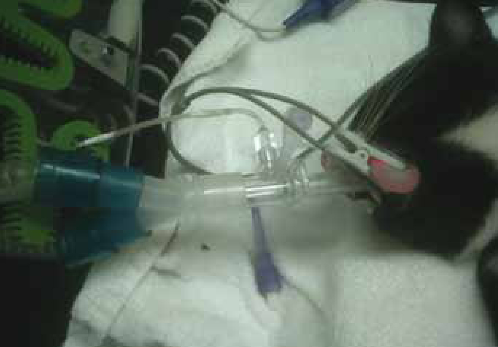
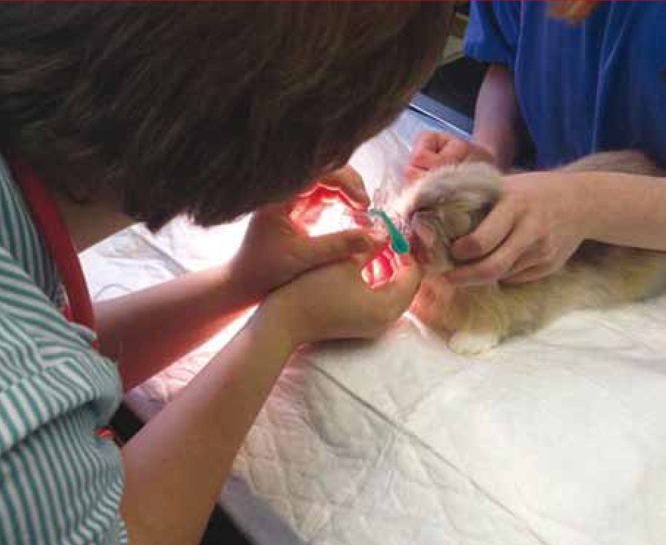
Photo 1 Cat with successful placement of the v-gel®.
A 2.95 kg, 6-month-old female domestic short haired cat was anaesthetised for routine ovariohysterectomy using a premedication of 0.14 mls medetomidine, 0.11 mls butorphanol and 0.17 mls meloxicam (given subcutaneously). The medetomidine was reversed after surgery with 0.07 mls of atipamezole injected into the quadriceps muscle.The cat was then placed on isoflurane via a mask, to achieve a surgical plane of anaesthesia. This was to prevent gagging when the v-gel® was inserted, thus decreasing the risk of laryngeal spasm. In addition the patient was placed on oxygen for a few minutes before device insertion. Achievement of a surgical plane of anaesthesia can also be done by induction with propofol (Abbott) for placement of the device. This cat required additional anaesthetic to achieve a surgical plane of anaesthesia as it was only sedated with the pre medication. It is the veterinary surgeon's preference to use mask induction at this surgery. It should be noted, however, that while using a mask there is the potential for environmental pollution with inhalation anaesthetics.The cat was placed on 2% isoflurane via a facemask for 2 minutes to achieve the required depth. The author has not seen any cats become unduly stressed by application of the facemask. The depth of anaesthesia was assessed by checking that the blink and pedal reflex were not present, and the position of the eye, which was in the downwards position.The v-gel® was attached to the capnograph and lubricated with KY Jelly to aid placement. Each side of the device is lubricated to aid placement but care must be taken not to get any of the lubrication gel in the lumen of the device as it could be aspirated by the patient or block the lumen of the v-gel® tube. The cat was held in right lateral recumbency for normal intubation as performed in this practice and the larynx desensitised with lignocaine (Intubeaze, CEVA). After checking there was no foreign material in the mouth that could be pushed into the larynx by the device, the v-gel® was then inserted. The end point of insertion is identified by the device dropping slightly over the base of the tongue, and by the shoulders of the device as they gently touch the pharyngeal arch.The v-gel® was supported using a stand to ensure that is remained in line with the direction of the mouth and was tied in place with WOW bandage. The cat remained stable throughout the anaesthetic.The recovery was smooth and the v-gel® was easily removed in one swift movement just before the cat began to swallow. In the author's opinion the use of the v-gel® reduced the coughing on recovery compared with the use with an endotracheal tube.
The larynx should be desensitised using an appropriate dose of local anaesthetic agent prior to placement. The v-gel® size is chosen by both checking the bodyweight guidelines supplied by the manufacturers and also measuring the device against the patient. When the airway opening is placed against the neck adjacent to the larynx, the connector should reach just past the incisors if correctly sized.
The v-gel® is then gently slid into the centre of the mouth and down into the pharynx. The easiest way to confirm correct placement is to check for capnograph traces, showing that the device is positioned over the larynx.
Because the large target of the pharynx is being aimed for, rather than the relatively small trachea, the skill needed to place v-gel® devices is relatively small compared with that needed for intubation. v-gels® are fast to insert, with average insertion times of 8 seconds in rabbits and 2.5 seconds in cats (Crotaz, 2013).
One study on cats at the University of Utrecht (van Oostrom et al, 2013) showed no leakage of anaesthetic agent from v-gels® in use making these devices much safer for staff than the use of face masks. A comparison trial between v-gels® and endotracheal tubes (van Oostrom et al, 2013) found significantly more upper airway stridor (respiratory noise) after recovery from endotracheal tube anaesthetics, suggesting that the recovery is more comfortable after v-gel® devices have been used.
With any airway system (face masks, endotracheal tubes or v-gels®) it is essential to support the circuit to prevent the airway device dragging on the patient or malpositioning during the procedure. The same company that makes the v-gels® also makes a useful circuit support — the D-grip, which allows rapid adjustable support of the anaesthetic circuit to any position that is required (www.docsinnovent.com).
Key points
- Endotracheal tubes are currently viewed as the standard technique for administering oxygen, nitrous oxide and volatile anaesthetic agents to patients under general anaesthesia or sedation.
- Supraglottic airway devices were developed for use in human anaesthesia to offer a simple and effective alternative to endotracheal intubation.
- Supraglottic airway devices allow the ventilation of patients by delivering anaesthetic gases/oxygen to a patient above the level of the vocal cords.
- Supraglottic airway devices function by forming an airway seal in the pharynx, rather than the trachea, while still allowing effective ventilation.
- The v-gel® device provides a patent airway for surgical procedures without the disadvantages of endotracheal intubation or face masking of patients.
The v-gel® spent many years in trial work both in general practice and referral practice. All the common issues with airway devices have been solved; for instance the inclusion of moulded ridges in the v-gel® to grip onto gauze ties and prevent slipping. The airway channel inside the v-gel® has been designed to be as large as the tracheal diameter of the patient. This means that the airway is not constricted and makes it much easier for the patient to breathe (Figure 5). Endotracheal tubes are 16 times harder to breathe through than the v-gel® (according to calculations using Poiseuille's Law), a critical factor for patients with compromised respiratory function.
Cost
It has been suggested that the v-gels® cost more than endotracheal tubes to use. In reality, because they are designed as re-usable devices, they cost between £3 and £4 per use, which is similar to other veterinary items such as intravenous fluid bags, and less than disposable endotracheal tubes if correctly used on a single patient use only basis.
Studies have shown an association between intubation and increased death rates in cats (Brodbelt et al, 2008). Use of anatomically designed SGADs removes many of the potential risks associated with intubation. In addition, the ability to use a device designed to be autoclaved between patients provides a cleaner airway solution without any risk of cross infection. It is the authors' experience that owners do not object to a small increase in price for the use of a cleaner and more advanced anaesthetic system.
Conclusion
SGADs were developed for use in human anaesthesia to offer a simple and effective alternative to endotracheal intubation. The human SGAD has been adapted to create a veterinary version, the v-gel®, resulting in an alternative method to endotracheal tubes to administer oxygen, nitrous oxide and volatile anaesthetic agents to these patients. Because these devices deliver anaesthetic gases/oxygen to a patient above the level of the vocal cords they may be a safer way of administering these agents to patients where endotracheal intubation may be viewed as being traumatic, i.e. cats and rabbits, and where poor technique during endotracheal intubation may potentially increase the patient's anaesthetic risk.