

The first step in the investigation of any skin problem involves the taking of a thorough general and dermatological history followed by a physical and dermatological examination. These procedures should be undertaken in every case, as they allow the clinician to formulate a list of differential diagnoses and an appropriate list of diagnostic procedures. Skin scrapes and hair plucks are important as part of the investigation for ectoparasites. However acetate tape impression smears, direct and indirect impression smears and fine needle aspirates are crucial to assess skin cytology. Stained swabs of ear discharge can help identify aural pathogens (Miller et al, 2013). Cytology can identify the cell types involved in the disease process to decide if the skin condition is inflammatory or neoplastic, as well as the presence of bacteria and yeast.
Diagnostic techniques
The choice of diagnostic technique will depend on the type of lesion that is presented. Where the lesion is superficial, such as an area of erythema, erosion, ulceration or primary lesions like papules or pustules, then acetate tape impression smears and direct and indirect smears are most useful. Where lesions are nodular then a fine needle aspirate is the diagnostic test of choice. Where the examination of otic cytology is needed samples of discharge can be taken from the ear using a swab. Basic investigative steps for cytology are low cost and quick and easy to undertake, however they consistently yield diagnostic information that can help with the management of the case (Sharkey et al, 2014).
Samples taken from the skin
Acetate tape impression smear
The best tape to use is a clear 5M Scotch tape as this holds its shape well when it is stained. The tape should be pressed firmly onto the area of skin to be examined. This may be an area of erythema or superficial erosion. Deeper areas of ulceration are probably best sampled by direct or indirect impression smears. To collect a representative sample the tape should be gently rubbed with a thumb nail to ensure good contact of the tape with the skin. A modified Wright's stain such as Diff-Quik can then be applied to the tape which is then carefully inverted with the adhesive slide downwards onto a microscope slide for examination. As the tape acts as a cover slip the sample can be examined under high power (x100 oil immersion) if necessary. This method can be used to identify surface bacteria and yeast, especially Malassezia spp.
Direct impression smears
This techniques is suitable for any exudative lesion, e.g. erosions, ulcers, papules and furuncules or from the undersides of crusts. Where the lesion is non exudative it can be picked with a sterile 25 gauge needle to express fluid. The microscope slide can then be gently pressed onto the lesion to collect exudate. The sample may be air dried or carefully warmed to fix it (e.g. a hair dryer or hand drier). The slide can then be stained using a modified Wright's stain (Diff Quik) and gently rinsed across the reverse of the slide and dried again. The sample should always be examined on low power (x10 objective) initially to select areas for close inspection subsequently on high power (x40) or under oil immersion (x100). Where pustules are present the same technique may be employed. The pustule should be picked with a sterile 25 gauge needle and the contents gently pressed onto the slide. It should be stained as above.
Indirect impression smears
These types of samples are best taken from areas of ulceration and exudation where a direct impression smear is difficult due to the location of the site. A cotton swab can be used to collect exudate. This may be from an exudative erosion or ulcer, e.g. lesion in a skin fold or from a sinus or from the ear canal. The swab should be gently rolled along the microscope slide, heat fixed and then stained as detailed above. Examination of the sample should follow a logical progression from low to high power.
Fine needle aspirates
Aspirates are most suitable for nodular lesions especially neoplastic and pyogranulomatous disease. Samples should be collected using a 21–23 gauge needle attached to a 2 or 5 ml syringe. The lesion should be sterilised with spirit and immobilised between finger and thumb. The needle may then be inserted into the lesion while pressure is applied to the plunger to create negative pressure. The needle can be gently repositioned within the lesion without removing it, to draw up as much material as possible. The pressure on the needle should be released before the needle is withdrawn. The sampling should be stopped if there is evidence of blood in the hub of the needle. The needle may then be removed and the plunger should be withdrawn to fill the syringe with air. The needle is then replaced and the contents of the needle expressed onto a clean microscope slide. The material may be gently smeared if necessary and stained as above. The slide should be scanned initially on a low power (x4 or x10) to identify areas of interest before using high power (x40) or oil immersion (x100).
Samples taken from the ear
Samples of otic discharge can be taken by inserting a cotton swab gently into the ear canal. The sample should be taken if possible from the junction of the horizontal and vertical canals. Once material has been absorbed onto the swab it should be gently rolled along a microscope slide. It may then be heat fixed using a hand or hair drier before being examined. Generally it is useful to examine the unstained sample for ectoparasites before staining the fixed sample. A Diff Quik will show basic cytological detail. The application of a cover slip allows the sample to be checked under oil immersion (x100) to look for the presence of an inflammatory infiltrate and potential pathogens (Chickering, 1988).
Interpretation of results
While veterinary nurses do not undertake the diagnosis of dermatological problems they are capable of taking diagnostic samples and reporting findings to the veterinary surgeon, that can then be verified to help in the selection of further tests or the institution of therapy. In order to interpret cytology it is important to have a basic knowledge of the most important cell types encountered on cytological samples and the different types of infectious organisms.
Inflammatory cell types
The following section describes the most common inflammatory cell types found on cutaneous cytology samples. Descriptions of cell characteristics are those seen microscopically under oil immersion lens when stained with a modified Wright's stain such as Diff Quik.
Neutrophils are the body's first line of defence against infection and are the most common cells found in acute inflammation. On cytology the neutrophil has a nucleus that stains dark blue/purple and is divided into two–five lobes. The cytoplasm stains pale pink. They are phagocytic cells and it is not unusual to find bacteria (cocci or rods) within their cytoplasm (Figure 1).
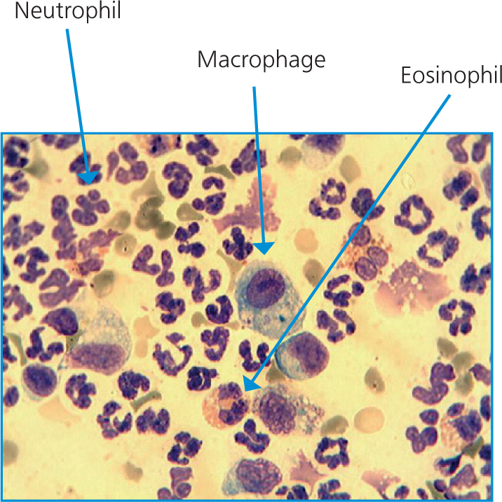
Eosinophils are usually seen on cytology samples from canine skin problems where there is a parasitic component to the inflammation, e.g. sarcoptic mange. In cats they are commonly associated with allergy. Eosinophils appear to be slightly larger than neutrophils on cytology, the nucleus is usually divided into two lobes rather than the two–five of the neutrophil. The cytoplasm is filled with bright red staining granules (Figure 1).
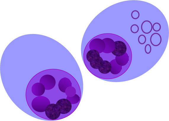
Lymphocytes are seen in chronic inflammatory processes and are generally associated with an immunological response to exogenous antigen. When seen in pure population they are indicative of neoplasia. They are small cells with minimal amounts of dark blue cytoplasm. The nucleus is round, fills most of the cell and is about the size of a red blood cell. Nucleoli are not prominent within the nucleus (Figure 2).
Plasma cells are specialised cells that are produced by the activation and differentiation of B cells after they encounter antigen. They are also a component of chronic inflammation but like lymphocytes when seen in pure population are suggestive of neoplasia. They are round to oval with variable amounts of deeply staining basophilic cytoplasm, occasionally they have a perinuclear clear area. The nuclei are medium sized, round and sit eccentrically in the cell. The nucleus has a ‘clock face; appearance due to the presence of granular to course chromatin and occasional small to medium sized nucleoli. Russel bodies, which are small vesicles containing antibody, can be visualised in the cytoplasm of some cells
Macrophages are another of the cells found in chronic inflammation. Their main function is to phagocytose foreign material which may be bacteria, fungi, cellular debris or extraneous material. These are large cells much bigger than eosinophils or neutrophils, often with foamy vacuolated cytoplasm. The nucleus is oval to round (Figure 1).
Mast cells can also be found in areas of chronic inflammation but are usually only found in small numbers. When identified in large numbers to the exclusion of other cell types the findings are suggestive of a mast cell tumour. They are round cells and are larger than neutrophils and eosinophils. They have a pale blue cytoplasm and a round nucleus which is often obscured by numerous dark blue granules that contain heparin and histamine. Granules will often ‘leak out’ into the section producing a stippled type of effect (Figure 3).
Pictorial representations of cell types and pathogens
| Neutrophils Neutrophils are important for host defence against infection. Most common of the inflammatory cells found in infection they are the first cells to arrive to fight bacteria. Distinctive multi-lobed nucleus usually 2–5 lobes seen and pinkish cytoplasm | |
| Eosinophils Eosinophils are important in the host defence against parasites. Commonly seen in allergy in cats. Slightly larger than a neutrophil they have a distinctive bilobed nucleus. Bright red staining granules are seen in the cytoplasm | |
| Lymphocytes Lymphocytes recognise and respond to exogenous antigens. Small cells with minimal cytoplasm, smudged chromatin and no nucleoli. Nucleus is round and the size of a red blood cell. Cytoplasm dark blue. Sign of chronic inflammation | |
| Macrophages Macrophages are found in tissue. They are a sign of chronic inflammation. They act to phagocytise bacteria, fungi, cellular debris and foreign material. Cells are usually large, often with foamy vacuolated cytoplasm. Nucleus oval to round | |
| Plasma cells Cells round to oval with variable amounts of deeply staining basophilic cytoplasm, occasional perinuclear clear area. Nuclei medium sized, round and eccentric with granular to coarse chromatin, occasional small/medium round nucleoli. Russell bodies can be seen in cytoplasm. | |
| Mast cells Round cells with a pale blue cytoplasm, larger than neutrophils/eosinophils. Round nucleus which is often obscured by numerous dark blue granules containing heparin and histamine, granules will often ‘leak out’ into the section |
Pathogens
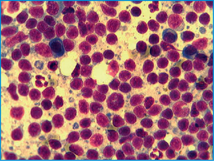
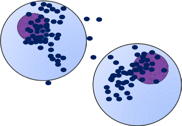
A coccus (plural cocci) is any bacterium that has a spherical, ovoid or generally round shape. It is one of the three distinct bacterial shapes, the other two being spiral-shaped cells and bacillus (rod-shaped). Cocci can be arranged in pairs, e.g. Neisseria spp., (which is not normally a skin pathogen), or long chains which is normal arrangement of Streptococcus spp., or in clusters e.g. Staphylococcus spp. (Figure 4). Bacillus appear as elongated rods when viewed under the microscope, they usually appear as single cells. Spiral shaped cells are not normally found on skin or ear samples.
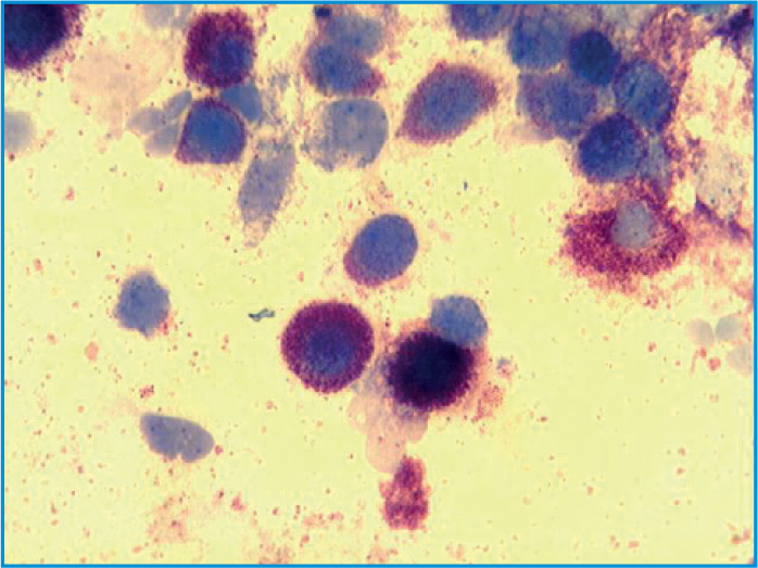
Yeast are much larger than bacteria on cytology and on cytology they can appear as budding organisms. Malassezia spp (Figure 5). is the principal yeast identified from the skin and ears in dogs. It appears as a dark blue/purple staining peanut shaped organism.
It is difficult to identify pathogens purely on the basis of cytology if a Diff Quik is used rather than a Gram stain. A Gram stain will stain Gram-negative bacteria red and Gram-positive bacteria blue. All bacteria and yeast will stain dark blue with a Diff Quik stain. While this is not problematic when cocci are identified, as all of the principal pathogens found on the skin are Gram-positive, it is more of an issue if rods are identified on cytology. Corynebacterium spp. which are Gram-positive rods look almost identical morphologically to Gram-negative organisms such as Pseudomonas spp. The differentiating colouring seen with a Gram stain is lost on the Diff Quik stain where all rods stain dark blue making precise identification impossible. This is particularly pertinent when it comes to the selection of therapy, as the spectrum of drugs used to treat Gram–negative infections such as Pseudomonas spp. is very different to those used to treat Gram-positive infection with organisms such as Corynebacterium spp.
Conclusion
The collection of samples for cytology can easily be undertaken by the veterinary nurse using a combination of different techniques. Cytology is a low cost diagnostic test that provides useful information to help in decision making on the need for further more advanced diagnostic tests or therapy.
Key Points
- Acetate tape impression smears, direct and indirect impression smears and fine needle aspirates are useful techniques to assess cutaneous cytology in canine and feline skin disease.
- Swabs taken from ears for cytology can help identify potential pathogens.
- Common cell types such as neutrophils, eosinophils, lymphocytes, plasma cells, macrophages and mast cells can be identified with practice.
- Bacteria cannot be definitively diagnosed on cytology alone, but cytology can help in decision making as to whether further tests such as culture and susceptibility are needed.
- Yeast can be identified on cytology without the need for culture.