
Babesiosis is a disease caused by the tickborne protozoan parasite Babesia species. There are a number of species within the Babesia family, including: Babesia canis canis, Babesia canis vogeli (from here on referred to as just Babesia canis and Babesia vogeli), Babesia gibsoni and Babesia (Theileria) annae. The two major tick species responsible for transmitting the disease are known to be Dermacentor reticulatus and Rhipicephalus sanguineus. D. reticulatus ticks are common throughout central and southern Europe, including some areas of the UK. R. sanguineus ticks are common in southern Europe but are not endemic in the UK where the climate does not suit their long-term survival outside, although imported ticks are reported to thrive in domestic environments in the UK under the right conditions. (Fisher and Halos, 2014; Stokes, 2015). Infection can lead to red cell lysis and immune-mediated disease, resulting in anaemia, icterus, lymphadenopathy, pyrexia, secondary renal and hepatic disease, and in severe cases, death.
Transmission and prevention
B. canis is transmitted to dogs when ticks take a blood meal. When a tick feeds on an infected host, B. canis stages penetrate the gut of the tick, multiply and migrate. Sporozoites are then transmitted to new hosts through the tick’s saliva. As B. canis is generally transmitted by ticks after 24 hours of feeding, it is important for pet owners to be vigilant and check for ticks on their pets every day, removing any ticks with a tick hook such as the O’Tom® tick remover (Figure 1). It is vital that owners are instructed by nurses how to remove ticks without stressing them and without leaving the head and mouthparts in situ. A simple ‘twist and pull action’ is required. This can also be performed with tweezers but they should be fine pointed and not blunt as crushing will stress the tick, causing it to regurgitate its stomach contents and increasing the risk of disease transmission. Traditional techniques to loosen the tick such as the application of petroleum jellies, freezing or burning will also increase this likelihood and are contraindicated. Pets will often be presented at nurse clinics to have ticks removed and this is the perfect opportunity for nurses to discuss safe removal as well as the risks of tick-borne disease transmission and good prevention practice for those at risk (ESCCAP, 2012).

No strategic control programmes have been developed for canine babesiosis (ESCCAP, 2012) and so the most effective preventative measure is a combination of appropriate tick prophylaxis with a product that repels, expels or rapidly kills ticks and prompt removal of any ticks found on the host.
Entry into the UK
Babesiosis is a common disease in dogs in mainland Europe, as well as in America, Africa and Asia. In February 2016, B. canis was reported to have been detected in three dogs from the Harlow area of Essex, UK (Swainsbury et al, 2016) and a fourth dog was found to be infected with babesiosis in March 2016 (Woodmansey, 2016). Although cases have been reported in the UK in the past, these have been in dogs that have travelled abroad or through association with travelling (Phipps et al, 2016). These four new cases are from four separate households in dogs which have not travelled abroad or been in contact with travelled animals (Woodsmansey, 2016). Tests by DNA barcoding identified the ticks found on the infected dogs as D. reticulatus and polymerase chain reaction (PCR) revealed infection with B. canis (Phipps et al, 2016).
It is unknown how the disease has entered the UK but it is speculated to have entered via infected D. reticulatus ticks. These may have arrived on an imported or travelled host dog or through vehicles from the continent contaminated with ticks. Following the removal of the mandatory tick treatment from the Pet Travel Scheme (PETS), there have been no regulations protecting the UK from exotic and infected ticks which carry exotic diseases such as Ehrlichia canis and Babesia spp. In 2012–2013, the Great Pet Travel Survey indicated that 28% of dog owner in the UK have travelled abroad with their pet (Morgan, 2014). Since the 2012 amendments to PETS, the number of owners importing and travelling abroad with their pets has risen, with approximately 164 836 dogs entering the UK under PETS in 2015 and non-commercial imports of dogs increased by 1614 between 2014 and 2015 (Kennel Club, 2016). Despite complying with the Pet Travel Scheme (PETS) requirements, the Great Pet Travel Survey 2012–2013 revealed that 36% of dog owners travelling to at risk areas took no specific precautions against their pet contracting exotic parasitic diseases (Bristol University, 2013).
The risk of an exotic disease establishing in the UK can be illustrated by the ecological theory of biological invasions whereby the chance of a disease establishing can be calculated by the propagule pressure (how often it is introduced) and the habitat suitability (both climate and vector availability) (Stokes and Wright, 2015). The vector, D. reticulatus, is already established in the UK although it is not as prevalent as some other endemic ticks such as Ixodes ricinus. With the UK climate and habitat highly suitable for D. reticulatus, the determining factor was how often the disease was introduced. This risk increased as the number of travelling dogs and imported pets entering the UK increased, resulting in a high risk of establishment in the UK. It is now widely considered that B. canis is an endemic disease in the UK with a very low likelihood of being able to eliminate or even prevent the spread of the disease across the UK (Woodmansey, 2016).
Know your ticks
It is important that ticks removed from pets are identified. This is useful, not only to monitor the spread of ticks capable of harbouring tick-borne diseases across the UK, but also so veterinary professionals and owners can be vigilant for the clinical signs of tick-borne diseases that the ticks in question may be carrying. Veterinary nurses can play a vital role in identifying ticks at the point of removal as well as identifying ticks brought in by the public.
Adult ticks can be examined by the naked eye to identify gross features. Anatomical features can be examined more closely under a dissecting/stereo microscope; x10 or x40 magnification, using ‘top’ incident light. The tick may be mounted on a small circle of Blu Tack for easy manipulation under the microscope.
Larval ticks and nymphs are smaller and will require microscopic examination for identification. They can be placed in a drop of water or liquid paraffin on a slide, under a coverslip. Features can then be examined under a x4 or x10 objective lens.
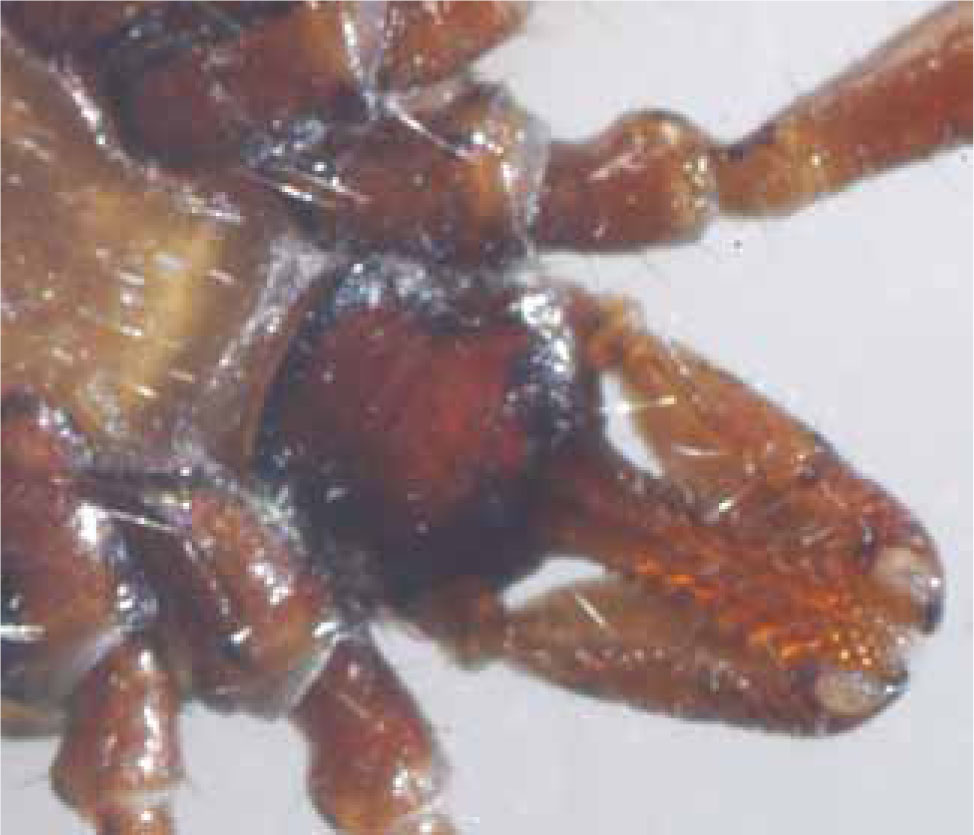
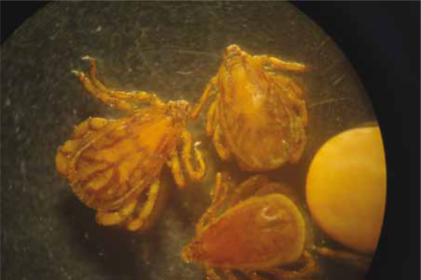
The first step in identification is to confirm that the ectoparasite is a tick and not a mite. Adults and nymphs are usually large enough to distinguish them on size. Larval ticks are similar in size but only have six legs and have mouthparts unique to ticks known as a hypostome (Figure 2).
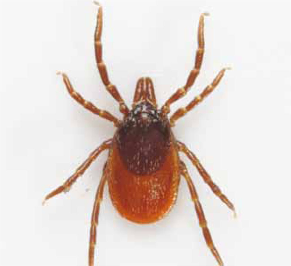
Male ticks can be distinguished from females by differences in the size of their dorsal plate or scutum. In males this covers the entire dorsum of the tick, where females have a scutum that only extends a short way dorsally behind the head (Figure 3). This allows the abdomen to expand during feeding.
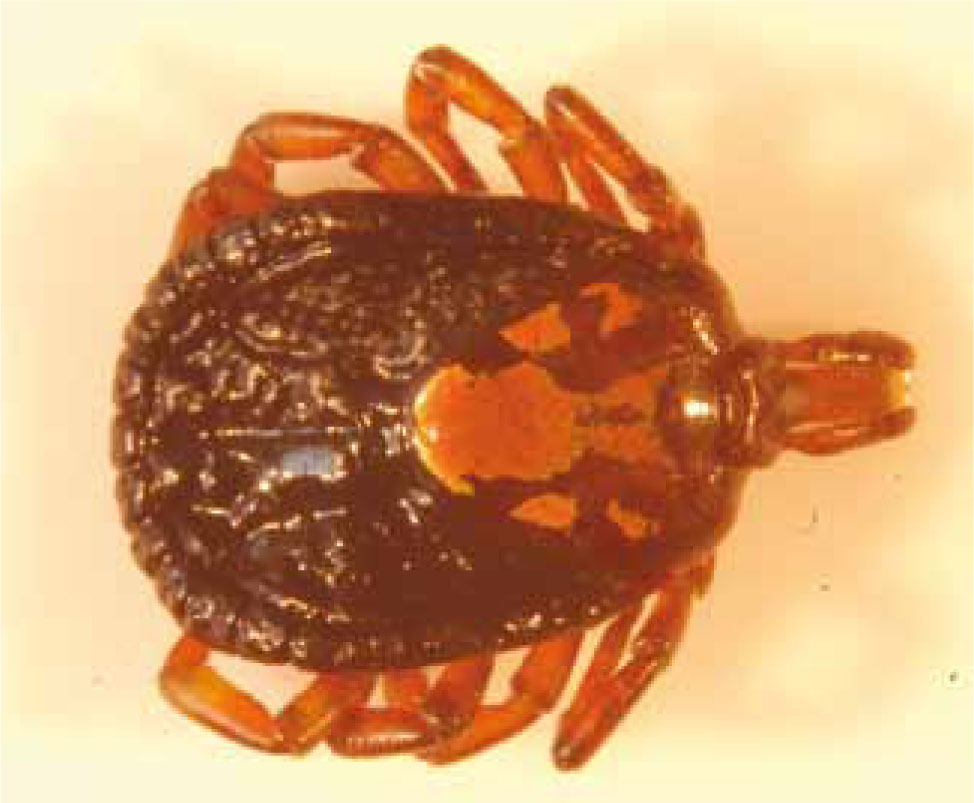
Both genera capable of transmitting B. canis have a groove posterior, and no groove anterior, to the anus. In addition, they have festoons, eyes and Dermacentor spp. have decoration on the scutum. Festoons are crimping around the edge of the abdomen giving a ‘Cornish pasty’ type appearance (Figure 4). Eyes are spots present symmetrically either side of the scutum. Ticks with these features should be sent away for identification if they cannot be identified in practice due to lack of experience or confidence. Public Health England are happy to receive ticks for identification, as are ESCCAP UK & Ireland. Ticks should be placed in a container with a secure lid and sent in an envelope marked ‘biological sample’. Samples should ideally be unfed or only partially engorged as features on the scutum and festoons may be partially obscured by engorgement.
Conclusion
Babesia canis represents an increasing risk to UK dogs, both in terms of exposure to the parasite abroad but also the risk of the parasite becoming endemic throughout the UK, as has recently occurred in Essex. Veterinary nurses play a vital role in controlling exposure of this parasite and reducing morbidity and fatality in dogs as a result. In giving accurate tick control advice and rapidly identifying exotic ticks, nurses will not only help to reduce disease in their clients’ pets but also help to limit the spread of this parasite and disease.
Key Points
Conflict of interest: none.