References

Aldrich J. Assessment and diagnosis of shock, 2nd ed. In: King L, Boag A (eds). Gloucester: BSAVA; 2007

Andrews A. The dyspnoeic cat. Veterinary Nursing Journal.. 2013; 28:(9)280-282 https://doi.org/10.1111/vnj.12062

Acute lung injury and acute respiratory distress syndrome. 2001. https://www.vetfolio.com/learn/article/acute-lung-injury-and-acute-respiratory-distress-syndrome (accessed 25 November 2020)

Acute respiratory distress syndrome in dogs and cats. Clinicians brief. 2019. https://www.cliniciansbrief.com/article/acute-respiratory-distress-syndrome-dogs-cats (accessed 18/06/20)

ARDS in Cats and Dogs', Clinicians Brief. 2008. https://www.cliniciansbrief.com/article/ards-cats-and-dogs (accessed 18/06/20)

DeClue AE, Cohn LA. Acute respiratory distress syndrome in dogs and cats: a review of clinical findings and pathophysiology. J Vet Emerg Crit Care.. 2007; 17:(4)340-347 https://doi.org/10.1111/j.1476-4431.2007.00247.x

Egleston S. The forgotten complication: aspiration pneumonia in the canine patient. Vet Nurs.. 2018; 9:(3)138-143 https://doi.org/10.12968/vetn.2018.9.3.138

Gray K. Management of tubes, lines and drains. The Veterinary Nurse.. 2018; 9:(10)525-531 https://doi.org/10.12968/vetn.2018.9.10.525

Haskey E. How to nurse the intensive care unit ventilator patient. The Veterinary Nurse.. 2013; 4:(2)108-113 https://doi.org/10.12968/vetn.2013.4.2.108

Hyndman P, Bray J. Temporary tracheostomy: a practical approach to the placement and management of a tracheostomy tube. Companion Anim.. 2017; 22:(8)471-479 https://doi.org/10.12968/coan.2017.22.8.471

King L, Waddell L. General approach to dyspnoea, 2nd ed. In: King L, Boag A (eds). Gloucester: BSAVA; 2007

Liew F, Martin D. Acute respiratory distress syndrome on the intensive care unit. Br J Hosp Med.. 2014; 75:(12)672-677 https://doi.org/10.12968/hmed.2014.75.12.672

Moore L. Systemic inflammatory response syndrome-an overview’. Veterinary Nursing Journal.. 2016; 31:(1)18-21

Tennant B. Acute lung injury may be triggered in dogs by transfusion of blood/plasma-containing products. Companion Anim.. 2014; 19:(6) https://doi.org/10.12968/coan.2014.19.6.282

ARDS: acute respiratory distress syndrome
02 March 2021
10 mins read
Volume 12 · Issue 2
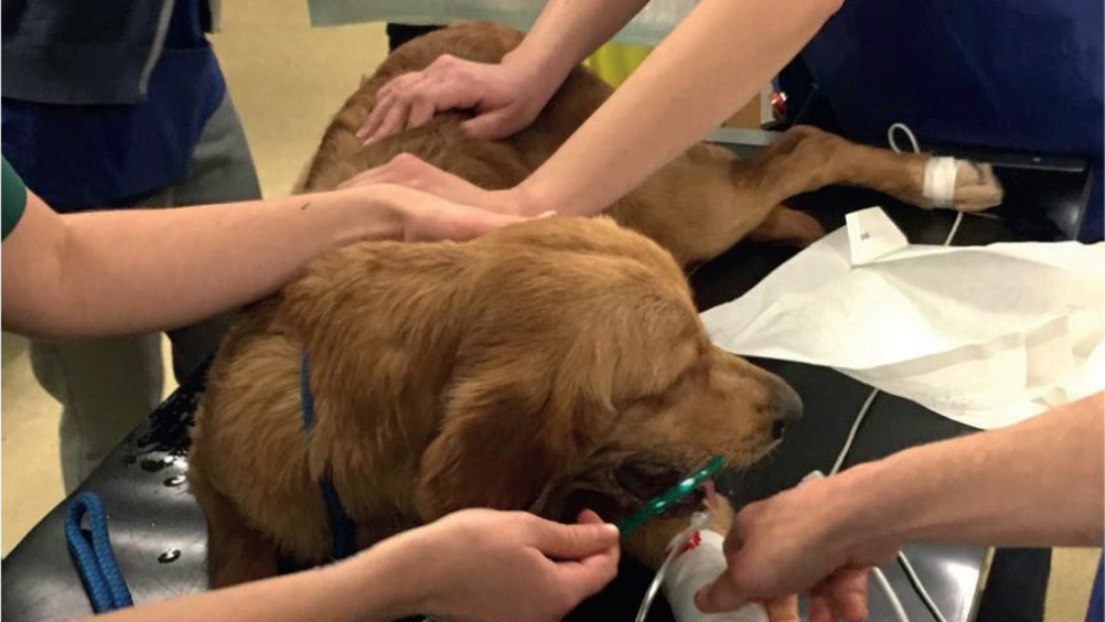
Figure 1. Patient receiving flow by oxygen while initial assessment is being carried out.

Abstract

Respiratory distress is a common presentation in an emergency and critical care setting. Acute respiratory distress syndrome (ARDS) is an acute onset condition where the lungs cannot provide the patient's vital organs with enough oxygen. ARDS can occur as a result of several underlying triggers. It is important that veterinary nurses know what to look out for in these patients, and how to appropriately nurse them to ensure they are not compromised further.

Acute respiratory distress syndrome (ARDS) is a severe respiratory disease that has not been well represented in veterinary medicine. Extensive research around this condition has been conducted in human medicine over the last 40 years, but in veterinary medicine the true incidence in animals is still unclear. Currently, this condition is much less common in animals than in people, however there are more cases published in the associated literature regarding dogs than cats. With the ever-increasing number of veterinary critical care facilities and the greater number of owners willing to pursue extensive treatment in critically ill animals, there will no doubt be an increase in the frequency of ARDS cases being identified and treated (Carpenter et al, 2001). ARDS occurs when there is a diffuse inflammation across the lung tissue. ARDS can develop as a result of a primary condition (e.g. pneumonia) or a systemic problem (e.g. trauma or sepsis) (Cavanagh, 2019).

Development of ARDS

In ARDS inflammation is triggered by either a primary condition, such as septic shock or pancreatitis, or by a severe pulmonary insult which could include aspiration pneumonia, pulmonary contusions, or smoke inhalation (King and Waddell, 2007). Other conditions that may increase a patient's chance of developing ARDS include inflammation, infection, systemic inflammatory response syndrome (SIRS), severe trauma, multiple blood transfusions, drowning/submersion injury or inhaled irritants (Cavanagh, 2019). Prevention of these conditions developing relies on treatment of shock and the underlying cause (Aldrich, 2007). ARDS is a secondary inflammatory response to injury leading to an inflammatory cascade and resulting in overwhelming pulmonary damage.

ARDS is a severe form of acute lung injury (ALI). In ALI, pulmonary inflammation is exhibited as vasculitis, interstitial and alveolar permeability oedema and infiltration of inflammatory cells (King and Waddell, 2007). Pro-inflammatory mediators damage the alveolar capillary membrane allowing fluid to leak from vascular space and into the alveoli (Moore, 2016). In ARDS there is the addition of a rapid increase in type II pneumocytes, hyaline membranes and eventually interstitial fibrosis (King and Waddell, 2007). This results in a reduction in lung capacity, increased dead space and right to left shunting (perfusion of non-ventilated lung space) (Tennant, 2014). The major difference between ALI and ARDS is the degree of hypoxaemia the patient is experiencing, with the more severe cases being termed ARDS (DeClue and Cohn, 2007). Patients experiencing shock are at major risk of developing ALI/ARDS (Aldrich, 2007).

Clinical signs

A patient may take between 1 and 4 days to develop clinical signs after the initial triggering event (Childers, 2008), which can include:

  • Progressive hypoxaemia
  • Tachypnoea
  • Respiratory distress
  • Cyanosis

Physical examination findings that can suggest a patient is developing ARDS can include:

  • Harsh lung sounds progressing to crackles
  • Orthopnoea
  • Use of auxiliary respiratory muscles (DeClue and Cohn, 2007).

Diagnosis

Diagnosis is made via the history and identification of an underlying trigger, in addition to the results of several clinical tests. Commonly used diagnostics include thoracic imaging (radiographs or computed tomography (CT)), airway sampling (bronchoalveolar lavage or transtracheal wash) and arterial blood gas sampling. Cavanagh (2019) outlines five criteria for diagnosing ARDS. It is suggested that four of the criteria must be met to diagnose a patient with ARDS.

The five criteria are as follows:

  • Acute onset respiratory distress (<72 hours)
  • Presence of known risk factors
  • Presence of pulmonary capillary leak not as a result of heart failure or fluid overload
  • Hypoxaemia and impaired gas exchange
  • An arterial blood gas sample with PaO2/FiO2 of <200 consistent with ARDS and 200–300 consistent with ALI
  • Evidence of diffuse pulmonary inflammation in an airway sample.

PaO2= partial pressure of oxygen, measurement of oxygen pressure in arterial blood

FiO2= fractional concentration of oxygen in inspired gas (King and Waddell, 2007).

Treatment and nursing care

Currently there is no specific treatment for ARDS, and management of cases is aimed at supportive care and treating the underlying cause. The main considerations in ARDS patients are:

  • Find and treat the cause
  • Minimise oedema accumulation
  • Provide supportive treatment as indicated
  • Avoid hypotension, volume overload, oxygen toxicity and infection (Carpenter et al, 2001).

The management of ARDS involves supportive measures, such as oxygen supplementation and ventilation, while the underlying cause of lung injury is treated (Liew and Martin, 2014). Patients that are suspected to have ARDS will be oxygen dependent, and will need to be treated in the same way as other respiratory compromised patients (Cavanagh, 2019). Veterinary nurses play a vital role in caring for these patients in order to prevent further complications arising, and to ensure the patient is as comfortable as possible throughout their hospitalisation.

While initial assessments are being carried out flow by or masked oxygen can be provided to facilitate physical examination and any procedures such as intravenous catheter placement or blood sampling (Figure 1). It is important in respiratory distress patients to allow them to rest briefly in an oxygen-enriched environment in order to reduce stress and prevent further respiratory compromise. It may be beneficial in these patients to consider some chemical restraint after triage and initial assessment to help patients calm down and facilitate further handling of that patient. The use of a staged approach in assessment and treatment allows the patient to rest in between in an oxygen-enriched environment to prevent on-going respiratory distress. Patients should always be minimally restrained, in a quiet and calm environment. EMLA cream should be used to facilitate venepuncture (Andrews, 2013).

Figure 1. Patient receiving flow by oxygen while initial assessment is being carried out.

Monitoring and supportive treatment

Oxygen therapy

Provision of oxygen is vitally important in patients experiencing respiratory distress (such as dyspnoea or tachypnoea) or patients that are hypoxaemic. Patients that have an SpO2 less than 93% or a PaO2 of less than 80 mmHg on room air should be provided with oxygen supplementation (Egleston, 2018). There are three main goals when providing oxygen therapy:

There are several different methods that can be used to provide oxygen supplementation to patients. Longer-term methods for oxygen therapy need to be considered in the hospitalised patient (Egleston, 2018).

Oxygen mask

Oxygen can be provided via a mask in any patient that is lying still, however, in some distressed patients it is poorly tolerated and repeated attempts to place the mask over the muzzle can further increase stress and as a result increase oxygen demand. With a tight-fitting mask and a flow rate of 5–6 litres/minute an FiO2 of 0.7–0.8 can be reached (King and Waddell, 2007).

If patients do not tolerate the mask, flow-by oxygen can be provided by holding the oxygen tubing near the mouth, and this is much less stressful for the patient (Figure 1) (King and Waddell, 2007).

Nasal prongs

Nasal prongs (Figure 2) are manufactured for human patients but can be easily placed in canine patients. A buster collar may need to be placed on the patient as the prongs can be easily removed by rubbing the face either on the bed or with a paw. Applying a small amount of local anaesthetic into the nares sometimes facilitates a patient's tolerance of prongs, however some patients just find them too uncomfortable.

Figure 2. Patient receiving oxygen supplementation through nasal prongs.

Nasal cannulas

Nasal cannulas (Figure 3) are useful in mobile patients, they are easy to place and can provide an FiO2of approximately 0.4. Some patients may require sedation for placement of the nasal catheter, and they can cause sneezing. Brachycephalic breeds are poor candidates for nasal oxygen supplementation because of their stenotic nares, and they commonly have increased vagal tone (King and Waddell, 2007).

Figure 3. Patient receiving oxygen therapy through a nasal cannula.

Nasal cannulas can be placed by veterinary nurses, below outlines the equipment needed and how they are placed:

Equipment:

  • Appropriate size cannula: 6–10 fg infant feeding tube
  • Local anaesthetic — proxymetacaine hydrochloride 0.5% w/v eye drops solution or lidocaine 2% solution
  • 3/0 metric non-absorbable monofilament suture material
  • 21g x 1” needle
  • Sterile lubricant.

Procedure:

  • Administer topical anaesthetic into the desired nostril/s and allow 5 minutes to take effect
  • Measure the infant feeding tube from the tip of the nose to the medial canthus of the eye and mark it with a permanent pen
  • Push the tip of the nose dorsally and insert the tube into the nostril pointing in a medioventral direction towards the opposing ear until the desired length has been achieved. If the patient does not tolerate, or resistance is felt, stop the procedure and alert the case veterinary surgeon
  • Secure in place with three sutures, one near the alar fold, one on the dorsomedial aspect of the nose and one on the dorsomedial surface of the head. Patients resent suture placement less if a 21g needle is inserted through the skin and the suture material threaded through the needle
  • Attach oxygen tubing and administer 100 ml/kg/minute of oxygen. The oxygen must be humidified. Reduce the flow of oxygen and instil more topical anaesthetic if the patient is showing signs of discomfort or sneezing.

Oxygen kennel

Oxygen kennels are the easiest way to provide oxygen therapy to patients and can provide an FiO2 of up to 0.9. However, as soon as the door is opened, the FiO2 drops down to room air, making examining and monitoring these patients difficult. Large breed dogs may also not fit into standard oxygen kennels and they are an expensive piece of equipment for a practice to purchase (King and Waddell, 2007).

All these methods should be used with a humidifier to prevent drying of the respiratory mucosa which can lead to infection (Egleston, 2018).

Mechanical ventilation

In many patients with ARDS mechanical ventilation is an essential component in the treatment (Liew and Martin, 2014). Mechanical ventilation is an expensive treatment and requires a dedicated nurse to intensively care for that patient (Figure 4). Mechanical ventilation aims to increase CO2 excretion, maximise alveolar oxygenation and ultimately reduce the patient's workload by breathing for them (King and Waddell, 2007). It must be emphasised that mechanical ventilation does not cure the patient but allows time to treat the underlying cause (Haskey, 2013).

Figure 4. Patient being mechanically ventilated.

Nursing care

Below outlines the nursing considerations that need to be accounted for when nursing a patient on a mechanical ventilator, however, they are still useful to all respiratory patients and can still be applied when nursing a patient with ARDS:

  • Eye care — ophthalmic lubricant should be applied every 2–4 hours to prevent eyes drying out and ulcer formation (particularly important in patients in an oxygen kennel). Fluorescein checks should be performed daily to check for corneal ulcers (Haskey, 2013)
  • Mouthcare — clean the mouth with a chlorhexidine mouthwash to reduce bacteria, which can cause further complications if they enter the lungs. Suctioning of the mouth and oropharynx every 4 hours prevents secretion build up (Haskey, 2013)
  • Tube care — suction down the endotracheal tube every 4 hours and deflate the cuff, untying the tube and repositioning at the same time to reduce the chances of pressure necrosis occurring (Haskey, 2013)
  • Positioning — like all recumbent patients, pressure sores, muscle atrophy, oedema and nerve damage are all big risks so repositioning patients every 2–4 hours is important, even if it is just very small changes. Sternal recumbency can help improve oxygenation and ventilation as it allows for maximal lung expansion (Haskey, 2013). It is important in patients that can mobilise to encourage them to do so as much as is tolerable (Figure 5)
  • Urination/defecation — it is important to try and keep patients clean and dry. If patients tolerate it (especially important in ventilator patients), and there are no contraindications, a urinary catheter with a closed collection system can help monitor urine output as well as aid in prevention of urine scoulding (Haskey, 2013)
  • Infection control — ARDS patients are at high risk of developing nosocomial infection as a result of the immune suppression effects of ARDS, and so should be appropriately barrier nursed. Catheters and any other tube/drain should be checked twice daily for signs of complications, and stoma sites should be cleaned as required (King and Waddell, 2007)
  • Nutrition — lack of nutrition can lead to muscle atrophy as well as impaire ventilator capacity. Feeding tubes should be considered or a provision of partial/total parenteral nutrition via a central line. Ideally nutrition should be provided at 50% resting energy requirement or more (Haskey, 2013)
  • Intravenous catheters — if patients are on a ventilator it will be more than likely that they will have a central line placed, however other respiratory distress patients will need an intravenous catheter in situ in case of emergencies. It is easier to have a patent catheter already in place than trying to place one as well as gain an airway if any complications arise. This will mean the catheter site will need checking twice daily and flushing at regular intervals to ensure patency (Gray, 2018)
  • Blood gas analysis — King and Waddell (2007) stated that blood gases should be measured at least every 6 hours and can be venous or arterial, depending on what the veterinary surgeon requires. The frequency enables a proper assessment of the progress that has been made to the patient's ventilation and enables the veterinary nurse to monitor for deterioration in pulmonary function or changes in acid-base status. If the patient is being ventilated, the ventilator settings should be adjusted in accordance with the findings of the blood result
  • Record keeping — patient parameters (heart rate, respiratory rate, blood pressure and temperature) should be recorded every 1–2 hours depending on stability of the patient and every 30 minutes for mechanical ventilation patients (including CO2 and SpO2) and clearly written so trends can be monitored (King and Waddell, 2007; Moore, 2016)
  • Analgesia/stress — for patients that are being nursed that do not need to be mechanically ventilated, creating a stress-free environment without visual stimulants is vital to keep stress and excitement to an absolute minimum. It is important to reduce stress in patients that are already respiratory compromised as we do not want to exacerbate their problems by causing them to breathe any harder or faster and to allow them time to calm down if they are finding things stressful. Some patients may need additional medical management to aid in controlling stress and anxiety. A veterinary surgeon should be consulted if there is a concern a patient is becoming over stimulated as analgesia or chemical sedation may be required. Providing appropriate analgesia will also aid in controlling patient comfort. Performing regular pain scoring provides a comparative means of assessing the patient's levels of discomfort (Hyndman and Bray, 2017).
Figure 5. Patient on nasal cannula mobilising just outside the kennel so he can still receive oxygen supplementation.

Conclusion

Supportive care remains the primary treatment in patients with ARDS (Carpenter et al, 2001), with mechanical ventilation being an essential component in the treatment. However, patients have a grave prognosis despite intensive care, although the underlying cause of the disease is major contributing factor to prognosis (Cavanagh, 2019), and recovery time in these patients is weeks to months. Patients that have suffered with ARDS and survived may be left with irreversible lung damage, fibrosis, and may develop ARDS again in the future. These patients can be rewarding to nurse — they require a holistic approach to their critical conditions and it is necessary to ensure the patient is as comfortable as possible throughout the hospitalisation.

KEY POINTS

  • Acute respiratory distress syndrome (ARDS) is under-represented in veterinary medicine and case frequency is likely to increase with the rise in number of critical care facilities.
  • ARDS is a severe form of acute lung injury and is a secondary inflammatory response to injury, triggered by a primary condition or a severe pulmonary insult.
  • Diagnosis is made by patient history and identified underlying triggers combined with clinical test results.
  • ARDS has no specific treatment, cases management consists of supportive care and treatment of the underlying cause.
  • Oxygen therapy can be given via oxygen masks, nasal prongs, nasal cannulas, oxygen kennels and mechanical ventilation.