References

Abrams K. Multiple punctate/grid keratotomy for indolent corneal ulcers. Clinicians Brief. 2008; 11:8-10

Banks KC, Stalla DE, Bunyak FE, White TA, Schultz LG, Giuliano EA. Comparison of two cleaning and sterilization protocols of diamond burr tips used in debridement for canine superficial chronic corneal epithelial defects. Vet Ophthalmol. 2019; 22:(5)614-622 https://doi.org/10.1111/vop.12632

Bayley KD, Read RA, Gates MC. Superficial keratectomy as a treatment for non-healing corneal ulceration associated with primary corneal endothelial degeneration. Vet Ophthalmol. 2019; 22:(4)485-492 https://doi.org/10.1111/vop.12617

Bentley E, Murphy CJ. Thermal cautery of the cornea for treatment of spontaneous chronic corneal epithelial defects in dogs and horses. J Am Vet Med Assoc. 2004; 224:(2)250-3 https://doi.org/10.2460/javma.2004.224.250

Bentley E. Spontaneous chronic corneal epithelial defects in dogs: a review. J Am Anim Hosp Assoc. 2005; 41:(3)158-165 https://doi.org/10.5326/0410158

Bhardwaj SD. Managing melting corneal ulcers with conventional and autologous serum therapy - a report of 2 pugs. Intas Polivet. 2016; 17:(2)399-400

Chen S, Mienaltowski MJ, Birk DE. Regulation of corneal stroma extra-cellular matrix assembly. Exp Eye Res. 2015; 133:69-80 https://doi.org/10.1016/j.exer.2014.08.001

Dawson C, Naranjo C, Sanchez-Maldonado B Immediate effects of diamond burr debridement in patients with spontaneous chronic corneal epithelial defects, light and electron microscopic evaluation. Vet Ophthalmol. 2017; 20:(1)11-15 https://doi.org/10.1111/vop.12337

Esson DW. Spontaneous chronic corneal epithelial defects (SCCED). Clinical atlas of canine and feline ophthalmic disease.: Wiley; 2015 https://doi.org/10.1002/9781118840801.ch70

Maximizing Individual Veterinary Nurses' Strengths'. 2017. https://www.vettimes.co.uk/article/maximising-individual-veterinary-nurses-strengths/ (accessed 16 April 2020)

Case of canine spontaneous chronic corneal epithelial defect. 2018. http://www.vettimes.co.uk/article/case-of-canine-spontaneous-chronic-corneal-epithelial-defect/ (accessed 19 January 2021)

Gosling AA, Labelle AL, Breaux CB. Management of spontaneous chronic corneal epithelial defects (SCCEDs) in dogs with diamond burr debridement and placement of a bandage contact lens. Vet Ophthalmol. 2013; 16:(2)83-88 https://doi.org/10.1111/j.1463-5224.2012.01026.x

Grinninger P, Verbruggen AM, Kraijer-Huver IM, Djajadiningrat-Laanen SC, Teske E, Boevé MH. Use of bandage contact lenses for treatment of spontaneous chronic corneal epithelial defects in dogs. J Small Anim Pract. 2015; 56:(7)446-9 https://doi.org/10.1111/jsap.12360

Guyonnet A, Desquilbet L, Faure J, Bourguet A, Donzel E, Chahory S. Outcome of medical therapy for keratomalacia in dogs. J Small Anim Pract. 2020; 61:(4)253-258 https://doi.org/10.1111/jsap.13118

Harvey L, Cameron K. Comparison of expectations between veterinarians and veterinary nurses in tasks and responsibilities in clinical practice. The Veterinary Nurse. 2019; 10:(6)327-331 https://doi.org/10.12968/vetn.2019.10.6.327

Hindley KE, Groth AD, King M, Graham K, Billson FM. Bacterial isolates, antimicrobial susceptibility, and clinical characteristics of bacterial keratitis in dogs presenting to referral practice in Australia. Vet Ophthalmol. 2016; 19:(5)418-26 https://doi.org/10.1111/vop.12325

Hung JH, Leidreiter K, White JS, Bernays ME. Clinical characteristics and treatment of spontaneous chronic corneal epithelial defects (SCCEDs) with diamond burr debridement. Vet Ophthalmol. 2020; 23:(4)764-769 https://doi.org/10.1111/vop.12772

Treating corneal ulceration in dogs part 1: superficial ulcers. 2017. https://www.vettimes.co.uk/article/treating-corneal-ulceration-in-dogs-part-1-superficial-ulcers (accessed 30 November 2020)

Maggs DJ, Miller PE, Ofri R Slatter‘s fundamentals of veterinary ophthalmology, 5th edn. St Louis: Elsevier Saunders; 2013

Mathews K, Kronen PW, Lascelles D Guidelines for recognition, assessment and treatment of pain: WSAVA Global Pain Council members and co-authors of this document. J Small Anim Pract. 2014; 55:(6)E10-E68 https://doi.org/10.1111/jsap.12200

Murphy CJ, Marfurt CF, McDermott A Spontaneous chronic corneal epithelial defects (SCCED) in dogs: clinical features, innervation, and effect of topical SP, with or without IGF-1. Invest Ophthalmol Vis Sci. 2001; 42:(10)2252-2261

O'Neill DG, Lee MM, Brodbelt DC, Church DB, Sanchez RF. Corneal ulcerative disease in dogs under primary veterinary care in England: epidemiology and clinical management. Canine Genet Epidemiol. 2017; 4:(1) https://doi.org/10.1186/s40575-017-0045-5

Ollivier FJ. Bacterial corneal diseases in dogs and cats. Clin Tech Small Anim Pract. 2003; 18:(3)193-198 https://doi.org/10.1016/S1096-2867(03)90016-8

Ollivier FJ, Gilger BC, Barrie KP Proteinases of the cornea and preocular tear film. Vet Ophthalmol. 2007; 10:(4)199-206 https://doi.org/10.1111/j.1463-5224.2007.00546.x

Pot SA, Gallhöfer NS, Matheis FL, Voelter-Ratson K, Hafezi F, Spiess BM. Corneal collagen cross-linking as treatment for infectious and noninfectious corneal melting in cats and dogs: results of a prospective, nonrandomized, controlled trial. Vet Ophthalmol. 2014; 17:(4)250-60 https://doi.org/10.1111/vop.12090

Sanchez RF. The cornea, 3rd edn. In: Gould D, McLellan GJ (eds). Gloucester: BSAVA; 2014

Sila GH, Morreale RJ, Lorimer DW A retrospective evaluation of the diamond burr superficial keratectomy in the treatment of spontaneous chronic corneal epithelial defects in dogs from 2006 to 2008. Vet Ophthalmol. 2009; 21

Spertus CB, Brown JM, Giuliano EA. Diamond burr debridement vs. grid keratotomy in canine SCCED with scanning electron microscopy diamond burr tip analysis. Vet Ophthalmol. 2017; 20:(6)505-513 https://doi.org/10.1111/vop.12460

Stanley RG, Hardman C, Johnson BW. Results of grid keratotomy, superficial keratectomy and debridement for the management of persistent corneal erosions in 92 dogs. Vet Ophthalmol. 1998; 1:(4)233-238 https://doi.org/10.1046/j.1463-5224.1998.00045.x

Startup FG. Corneal ulceration in the dog. J Small Anim Pract. 1984; 25:(12)737-752 https://doi.org/10.1111/j.1748-5827.1984.tb00444.x

Turner SM. Essential ophthalmology. Vet Rec. 2008; 163:693-693 https://doi.org/10.1136/vr.163.23.693-a

Obvious ophthalmology: corneal ulceration. 2014. https://vetcpd.co.uk/wp-content/uploads/2016/08/OPHTH-ULCER-preview.pdf

Wu D, Smith SM, Stine JM Treatment of spontaneous chronic corneal epithelial defects (SCCEDs) with diamond burr debridement vs combination diamond burr debridement and superficial grid keratotomy. Vet Ophthalmol. 2018; 21:(6)622-631 https://doi.org/10.1111/vop.12556

Corneal ulcers part 1: aetiology and management of superficial corneal ulcers
02 June 2021
9 mins read
Volume 12 · Issue 5
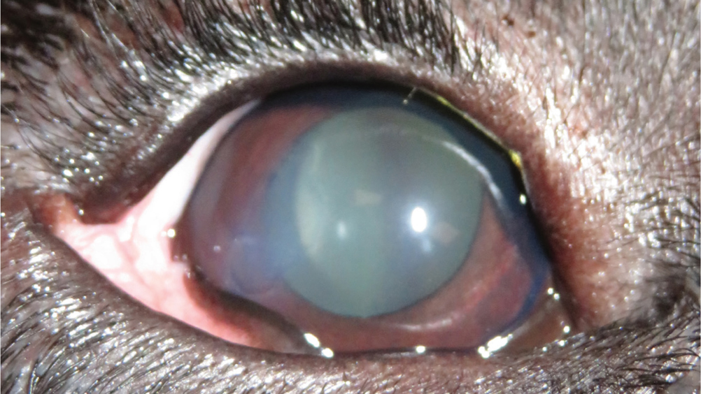
Figure 1. Superficial corneal ulcer.

Abstract

Corneal ulceration is one of the most common ocular problems presented in first opinion practice. Ocular diseases in dogs can be distressing for both the patient and the owner, registered veterinary nurses can provide advice to distressed owners and ensure that patients are provided with evidence-based and holistic care. Part one of this two-part series discusses the aetiology and management of superficial corneal ulcers. The second part of this article will discuss deep corneal ulcers and will explore the role of registered veterinary nurses within ocular diseases.

Corneal ulceration is one of the most common ocular disorders presented in first opinion practice (Startup, 1984; O'Neill et al, 2017). Corneal ulceration is described as a break in continuity of the corneal epithelium with exposure of the underlying stroma; they can be classified according to depth and extent (Maggs, 2013). Anecdotally, registered veterinary nurses (RVN) are often underutilised in veterinary practice and the author has noticed a particular underutilisation in ophthalmic nursing in first opinion practice (Gerrard, 2017; Harvey and Cameron, 2019). RVNs can be well utilised in corneal ulceration cases — their role extends from the recognition and understanding of corneal ulcers, ensuring appropriate handling and restraint, performing routine ophthalmic tests, recognising ophthalmic pain and supporting and educating owners.

The first part of this two-part series looks at the aetiology of superficial corneal ulcers and spontaneous chronic corneal epithelial defects (SCCED), the associated complications and the medical and surgical treatment options. The second part will discuss the aetiology, diagnosis and treatment of deep corneal ulcers and descemetoceles, and will explore the role of veterinary nurses in ophthalmology including: routine ophthalmic tests, patient handling and client education.

Register now to continue reading

Thank you for visiting The Veterinary Nurse and reading some of our peer-reviewed content for veterinary professionals. To continue reading this article, please register today.