
The key to successfully treating fish diseases is to recognise the earliest signs of diseases in fish and then arrive at a correct diagnosis. If a health problem arises, sick fishes will show a range of changes in their behaviour, feeding activity, and general physical condition. The good news is that no matter which of the >40 000 species of fish that may be seen in practice, the clinical signs presented are very similar. But the bad news is that the clinical signs displayed are often non-specific, and are insufficient to arrive at a diagnosis.
Healthy fish
Before clinical signs of illness are discussed, it is important that veterinary practitioners are familiar with the appearance of healthy fish. The following are characteristics displayed by a healthy fish:
Sick fish
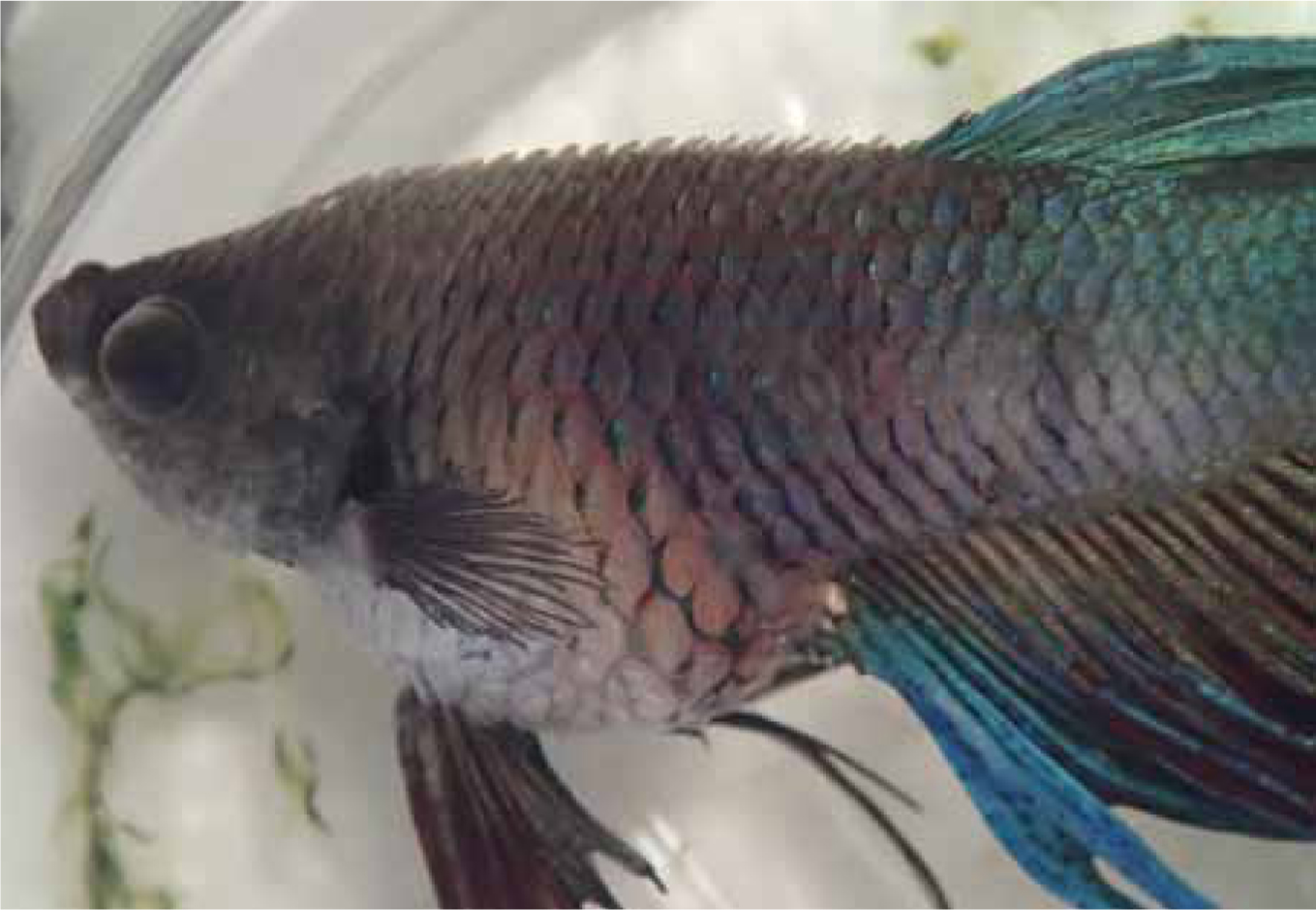
When fish are unwell, they may display all or a combination of the following behavioural changes (Figure 1):
Some of these behavioural changes provide the observer with some clues. One of the earliest signs of disease is inappetance. Inappetance may be a result of incorrect environment (e.g. water temperature being too high, or too low), a range of infectious diseases and more. Fish that are piping at the water surface are likely to be suffering from oxygen deprivation or carbon dioxide toxicosis (Loh and Landos, 2011; Loh, 2014). This may be the result of changes in dissolved gases, or if there is compromise to their respiratory system (e.g. parasitic infections of the gills, nitrite toxicosis and acidic water). These affected fish would also present with increased respiratory rate and effort. Fish that flash (scrape their bodies against objects) are reacting to skin irritation which can occur with parasitic infestations (Wildgoose, 2001; Loh and Landos, 2011), such as fish lice (Argulus), skin flukes (Gyrodactylus spp.) and a range of protozoa (e.g. white spot disease). All these same agents also cause fish to hold their fins clamped (Loh and Landos, 2011; Loh and Landos, 2014).
Sometimes, there may be the obvious presence of pathogens. Fish lice (Argulus) and anchor worm (Lernaea) and white spot disease (Figure 2) are perhaps the only conditions where gross findings are pathognomonic for those conditions. Even so, some fish affected with lymphocystis (Figure 2) can be confused with Ichthyophthirius. In fact, the majority of cases presented to the veterinarian are more challenging.
As disease progresses, the following may be observed:
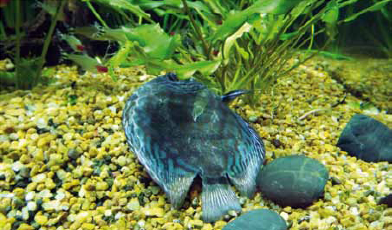
When stressed, discus fish turn a solid dark colour, while fish like oscars turn pale, and yet others like the koi develop diffuse skin congestion. Excess mucus (or slime) is contrasted well against the body of darkly pigmented fish, so it is useful to examine black fish. Note that this excess slime is more easily observed while fish remain in the water. This slime is often produced in response to skin irritation such as can occur with a range of ectoparasitic diseases and incorrect water conditions (e.g. low pH). Fish can develop tumours which need to be differentiated from neoplasia, inflammation and cystic structures (e.g. parasitic cysts). They can also develop ulcers which can be extensive and deep, exposing muscle and even bone. Ulcers in fish are not always a direct result of primary bacterial infection. More often than not, the inciting cause is parasitic (e.g. due to skin fluke infestation). Fish with ulcers commonly develop dermato-septicaemia, and therefore prompt treatment targeting the primary cause is necessary.
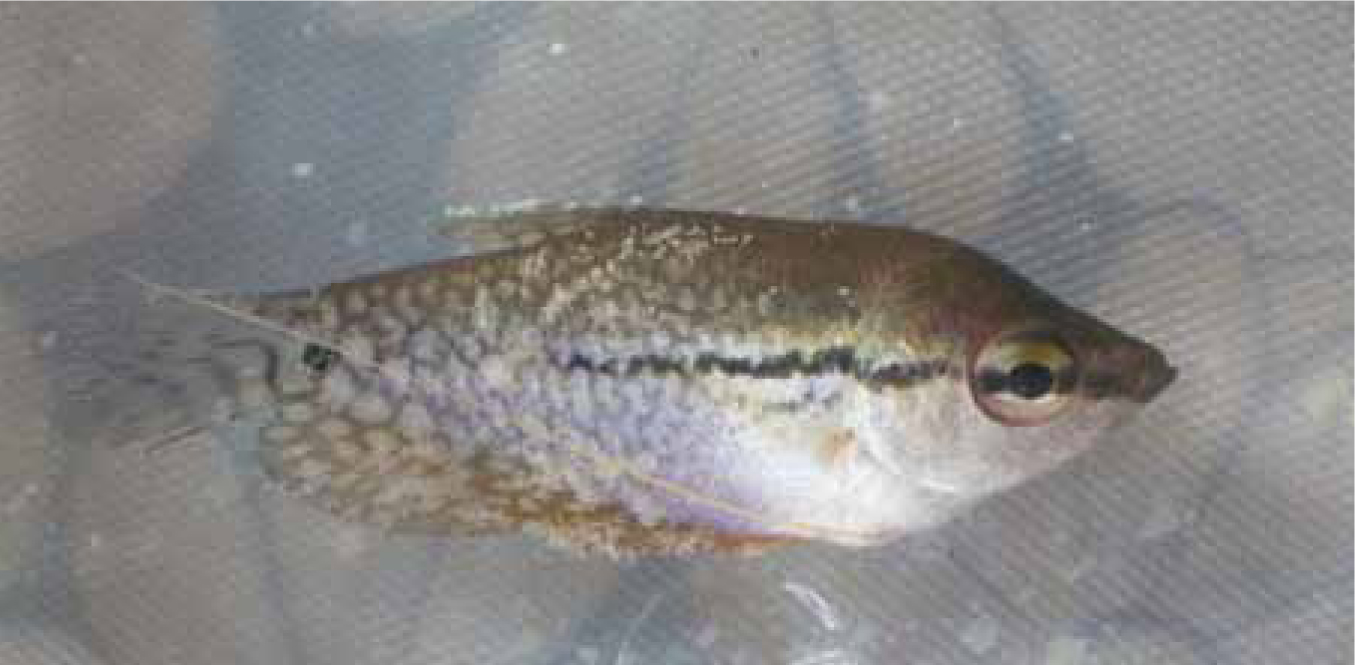
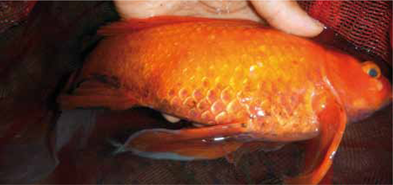
The terms ‘dropsy’ and ‘pop-eye’ are often used by hobbyists to denote bacterial septicaemia with consequent ascites, scale protrusion and exophthalmia. While this may be true in some cases, it is not always so. In fact, dropsy is a non-specific sign of fluid accumulation due to failure of fluid balance (or osmoregulation). Not all fish with bacterial infections end up with dropsy and not all conditions presenting as dropsy are due to primary bacterial infections (Loh and Landos, 2011; Loh, 2014). This fluid build-up may occur for several reasons including the failure to keep water out, the failure to excrete fluid or the over-production of fluid. To simplify things a little, the fish maintains its fluid in balance by properly functioning skin/mucus barrier, gills, kidneys and cardiovascular system. If there is damage to any of these systems (e.g. skin ulcers from fungal infections in barramundi, bacterial gill disease in guppy, kidney cysts in goldfish, ovarian cancers in koi), then fluid can accumulate in the fish (Loh and Landos, 2011; Loh, 2014).
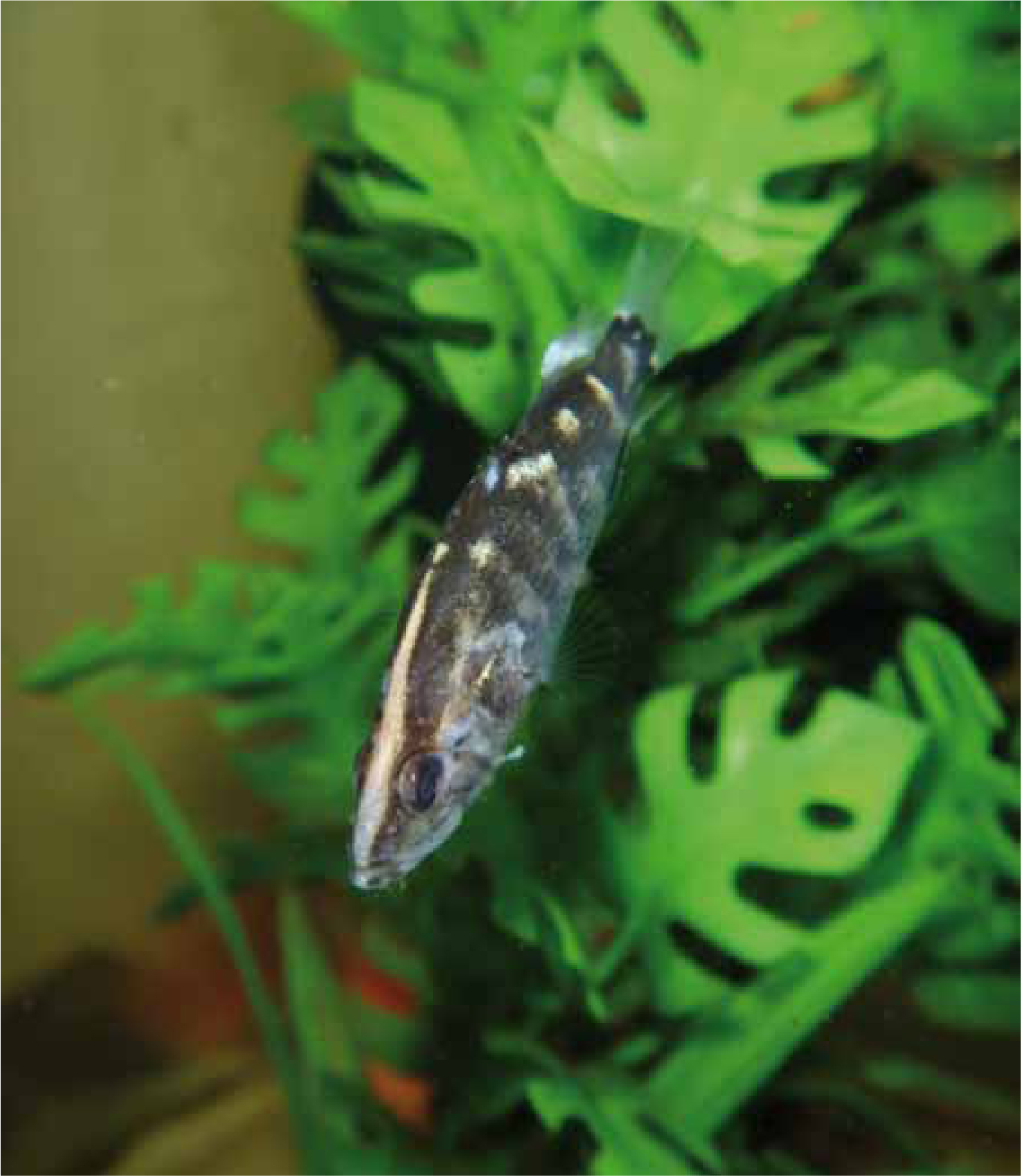
In contrast, fish can present in thin condition. Fish that are malnourished often present with disproportionately larger heads, and their bodies are long and slender. In the salmonid industry, these fish are called ‘pin heads’ (Loh and Landos, 2011). Fish with chronic diseases such as mycobacteriosis, or zebrafish with Pseudoloma infections also present as thin (Noga, 2010).
Gills are worth examining grossly. Acute irritation can lead to excess mucus, whereas chronic irritation leads to hyperplastic changes such as occurs with myxosporean infections in a disease known as ‘hamburger gills’. Gill colour should be noted; pale gills are diagnostic for anaemia and dark brown gills are consistent with nitrite toxicosis (Noga, 2010; Loh and Landos, 2011).
Fish with enteric disorders often present with long faecal trails and sometimes these may be empty. It is common for fish with hexamitiasis to present in this way, or to produce white faeces, or empty faecal casts (Loh and Landos, 2011). Some viral diseases also present this way (Noga, 2010).
Morbidity and mortality
Eventually, if the disease is not treated promptly, mortalities result. Some epidemiological history on the time course of mortality events can be helpful. For example, high mortalities (>40%) in a short period make the veterinary practitioner suspect environmental (toxins, low DO) and viral diseases. Moderate mortalities and the presence of sick fish over a period of 1 week may suggest other less lethal infectious diseases (e.g. bacterial, viral) and slowly fatal environmental conditions (low pH). In cases where there is low mortality (<20%) over extended periods (>1–2 weeks), chronic infectious diseases (e.g. parasitic), environmental degradations (e.g. high nitrate) and nutritional deficiencies may be more likely.
For mortality events where a range of species is involved, it is more likely caused by an environmental problem (such as toxicity or oxygen depletion). Deaths limited to one species (where other species are also present) is more likely a result of an infectious agent.
Role of the veterinary nurse
As the veterinary nurse, it is important to perform a triage to help the veterinarian on duty to prioritise patients. For fish that are to be admitted to the clinic, it would be important to know the clinical signs, and the morbidity/mortality rate, in order to provide time-critical intervention. For example, if fish present with respiratory signs of illness, then the fish patient will need oxygenation/aeration and plenty of dechlorinated water on arrival, and they would need to be seen promptly. If fish require surgical intervention, then the client would be advised to bring sufficient original tank water (provided the parameters are suitable) for the anaesthesia, recovery, and for their homeward journey.
In preparation for the consultation, the veterinary nurse may record a complete history such as what species are kept, which fish are affected (species, size and age), whether there had been recent introductions (newly introduced fish and plants may carry diseases), information about the tank (volume, filtration), water quality parameters (temperature, ammonia, nitrite, nitrate, pH, alkalinity, general hardness, salinity), medicines used previously, and more. The nurse may also perform water quality testing, record their observations of clinical signs displayed by fish, and report these to the veterinarian.
Conclusion
Observing fish clinically and grossly is helpful in early detection of fish diseases. This is integral to early intervention; athough the cause may not always be obvious and a definitive diagnosis is often only achieved after taking a complete history and running routine examination of water quality, the aquarium and filtration system, and of the animal.