
When using neuromuscular blocking agents (NBA) Bowman (2006) states that it is in the skill of the person in charge of the anaesthetic to keep the patient unconscious, so as not to allow the patient to become awake and paralysed, a particularly heavy burden for a nurse that has not had specific training in NBA use. This article will look at the history and relevant areas to nursing care that are required through procedures using NBAs.
History of NBAs
Anaesthesia protocols used in veterinary medicine have certain muscle relaxation qualities, however, in order to provide marked muscle relaxation a significantly higher dose of sedative/anaesthetic agent is required, thus causing an increasing risk of morbidity to the patient due to their inherent cardiovascular side effects. The triad of anaesthesia was introduced partly due to the discovery of muscle relaxants. The three points of the triad are hypnosis, analgesia and muscle relaxation (Raghavendra, 2002; Flaherty, 2009). NBAs are required in addition to normal sedative/anaesthetic protocols in order to prevent all skeletal muscle movement during intricate or more complicated surgeries (Bowman, 2006; Murrell and Ford-Fennah, 2011).
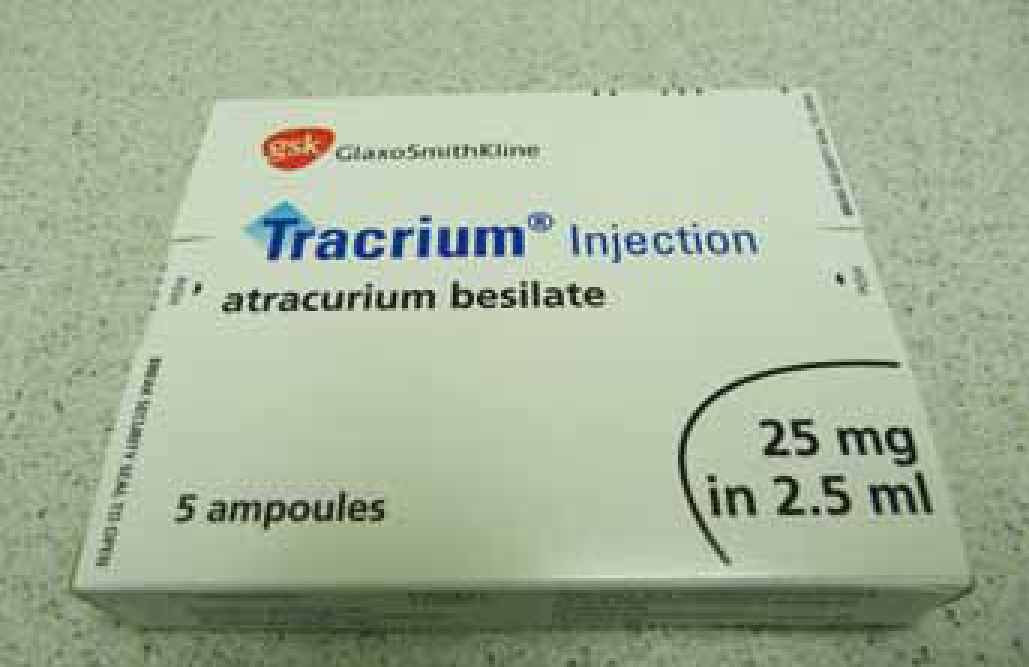
Curare is a plant-based substance that has been utilised for hunting by indigenous South Americans since the 16th century, its poisonous properties were transferred to hunting arrows causing death by paralysis (Bowman, 2006; Archer, 2012). An explorer (Richard Gill) who suffered with multiple sclerosis found it helped his spastic paralysis and imported some to the USA where drug companies realised its usefulness in anaesthesia. In addition its use as an NBA also became apparent. The synthetic forms of NBAs, based on curare, such as atracurium (Figure 1) have been utilised in human anaesthesia for around 30 years. Primary veterinary practices rarely find use for NBAs, however referral practices, specifically ophthalmology and soft tissue specialists, may use these drugs more regularly. Veterinary nurses need to have prior knowledge on the physiology of these drugs before they assist with the safe anaesthetic and recovery of patients undergoing admission of NBAs (Clutton, 2007; Archer, 2012).
NBAs work at the neuromuscular junction where a motor nerve connects with muscles (Murrell and Ford-Fennah, 2011). Acetylcholine (Ach) release is triggered by an action potential reaching the neuromuscular junction via the motor nerve. Ach received at the postsynaptic membrane of the neuromuscular junction causes the opening of an ion channel. Sodium ions move through to the muscle fibre causing depolarisation stimulating muscle contraction. NBAs act to prevent this depolarisation at the postsynaptic membrane therefore preventing muscle movement.
NBAs
NBAs (Table 1) can be divided into two different categories, these are:
| Dose required/duration of action | Contraindications | Additional information | |
|---|---|---|---|
| Non-depolarising | |||
| Pancuronium | Dose — 0.07 mg/kg |
Undesirable cardiovascular effects (tachycardia) and histamine release Contraindicated in renal failure as excreted via the kidneys | Long onset of action, slow recovery. Oldest drug, rarely used |
| Vecuronium | Dose — 0.1 mg/kg |
Metabolised by the liver, excreted by kidneys and bile, contraindicated in liver or kidney failure | Widely used Can be topped up Powder has to be reconstituted with water, the vial then lasts 24 hours |
| Rocuronium | Dose — 0.35 mg/kg. Most recently developed |
Fastest onset of non depolarising agents |
|
| Atracurium | Dose — 0.2-0.5 mg/kg |
Temperature and pH dependent reactions. Patients suffering from hypothermia or acid base abnormalities may have increased duration of action |
Broken down by hoffman degradation (PH and temperature dependent reaction), so not reliant on the renal system Can be topped up when 1 twitch reoccurs |
| Mivacurium | Dose — 0.08 mg/kg |
Not to be used in dogs |
Not regularly used |
| Depolarising | |||
| Suxamethonium (depolarising) | Dose — 0.1–0.3 mg/kg. Rapid onset Duration of action — 2–6 minutes in the cat, 20 minutes in the dog | Cardiovascular effects, malignant hyperthermia, increases intraocular pressure | Main use — rapid intubation in cats Must refrigerate |
Non-depolarising agents can be further divided into:
These drugs are not licensed for use in dogs and cats but are widely used in veterinary medicine (Murrell and Ford-Fennah, 2011; Archer, 2012). Before use dosages should be double checked by colleagues to ensure incorrect dosing is prevented.
Clutton (2007) theorises that due to the discovery and further authorisation of sugammadex (rocuronium antagonist) in human medicine in 2008 (European Medicines Agency, 2008), rocuronium may become the drug of choice in future for veterinary practices. Rocuronium's quick onset of action and limited side effects could take over the popularity of atracurium, avoiding its pH and temperature dependent side effects and potential histamine release.
Monitoring aids
It is important to remember that using NBAs under anaesthesia makes assessing depth more difficult. The eye will rotate into the central position and in some cases, such as during ophthalmic surgery, may not be available for assessment; lubrication should be applied to the eyes where necessary to protect them from damage. Ventilation will need to be controlled by intermittent positive pressure ventilation (IPPV) as intercostal muscle/diaphragm movement will cease and increases in respiration will therefore not be seen, the palpebral reflex will not respond and there will be no jaw tone or any twitches to indicate inadequate anaesthetic. The patient should be monitored by pulse oximeter, electrocardiogram (ECG), capnograph and blood pressure in order to recognise any hypertension, hypercapnia or tachycardia signalling increased cardiac output, an awake paralysed patient is not an option (Murrell and Ford-Fennah, 2011; archer 2012). The use of a peripheral nerve stimulator or train-of-four (TO4) is vital in order to indicate the effect of NBAs and requirement for additional doses.
Peripheral nerve stimulator
Peripheral nerve stimulators are extremely important when using NBAs to assess neuromuscular transmission. Neuromuscular transmission needs to be assessed regularly in order to confirm that the NBA in use is working sufficiently. Effective NBA admission = no muscular response.
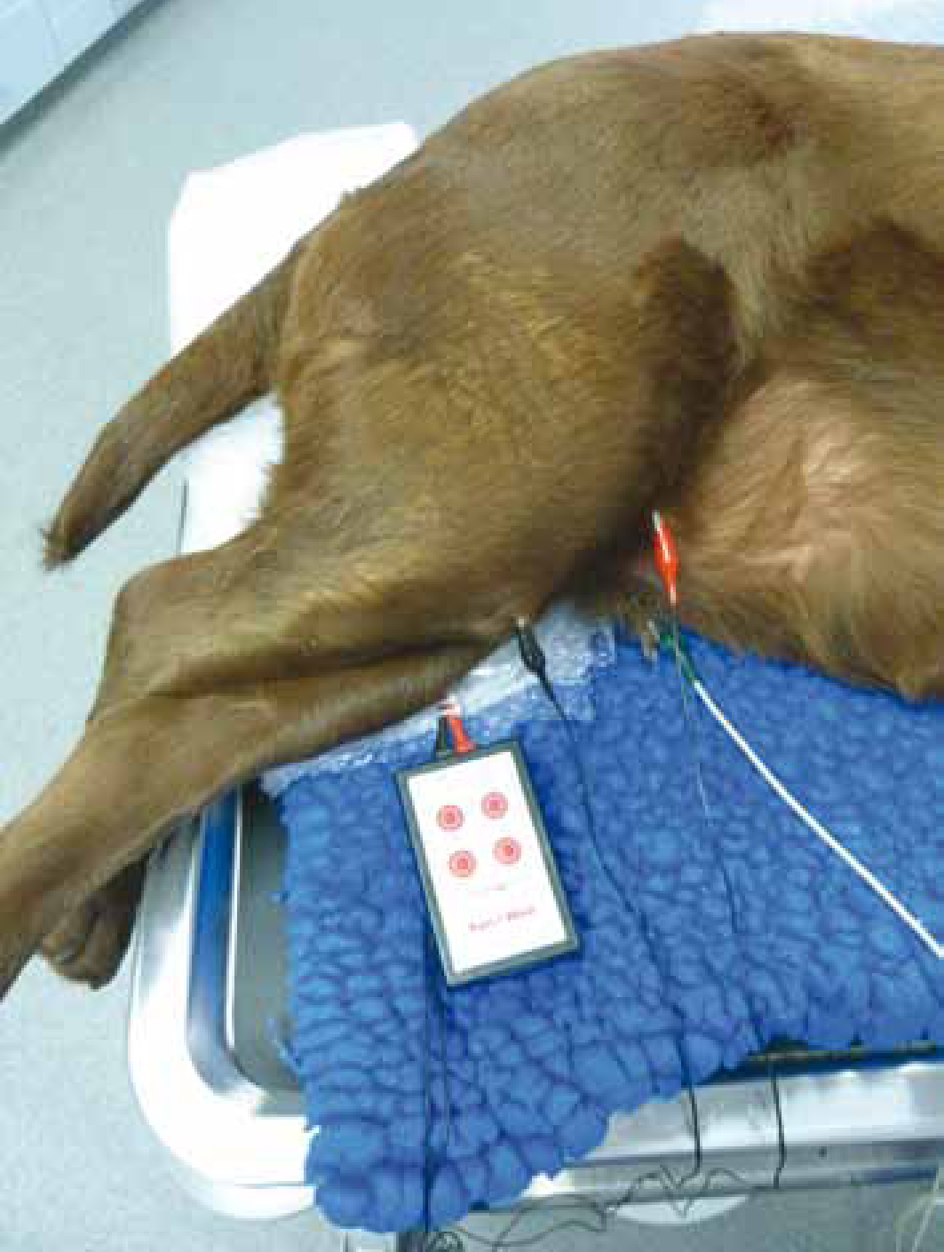
The peripheral nerve stimulator (Figure 2) can produce two patterns of muscular responses: the TO4 pattern and the double burst stimulation (DBS) pattern. These muscular contractions in these patterns are produced via a current that is sent from one electrode to another (attached by crocodile clips to the skin), causing the motor nerve it is attached to to respond with muscle contractions. Nerves that are commonly used to assess transmission of muscular movement are the facial nerve, ulnar nerve (Figure 3) and the deep peroneal nerve. The TO4 pattern should produce four equal strength muscle contractions (when NBA is not in use), produced over 2 seconds. Muscular contractions fade after administration of NBAs; the fourth will be the first to fade, continuing with the third, second and finally the first. The TO4 pattern should be monitored by sight and touch every 5 minutes during surgery to ensure NBA use is adequate for the surgery taking place. The first muscular contraction will be the first to reappear once the NBA is wearing off; when the first muscular response is seen the veterinary surgeon should be notified and administration of more NBA may be required to continue surgery (Archer, 2012).
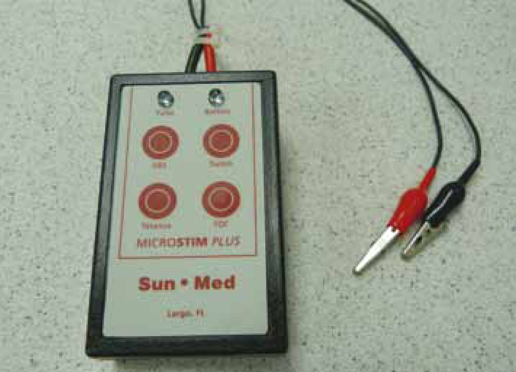
Alternatively the DBS may be used. This works by giving two bursts of muscle contractions, unlike four in the TO4, making it more accurate in defining the fading of muscular response.
The clips of the stimulator should be placed before NBA admission and when the patient is in position ready for surgery to take place. Recording of baseline values such as four equal strong kicks seen and felt is extremely important prior to NBA administration, to indicate what is expected to be observed when the NBA wears off in order to start off recovery once surgery is finished (Flaherty, 2009; Archer, 2012).
At the end of anaesthesia the DBS should be tested and should produce two equal strength muscular contractions a ratio of 0.9–1.0 (almost identical responses between the two contractions), however it has been shown that observations via the human eye or touch are unable to determine that contractions are equal beyond 0.6 (Archer, 2012). If the nurse is unsure of the equality of the contractions, they should be retested after 5 minutes and the veterinary surgeon should be informed because if they are not equal or similar to what was seen prior to NBA admission then the NBA may be still affecting the patient's response, and more time may be required to ensure the NBA has worn off or reversal of the NBA may be required (Archer, 2012).
Playfor (2002) deems the TO4 untrustworthy in some instances of hypothermia, as studies showed that the slightest variation in temperature alters the TO4 response to stimulation. Temperatures of 32°c or more showed little reduction in activity or muscular contraction responses, whereas 32°c or less showed extensive reduction in muscular responses.
The position of the TO4, usually connected at the ulnar nerve, has limited evaluation of diaphragmatic control, as blockade and recovery of these occur at different times. This indicates that the neuromuscular status of the patient should not be assessed solely on the TO4 activity.
IPPV
The use of a ventilator is ideal when using NBAs as the intercostal muscles and the diaphragm will not be controlled by the patient, so the patient will need to be artificially ventilated. Ventilators should be checked and have their settings ready prior to patient induction. Capnography should be used in conjunction with the ventilator as capnography can indicate numerous changes in patient condition, such as lightening of anaesthesia/neuromuscular blockade deficiency (bucking of the ventilator), tube obstruction (certain surgical procedures may require the head to be in a position that may compromise the endotracheal tube causing a blockage) and increase in end tidal carbon dioxide (CO2) (increased cardiac output) (Clutton, 2007; Bowcott, 2011; Taylor, 2011; Archer, 2012).
If a ventilator is not available then one nurse should have the sole responsibility of giving IPPV to the patient. A pressure gauge and Wright's spirometer can be used to help ensure adequate ventilation, another nurse would then be responsible for assisting with all other aspects of the anaesthetic. This situation is not ideal as it occupies two nurses (Clutton 2007; Taylor, 2011), while only one is required if a ventilator is present. On termination of the procedure and return of good muscle contraction via TO4, the patient should be able to ventilate themselves. They should be taken off the ventilator before the anaesthetic agent is turned off. Chest excursions should be observed in sternal recumbancy for good movement (make sure not abdominal breathing) and capnography observed for hypoventilation and maintenance of end tidal CO2 levels. Breaths may be shallow and ineffective and end tidal CO2 may be low, these are all indications of continuing NBA effects, the patient should be placed back onto the ventilator or manually ventilated and the veterinary surgeon should be contacted, the decision may be made by to give the reversal agent (see below) (Archer, 2012).
Reversal use
The short acting effects of NBAs in veterinary practice are normally timed to provide sufficient time to perform the surgery required; reversal of NBAs may be needed in situations where the muscular responses are not similar to the response given prior to NBA use, or if ventilation is not efficient when the patient is removed from the ventilator (inadequate chest exertions and unable to maintain CO2 at an acceptable level). Further relaxation can occur if there is poor muscle function when the reversal agent is given, as the reversal agent may be eliminated quicker than the muscle relaxant; monitoring needs to be increased in cases where all four muscle contractions have not returned (Flaherty and Auckburally, 2007; Murrell and Ford-Fennah, 2011; Archer 2012; Hunter, 2012).
Reversal agents are anti-acetylcholinesterase drugs; they prevent acetylcholinesterase breaking down acetylcholine (Ach), causing a rise in Ach, and increased competition to occupy the receptors at the neuromuscular junction. This can cause adverse side effects due to the increased Ach in circulation, including increased salivation, cardiac arrhythmias, bronchospasm, and bradycardia, so an anticholinergic drug such as atropine or glycoparalate should be given before hand to counteract the side effects. The rapid onset and short duration of action produced by atropine and endrophonium (reversal agent) makes them an ideal combination, alternatively the slow onset, long duration properties of neostigmine (reversal agent) and glycopyrronium also make them well matched (Flaherty and Auckburally, 2007; Murrell and Ford-Fennah, 2011; Archer, 2012).
Recovery
The percentage of patient deaths is higher in the recovery period than at any other stage (Crompton and Hill, 2011), this is believed to be due to the reduction in monitoring, extubation and hypothermia. One nurse should be performing one to one care during recovery, this allows the nurse to become accustomed to any small but significant changes that may occur. Vitals including temperature, pulse and respiration rate, should be recorded every 10–15 minutes until the patient lifts their head (Crompton and Hill, 2011).
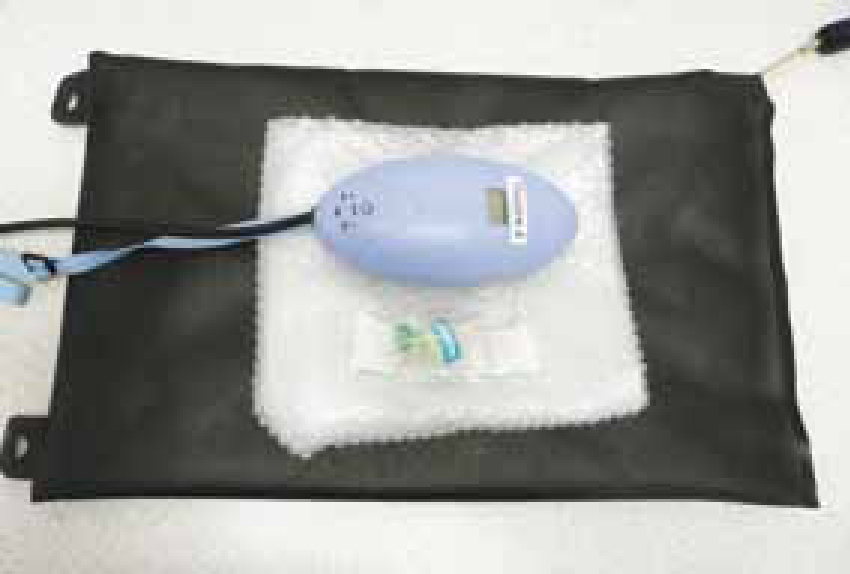
Oxygen delivery should continue until extubation to the point where the patient can swallow (consciousness deemed acceptable), intubation equipment should be prepared ready for re-intubation if required and appropriate induction agent available in the unlikely event of returning paralysis as laryngeal muscles are extremely sensitive to NBA effects (Archer, 2012). The patient is best recovered in sternal recumbancy for full inflation of the lungs. SPO2 should be maintained without oxygen delivery above 95% (Devey and Crowe, 2007; Crompton and Hill, 2011). Heating aids (Figure 4) such as fluid warmers, inditherm beds, hotties and bubblewrap used in surgery should also be utilised during recovery.
Dugdale (2010) noted that in mild hypothermia NBAs seem to become antagonised, but in severe hypothermia the opposite effect is seen making them re-relax, this shows the importance of continual monitoring of temperature and of the TO4 until fully recovered.
Contraindications
NBAs are contraindicated for use (Table 1) in certain patients, for example those patients that are concurrently have myasthenia gravis, as these patients will be more sensitive to NBA effects.
Thorough cannula flushes before recovery and removal of the peripheral nerve stimulator are important to ensure no residual NBA is left in the cannula (preventing further relaxation when recovered). Syringe labels are ideal for use in veterinary practice to help prevent incorrect drug administration, this became a requirement in 2003 in critical care areas (Carter, 2004; Arnot-Smith and Smith, 2010; Archer 2012).
Conclusion
A relevant induction programme should be implemented by a fully competent member of staff to nurses that will be exposed to patients under NBAs, or continuing professional development (CPD) time should be provided so knowledge can be sought out. The way in which each patient receiving neuromuscular blockade is handled follows a general protocol however the differences experienced are dependent on patient reactions and nurse interpretation of muscle activity. Preparation of drugs and equipment pre and post surgery is vital to ensure patient safety (Archer, 2012).