Summer poisoning hazards to pets
02 June 2022
17 mins read
Volume 13 · Issue 5
Box 1.

Abstract

As the spring turns to summer, owners and their pets will spend even more time out of doors. Some venomous animals are more active in the warmer months and there is risk of adder bites or stings from bees, wasps and hornets. Adder bites can result in significant morbidity but low mortality. Insect stings commonly cause local reactions and although these are generally mild, stings involving the airway are more hazardous since there is risk of respiratory obstruction. In addition, there is also a risk of anaphylaxis in sensitive individuals (just as in people) and multiple stings can cause multiorgan damage. Slug and snail killer products are more commonly used in the summer and are therefore more accessible to pets. These commonly contain ferric phosphate rather than metaldehyde which has been banned in the UK, and are less hazardous. Harmful summer plants include those containing cardiac glycosides such as foxglove and oleander. Some plants such as hogweed contain compounds that cause skin damage following dermal contact in combination with exposure to sunlight, and are therefore a particular risk on sunny days.

Adder and stinging insects are more active in the warmer months, so bites and stings are a potential risk as people and pets spend more time outdoors in the warm weather. Slug and snail baits are more commonly used in summer months to control these garden pests, and pets may be exposed to scattered pellets or chewing inadequately stored containers. Algal blooms are also a potential risk, particularly in summer months and these may be more common in some years. Common flowers may also pose a risk in gardens or on walks.

This article briefly discusses some of the seasonal summer poisoning hazards for pets. If more detailed information is required when managing a case, consult a poisons information service (Box 1).

Box 1.Veterinary poisons information services

  • Veterinary Poisons Information Service (VPIS) https://www.vpisglobal.com/
  • ASPCA Animal Poison Control Center https://www.aspca.org/pet-care/animal-poison-control
  • Specialist veterinary poisons information services are available, but some human poisons centres also answer enquiries about animal poisoning. Check with your local centre.

Adder bite

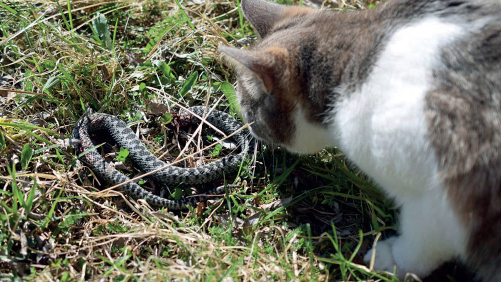
The European adder, Vipera berus berus is the only venomous snake native to the UK where it is a protected species. The adder generally only bites when provoked (Figure 1), and bites rarely occur during the winter when the snake is in hibernation but are frequent during the summer months usually when dogs explore while out on a walk. The adder is most commonly found on dry, sandy heaths, sand dunes, rocky hillsides, moorlands and woodland edges.

Figure 1. A cat hunting an adder.

Adder envenomation causes significant morbidity but low mortality. Not all bites result in envenomation (a snake can inflict a ‘dry bite’). The exact composition of adder venom is unknown and there are seasonal, individual and geographical differences. It is a complex mixture of proteins, mainly proteases, peptide hydrolases, hyaluronidase and phospholipases (Siigur et al, 1979; Calderón et al, 1993; Samel et al, 2006), that produce effects on multiple organ systems.

Clinical effects

All symptomatic animals develop local effects at the bite site with painful swelling that may occur within minutes of the bite and in most cases is present within 2 hours (sometimes up to 6 hours). Puncture wounds may be visible (two punctures about 1 cm apart) and may weep bloody exudate. The swelling can last several days and gradually spreads sometimes becoming haemorrhagic (Kängström, 1989). Without antivenom swelling will increase (Lervik et al, 2010) and reach a maximum after about 48 hours (Kängström, 1989). Ventral oedema may also occur (Kängström, 1989). If the bite is to the face the swelling may affect the animal's ability to eat, drink and thermoregulate. Severe oedema of the neck is not likely to result in suffocation (Kängström, 1989). Swelling can last several days. Lameness may occur with bites to a limb.

Systemic effects include shock, collapse and hypotension, mental status changes, pain, tachycardia or bradycardia, pyrexia or hypothermia, bruising, pale mucous membranes, vomiting and diarrhoea, panting and tachypnoea.

Arrhythmias, coagulopathy, haematological, renal and hepatic effects can also occur. Death from adder bite is uncommon (3–4.6% of cases) (Kängström, 1989; Hume, 2000; Sim, 2000; Sutton et al, 2011) and can occur after 5–7 days in dogs that receive no antivenom (Bratberg and Flesja, 1973).

Treatment

Observation is advised for any animal with clinical signs after an adder bite for a minimum period of 12 hours with monitoring of vital signs and the spread of the local swelling. In severe cases renal and hepatic function, creatine kinase, haematology, cardiac troponin I and markers of anaemia and coagulopathy should be monitored. The most important interventions are intravenous (IV) fluids and antivenom administration. IV fluids can help protect the kidneys, ensure adequate hydration, and correct hypovolaemia and haemoconcentration. The electrocardiogram (ECG) should be checked on admission and at 12 hours and 24 hours, if possible.

Antivenom is the most specific treatment in snake bite; it is expected to reduce morbidity, stop the progression of swelling and reduce hospitalisation time. Indications for administration of antivenom are given in Box 2. Analgesia should be given as required, but opioid analgesics are preferred over non-steroidal anti-inflammatory drugs because of the risk of renal impairment after adder envenomation.

Box 2.Indications for the administration of antivenom in dogs with adder bite

  • In all cases where the bite was to the facial region, as severe swelling may impair thermoregulation.
  • In all cases where the swelling is significant or has spread beyond the next major joint proximal to the bite.
  • In any animal with hypotension unresponsive to intravenous fluid therapy.
  • In any animal with evidence of coagulopathy.
  • In any animal with any electrocardiogram abnormality.

Routine use of an antibiotic is not required as bacterial infection is not common with snake bite. In a review of 53 cases of canine adder bite, 19% received antibiotics but none developed signs of infection (Lervik et al, 2010). A prophylactic broad-spectrum antibiotic should be considered in any dog with widespread necrosis.

There are no studies evaluating the effect of antihistamines (e.g. chlorphenamine) on the outcome of adder bite in dogs. They have been used in many cases and are theoretically of benefit as the venom causes a release of endogenous histamine. Antihistamines also have a sedating effect that may be beneficial.

Although frequently used, there is no role for steroids in the treatment of adder bites in dogs (except in rare cases of anaphylactic reactions to antivenom) (Warrell, 2011 personal comminication). The initial swelling is not an inflammatory response but a result of the cytotoxic effect of the venom. Steroids can also slow and diminish the response to antivenom and increase the risk of infection.

In dogs with severe anaemia, coagulopathy or bleeding, packed red cells or blood transfusions may be required. In most cases arrhythmias resolve without specific treatment (Kängström, 1989; Harjen et al, 2020); antivenom is the most specific treatment but antiarrhythmics may be used, if required.

As some signs may be delayed (e.g. cardiac signs, haematological changes) follow up at 1–3 days is suggested in animals discharged before this time. Follow up should include physical examination and assessment of bite site, an ECG and blood tests for renal function, liver enzymes, creatine kinase, haematology, cardiac troponin I and markers of anaemia and coagulopathy.

Bee, wasp and hornet stings

Female hymenoptera (bees (Figure 2), wasps and hornets) have stinging apparatus at the end of the abdomen. Bees have a barbed stinger with lancets that firmly attach to the skin of the victim during the attack. Stinging results in the tearing off of the apparatus and death of the insect. The reflex action of the stinger muscles, however, continues to inject more venom into the victim. Wasps and hornets have unbarbed stingers and can inflict multiple stings. Wasps are much more aggressive than bees, which are usually docile (except for Africanised honeybees in the Americas).

Figure 2. Bee stings can result in local or systemic effects and anaphylaxis.

Clinical signs

Hymenoptera stings may result in localised pain and swelling (Figure 3), and only very rarely cause severe toxicity. Death can also occur from severe local reactions, particularly if the airway is involved resulting in respiratory obstruction (Fitzgerald and Flood, 2006), but most deaths occur as a result of immediate anaphylaxis (Fitzgerald and Flood, 2006), which is not dose related.

Figure 3. Swelling round the eyes and slight swelling of the muzzle in a dog with a wasp sting in the mouth.

Local reactions include immediate pain, erythema, swelling, inflammation and pruritus. These effects are usually self-limiting and resolve within about 24 hours. In some cases, there may be a larger, more regional reaction which spreads out from the site of the sting with erythema, local oedema and cellulitis (infection is rare). This reaction may not develop until 24 hours after the sting (Fitzgerald, 2012).

As in people, in sensitive individuals a single sting may produce serious, potentially fatal, anaphylactic reactions usually within 30 minutes. Death can occur from a single sting and can occur within 1 hour (Peterson, 2007). Clinical effects include urticaria, vomiting, angioedema, collapse, hypotension, bronchoconstriction, wheezing and dyspnoea. Local airway oedema can cause congestion of the larynx and surrounding tissue.

Animals with multiple stings become unwell immediately with local effects but may not present until 24 hours or longer with more severe signs (Noble and Armstrong, 1999). In animals with multiple stings there are local effects and multi-organ damage with depression, pyrexia, red-brown urine and vomitus, melaena, rhabdomyolysis, acute renal failure, myoglobinuria, elevated liver enzymes, disseminated intravascular coagulation (DIC), pulmonary oedema, respiratory distress and convulsions. Haematological changes show inflammation with evidence of haemolysis. Severity of poisoning can be related to the number of bee stings in dogs (Table 1).


Table 1. Toxic dose of bee stings in dogs
Number of stings Possible outcome
1 sting Often mild effects and rapid recovery, but death can result if anaphylaxis occurs
<14 stings/kg Survival
14–24 sting/kg Critical
>24 stings/kg Fatal

(Peterson, 2007)

Delayed reactions from insect stings are rare and occur within 3 to 14 days after the sting and include rash, serum sickness, pyrexia, general malaise and lymphadenopathy (Fitzgerald, 2013). The delay between the sting and onset of signs may result in the diagnosis being missed.

Treatment

An animal with anaphylaxis must be stabilised before removal of bee stings is attempted. Any bee sting still embedded in the skin should be promptly removed. The emphasis is on speed of removal without concern for the method used (Vissher et al, 1996). If multiple bee stings are suspected the animal should be carefully examined to locate all the bees. This should include examination of the mouth, because pets will often snap at flying insects and can easily be stung in the mouth.

Prompt treatment is required for animals with anaphylactic reactions and adrenaline (epinephrine) is recommended for severe reactions. After apparent recovery owners should be advised to return if the animal becomes unwell over the following few days (e.g. vomiting, weakness, depression, dark urine).

Treatment of a local reaction from a sting is symptomatic with antihistamines, steroids and analgesia, if required. A cold compress may also be help alleviate discomfort (Reisman, 1994). Routine use of antibiotics is not required for a single sting, as infection following insect stings is rare (Fitzgerald, 2013). Animals should be monitored for 30–90 minutes for signs of anaphylaxis (Osweiler et al, 2010) and any animal with marked depression or weakness on admission or in the days following the sting(s) should be assessed as for multiple stings.

Any animal with multiple stings should be admitted for observation and management for at least 24 hours (Fitzgerald, 2013). Management is aimed at correction of hypovolaemia, prevention of vascular stasis (Cowell et al, 1991) and maintenance of urine output (Wysoke et al, 1990). Monitoring should include vital signs (cardiac, respiratory), liver and renal function, packed celled volume (PCV), haematology and coagulation parameters. Antihistamines, corticosteroids and analgesics can be given for local effects. IV fluids should be given for hypotension and to maintain hydration, but care should be taken in animals with suspected pulmonary oedema, as overzealous use of fluids will result in exacerbation. If hypotension persists despite IV fluids, dopamine, noradrenaline (norepinephrine) or adrenaline (epinephrine) may be required (Osweiler et al., 2010). Aggressive airway management may be required in severe cases and fresh frozen plasma or a blood transfusion may be required for coagulopathies or severe anaemia.

Blue-green algae

Blue-green algae (cyanobacteria) are a group of bacteria found in fresh, brackish and marine water bodies. They often, but not always, have a blue-green colour (can also be red, brown and black). Blue-green algae are either floating (planktonic) or bottom-dwelling (benthic). Under favourable conditions blue-green algae may form massive growths or visible growths or blooms (most commonly in late spring, summer and early autumn). Many blue-green algae contain or produce a variety of toxins, although not all species are toxic. Exposures in animals usually occur when they drink water from ponds or water bodies where the algae have bloomed (Figure 4). In dogs, exposures have occurred as a result of the animals swimming in infested water bodies and also from grooming after skin exposures. The toxins produced by the blue-green algae have a high acute toxicity and exposures frequently result in fatality. Cyanobacterial blooms occur worldwide, and it has been recognised for over a century that they are responsible for deaths in livestock, birds, fish, wildlife and dogs (Stewart et al, 2008; Smith and Daniels, 2018).

Figure 4. Blue green algae (cyanobacteria) produce various toxins, but not all algal blooms are hazardous.

The mechanisms of toxicity vary between cyanobacteria species. The two main toxin groups are hepatotoxins and neurotoxins, which are released when cells die.

Hepatotoxic compounds are the most commonly encountered poisoning with cyanobacteria (Carmichael, 1992; Tyagi et al, 1999). These compounds disrupt normal cytoskeletal structure (Falconer and Yeung, 1992; Dawson, 1998) leading to hepatic necrosis and failure. Fatalities resulting from exposure to this type of toxin normally occur as a result of hepatic necrosis, haemorrhage and hypovolaemic shock (McDermott et al, 1998).

Neurotoxic compounds are produced by several cyanobacteria. Anatoxin-a (previously called very fast death factor) mimics the neurotransmitter acetylcholine and binds to acetylcholine receptors but is not degraded by acetylcholinesterases (Tyagi et al, 1999) leading to neurological effects.

In addition, the cell walls of many of the blue-green algae species contain endotoxins which are irritant to mucous membranes and skin, which can cause gastrointestinal and dermal effects.

Clinical effects

Gastrointestinal effects are often the primary presenting sign after cyanobacteria exposure (Corkill et al, 1989; Elford et al, 2012). Dermal effects such as pruritus and urticaria may occur (Puschner et al, 2017), but are rare in dogs possibly because their hair provides some degree of protection from direct skin contact.

Dogs are most commonly exposed to hepatotoxic cyanotoxins, and liver enzymes usually increase within 24 hours of exposure (Harding et al, 1995). Initial signs of hepatotoxic cyanotoxin ingestion are non-specific with vomiting, diarrhoea, anorexia and lethargy. This is followed by evidence of haemorrhage, hypotension or hypovolaemic shock and jaundice. Death is a result of hypovolaemic shock with hepatic haemorrhage or hepatic insufficiency.

Onset of clinical effects is usually very rapid if a neurotoxic species of cyanobacteria is ingested and often occurs within 15 minutes. The animal is likely to present within 1 hour (Elford et al, 2012). There are several neurotoxic cyanotoxins and signs of exposure can include hypersalivation, muscle rigidity and tremors, paralysis, diarrhoea, bradycardia, cyanosis, convulsions, collapse and respiratory failure.

Treatment

It will usually not be possible to determine if the cyanobacteria involved in a case is toxic within a clinically relevant time, so all exposures must be assumed to be toxic.

Emesis should only be induced if the animal is asymptomatic, and ingestion was very recent (<1 hour). Adsorbents should be given if practicable. If hepatotoxicity is suspected or confirmed, colestyramine, if available, is recommended instead of activated charcoal (Rankin et al, 2013). Thorough dermal decontamination is essential to prevent or reduce exposure from material on the coat. In critically ill animals, the dog should be stabilised first and then washed. Protective clothing (e.g. gloves) should be warn to protect staff from dermal contact with blue-green algae during decontamination of exposed animals.

All asymptomatic animals should be observed for at least 6 hours post-exposure in the veterinary surgery. Liver enzymes should be measured to establish a baseline. If normal and the animal is asymptomatic after 6 hours, then observation can continue at home for another 24 hours and the owners advised to return if the dog becomes unwell. Signs of neurotoxicity can occur rapidly within the first 6 hours and if the animal is well after this time, it is reasonable to assume there will be no neurotoxicosis. Any further treatment of a symptomatic animal should be aimed at monitoring and management of liver toxicity.

In symptomatic animals, the liver enzymes, clotting parameters, renal function and vital signs should be monitored. Liver enzymes should be measured to establish a baseline when the animal is brought in even if asymptomatic. If abnormal or the animal is symptomatic the liver enzymes should be measured again at 24 and 48 hours. If hepatotoxicity is suspected or confirmed colestyramine (cholestyramine) should be given instead of activated charcoal. If activated charcoal has been given, then give colestyramine when the next dose is due. Colestyramine may still be useful several days after exposure (Rankin et al, 2013).

Liver protectants such as acetylcysteine and/or S-adenosyl-L-methionine (SAMe) should be considered. In animals with hepatic failure vitamin K therapy can be given, and blood transfusion and correction of electrolyte imbalance is recommended (Stewart et al, 2008). Other treatment is mainly supportive with IV fluids and an antiemetic if required, to ensure hydration.

Management of animals with neurotoxicity from cyanobacteria exposure is supportive. Assisted ventilation may be required for respiratory depression. Diazepam is not always effective for control of severe muscle twitching or convulsant activity and propofol or isoflurane may be used. Atropine may be considered, if the animal is exhibiting cholinergic signs, such as hypersalivation and bradycardia (Beasley et al, 1989).

Slug baits

Products to controls slugs and snails are widely used by gardeners to reduce damage to plants. Slug baits are typically blue-green pellets and numerous products are available (Figure 5).

Figure 5. Various products are available for slug and snail control in the UK, but most now contain ferric phosphate.

Metaldehyde has long been a common ingredient of molluscicide preparations, but the outdoor use of metaldehyde slug baits was banned in the UK from 31 March 2022, in a move to protect wildlife and the environment (Defra, 2020). It is likely to be available in homes for some time, however, so cases are likely to continue. Methiocarb is a carbamate pesticide and although it is used as an insecticide (often in seed dressings), use as a molluscicide was banned in the UK in 2015. Following the banning of metaldehyde and methiocarb molluscicides, products now commonly contain ferric phosphate (usually 1 or 3%).

Clinical effects

With metaldehyde, onset of clinical signs after ingestion may be very rapid, often within 30 minutes of ingestion, but may be delayed for up to 3 hours. Common signs of metaldehyde toxicosis are vomiting, diarrhoea (may be discoloured blue-green), ataxia, tremor, twitching, muscle spasms or fasciculation, seizures, opisothotonus and pyrexia. Respiratory effects and cardiac effects are less common (Bates et al, 2012). Metaldehyde-induced seizures can last up to 72 hours but on average last 15 hours (Bates et al, 2012). Complications of prolonged seizure activity include severe pyrexia, rhabdomyolysis, disseminated intravascular coagulation (DIC) and multi-organ failure. The fatality rate in dogs with metaldehyde poisoning is approximately 14–17% (James, 1955; Studdert, 1985; Robertson et al, 1992; Yas-Natan et al, 2007; Bates et al, 2012).

In ferric phosphate products (1 or 3%) the other 99 or 97% is usually bran or wheat which can cause gastrointestinal signs with diarrhoea, vomiting, lethargy, abdominal discomfort and coloured stools. Occasionally, haematemesis and melaena may occur (Buhl et al, 2013; Tizzard and Bates, 2018). Iron poisoning is less of a risk with ferric phosphate-containing slug and snail killers, compared with iron-containing pharmaceuticals, because of the low oral bioavailability of iron in this form.

Treatment

If the dog is presented to the surgery within 1 hour of metaldehyde exposure and is asymptomatic then induction of emesis may be considered. Administration of an emetic after this time is best avoided because of risk of seizures. Gastric lavage should be considered in metaldehyde toxicosis, particularly if the dog is thought to have ingested a significant quantity, or has developed moderate to severe signs soon after ingestion. Activated charcoal is probably best avoided in conscious animals because of the risk of rapid onset seizures.

The mainstay of treatment of metaldehyde toxicosis is control of seizures and prevention of complications of prolonged seizure activity. Multiple drugs may be needed to control seizures (Bates et al, 2012) and it is important to be prepared to escalate therapy quickly if the animal shows signs of deterioration.

Diazepam is an appropriate first-choice drug to use in any animal with twitching or seizures, and may be considered for pets with mild signs. Propofol should be used immediately for any case presenting with moderate or severe signs. Animals may need to be on propofol for some time, so it is more practical to use a constant rate infusion (CRI). Midazolam, isoflurane, barbiturates or methocarbamol (Richardson et al, 2003) can also be used.

Ideally animals should be kept under anaesthetic for about 6–12 hours and then the dose slowly tapered off and the animal observed closely to see if seizures recur. If they do then the dosing should be resumed as before. Animals with metaldehyde toxicosis may be sedated or comatose for a prolonged period and will need appropriate monitoring and care.

Where possible, blood gases and body temperature should be monitored. Cooling measures should be instituted for any animal with pyrexia, however the temperature should fall once seizures are controlled. Acidosis is managed by restoring tissue perfusion through administration of IV fluids containing a bicarbonate precursor (such as lactate) or, if required, sodium bicarbonate therapy. Once signs are controlled, acidosis may resolve without the need for bicarbonate therapy.

Where possible baseline liver enzyme activities should be measured on admission and repeated at 24–48 hours, and beyond as required. These abnormalities usually resolve with supportive care only and require no specific intervention.

Metaldehyde is not lipophilic (the log P is very low, only 0.12) and therefore it is unlikely that lipid emulsion would be useful. Although there are a few reports of the use of lipid emulsion in the management of metaldehyde toxicosis (Lelescu et al, 2017; Bergamini et al, 2020), these animals received multiple therapies and it is impossible to determine the contribution of lipid emulsion to the successful outcome of the cases.

Haemodialysis has been shown to remove metaldehyde from canine plasma in vitro (Mauser et al, 2016) and is effective in clinical cases (Teichmann-Knorrn et al, 2020), but availability will restrict its use.

Treatment of dogs that have ingested ferric phosphate molluscicide is symptomatic and supportive, with management of gastrointestinal signs. Iron is not adsorbed by activated charcoal and is not recommended.

Summer plants

Cardiac glycoside-containing plants

Cardiac glycosides are found in numerous plants. Most cases involve foxglove (Digitalis purpurea, Figure 6a), oleander (Nerium oleander, Figure 6b; Trautvetter et al, 1969; Page and Murtaugh, 2015) and lily of the valley (Convallaria majalis;Moxley et al, 1989; Atkinson et al, 2008).

Figure 6. Cardiac glycosides are found in (a) foxglove and (b) oleander.

Some plants such as foxglove are bitter and so may not be eaten readily. The toxic dose of these plants is difficult to determine and in most clinical cases the quantity ingested is usually unknown. Even a relatively small quantity can cause severe signs of poisoning, although severe cases in cats and dogs are not common. Foxglove ingestion in guinea pigs, however, commonly causes severe poisoning (Blackett and Bates, 2018).

Clinical signs usually occur within 6 hours with gastrointestinal effects, cardiac arrhythmias and electrolyte imbalance. The earliest signs of poisoning are vomiting, abdominal discomfort and diarrhoea (or constipation). Animals may appear disorientated with weakness, depression and dullness. There is collapse, recumbency and convulsions in severe cases. Cardiac arrhythmias are very variable, and may be difficult to manage. The risk of poisoning is increased in animals with pre-existing renal impairment or cardiac disease.

Emesis and repeat dose activated charcoal should be given following ingestion of plant material containing cardiac glycosides, as they undergo enterohepatic recirculation.

Treatment is supportive. If possible, electrolytes, particularly potassium should be checked. Rehydration may be required, but calcium-containing fluids should be avoided as elevated serum calcium concentrations can increase the effect of cardiac glycosides on the myocardium. The blood pressure, heart rate and ECG should be monitored, if possible. Insulin and dextrose can be used for marked hyperkalaemia.

Atropine (0.01–0.03 mg/kg IV) can be used for bradycardia. Lidocaine (lignocaine) or procainamide have been used in the management of ventricular arrhythmias, and pacing has been used in a dog with refractory AV block after ingestion of lily-of-the-valley (Atkinson et al, 2008), but this will be impractical in most cases owing to cost and availability.

Digoxin antibody Fab fragments (DigiFab®, Protherics Medicines Development Ltd) are an option in cardiogenic glycoside poisoning, but this drug is very expensive (>£750/vial) and it may be difficult to obtain. It has been used in clinical cases (Pao-Franco et al, 2017) of cardiac glycoside poisoning, particularly with N. oleander, in dogs. Digoxin antibodies should be considered in cases of severe poisoning, e.g. hyperkalaemia resistant to correction, tachyarrhythmias or bradyarrhythmias unresponsive to drug therapy or associated with hypotension. In humans 5–10 vials is recommended for cardiac glycoside poisoning, but in veterinary medicine it is recommended that 1 or 2 vials are given initially, and the effects observed (Gwaltney-Brant and Rumbeiha, 2002). More vials can be given if there is some clinical improvement.

Phytophotodermatitis

Many plants can cause dermal reactions, particularly with repeated exposure but some produce effects from contact in combination with sunlight. This occurs with hogweed. Heracleum mantegazzianum (giant hogweed,) is a very tall plant found throughout Britain and Ireland, often beside water. The native Heracleum sphondylium (cow parsnip, hogweed, keck) is a smaller plant but it also found throughout Britain and Ireland.

These plants contain psoralens which sensitise the skin to sunlight (they are phytophototoxins). Skin reactions occur from contact with the plant in combination with exposure to long-wave ultraviolet light, that is, sunlight (Klaber, 1942). Skin damage can result from any activity that involves touching the plants, in combination with exposure to ultraviolet light. Phototoxic effects are the result of ultraviolet light-induced binding of psoralens into nuclear DNA, resulting in cross-links and subsequent cell death. This causes skin damage (phytophototoxicity), which may be severe (Song and Tapley, 1979). Even close contact with animals which have been in contact with hogweed may result in dermatitis in humans (Camm et al, 1976), so care must be taken when handling animals exposed to hogweed.

Dermal effects from contact with hogweed have been reported in dogs (Hintermann, 1967; Dolowy, 1996; Pötz, 2015), often when they have been playing and run into a stand of hogweed plants. Cases are occasionally reported on social media. The fur may provide some protection and effects can be more pronounced on areas with less hair cover. Gastrointestinal signs and ulceration around the mouth have been reported from ingestion of hogweed in animals, however, most cases involve dermal exposure, with severe erythema, alopecia, oedema, scaling, blistering, ulceration and dermatitis. Dermal signs can progress over 24–48 hours and recovery can take weeks; there is also the risk of secondary infection of affected areas.

If exposure was recent, the area should be washed thoroughly with soap and water as soon as possible. It is important to keep the animal out of sunlight for at least 48 hours post exposure, even if they are asymptomatic. If dermal signs develop, management is symptomatic with analgesia, antihistamines for erythema or blisters, and systemic or topical steroids to relieve irritation (Pötz, 2015; Baker et al, 2017). Antibiotics may be required in cases of secondary infection of dermal lesions.

If a dog presents with skin lesions and no apparent explanation, owners should be questioned about possible exposure to hogweed.

KEY POINTS

  • Adder bites and stings from bees, wasps and hornets are a risk in warmer months.
  • Adder bites cause significant morbidity but low mortality.
  • Bee, wasp and hornet stings commonly cause local reactions but can also cause anaphylaxis; multiple stings can result in systemic effects.
  • Poisoning with slug bait is common in summer but cases of metaldehyde poisoning in the UK should decline following its ban.
  • Summer plants may also be hazardous, particularly those containing phototoxins that cause dermal effects in combination with exposure to sunlight.

Conclusion

In the summer months people and pets may spend more time outdoors where they may be at risk of bites and stings from animals that are active during the summer months such as adders, bees, wasps and hornets. Algal blooms are also common in the summer and dogs may be exposed when swimming in or drinking from water bodies in warm weather. Ingestion of slug bait is also common at this time of year and some plants are particularly hazardous in the summer as the toxic chemicals they contain cause dermal reactions in combination with exposure to sunlight.