Electromyography is a performance evaluation tool commonly employed to assess efficacy of training and rehabilitation regimens in humans (Hanon et al, 2005). However limited research has been conducted in the horse and most studies that have been published have been restricted to the laboratory environment (Peham et al, 2001; Peham and Schobesberger, 2006; Licka et al, 2009; Zsoldos et al, 2010). Surface electromyography (sEMG) systems are now available which can assess muscle activity by measuring motor unit action potentials (MUAP) in ‘real-time’ via telemetric monitoring thus facilitating objective analysis of equine performance and training practices outside of the laboratory. Analysis of sEMG data can be used to establish onset and offset of muscle contraction and also determine muscle performance via analysis of signal amplitude (a measure of magnitude of MUAP) (Hanon et al, 1998; Hanon et al, 2005; Richards, 2008). Analysis of the mean EMG frequency (mEMGF) can provide an objective measure of fitness levels (Duc et al, 2005; Hikari et al, 2006) while a left shift in the median EMG frequency (MeEMGF) over time can indicate fatigue during isometric testing (Hanon et al, 2005).
Horse racing is a traditional sport and many equestrian training regimens are currently underpinned by historic and anecdotal good practice (Smith et al, 1999; Ely et al, 2010). Equine training generally aims to condition a horse so it has sufficient musculoskeletal and cardiovascular fitness for the level of performance required, while preventing the accumulation of injury; a secondary aim, is to promote a ‘balanced’ athlete to optimise performance and reduce risk of injury. The aim of training racehorses is to improve the horse's endurance, speed, stride, balance and coordination to postpone fatigue and maximise the physiological potential of the individual horse (Ferrari et al, 2009). Training to date in racehorses has concentrated on cardiovascular assessment and changes in stride parameters neglecting assessment of adaptation in the musculoskeletal system (Ferrari et al, 2009) despite musculoskeletal injury being a major cause of wastage and days lost from training and racing in the UK racing industry (Williams et al, 2001; Stover, 2003). National Hunt racehorses have been consistently shown to be more at risk of musculoskeletal injury than their flat racing counterparts (Williams et al, 2001; Parkin et al, 2004) with most injuries occurring in training rather than during racing (Pickersgill et al, 2000). Gait changes are often observed as a precursor to injury in racehorses suggesting that musculoskeletal health could be integral to reducing injury levels (Rogers et al, 2005).
In flat racehorses, who perform at maximum capacity, maximum heartrate (HRmax) has been shown to correlate with maximum rate of oxygen uptake at the muscles (VO2 max) and exercise capacity, and is used as a physiological measure of fitness and athletic potential (Gramkov and Evans, 2006; Gur and Matur, 2012). National Hunt racehorses race longer distances than flat horses and are also required to jump; therefore horses need to predominately possess aerobic endurance capacity, approximately 90%, with some anaerobic power required at the obstacles and for the concluding stages of the race (Evans, 1994). Traditionally National Hunt horses train at submaximal levels to build aerobic endurance however only one study to date has evaluated the efficacy of training practices in this sphere; Ely et al (2010) found that horses that had amassed longer canter distances in training in the 30 days preceding a race were more successful. The accumulation of distance at submaximal exercise levels could be improving performance via musculoskeletal conditioning as well as developing aerobic capacity. Musculoskeletal strength and conditioning are intrinsically influential on racing success, therefore a system that could objectively analyse muscular ‘fitness’, such as sEMG, could inform performance and help identify precursory events to injury. This study aimed to establish if sEMG could be utilised to assess performance of the Gluteus superficialis muscle during canter interval training in National Hunt racehorses of variable fitness.
Materials and methods
Nine National Hunt Thoroughbred racehorses of variable fitness and work level were actively engaged in canter interval training, conducted on an all-weather gallop, silica sand mix. These horses were selected by convenience sampling in consultation with their trainer. All horses that participated were declared sound by the trainer, were clipped, were subjected to similar management regimens and showed no distress by the experimental protocol. Ethical approval for the study was granted by the UWE Hartpury Ethics Committee.
Experimental protocol
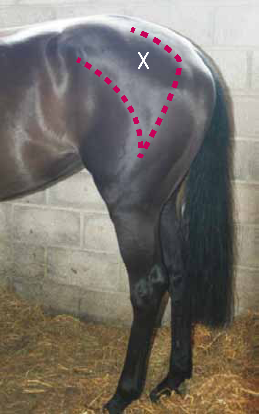
The Gluteus superficialis muscle was selected for this preliminary investigation. This was chosen as it is acknowledged to generate a large proportion of the power during cantering (Clayton et al, 2002; Dutto et al, 2004), was easy to access to verify sensors remained in situ during the experiment and could be readily palpated to identify the main muscle body to reduce cross talk. The origin of the Gluteal superficialis occurs at the Tuber coxae, with the insertion on the 3rd trochanter of the femur; the left and right Gluteal superficialis of nine racehorses were palpated utilising these anatomic landmarks enabling identification of the maximum muscle body and definition of a suitable test area for sensor placement (Figure 1). This was conducted by a single experienced experimenter to reduce placement error. All horses had been previously skin-clipped and the test areas were prepared using alcohol wipes to remove skin debris and grease to ensure good EMG signals (Konrad, 2005; Rouffet and Hautier, 2008). Sensors housing bipolar sEMG electrodes (Delsys Trigno™; Boston, USA) were secured bilaterally, with the electrode bars aligned parallel to the muscle fibre direction using doubled-sided adhesive and secured further with duct tape. Horses then underwent their normal warm up routine; subjects paused at the base of the gallops to enable sensors to be turned on and then commenced their normal canter interval training regimen.
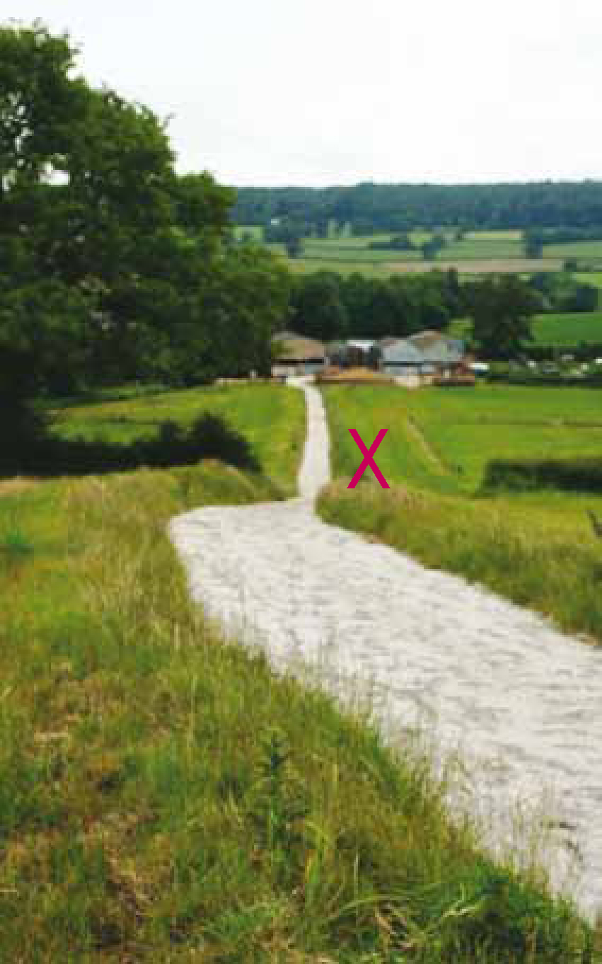
Measurements were obtained via an EMG telemetric system (Delsys, Trigno™) at a sampling rate of 4000 Hz, 16-bit resolution and a gain of 300; the system was located alongside the data collection area and powered by a generator. The gallops were 847 m in length and comprised a straight with an increasing elevation of 347 m from the start to a subsequent oval flat circuit of 500 m (Figure 2). A pilot study determined that telemetric data collection was limited to a field of approximately 15 m. A standardised 10 m length was selected for the maximum incline (top) of the gallops as this represented the period of maximum muscle activity and the point where approximately 60% of canter work had been undertaken. Data were collected using the Delsys, Trigno™ EMGworks acquisition program for each of the runs undertaken within the horse's training regimen. The number of runs conducted varied between each horse in accordance with the level of work predetermined by the trainer (2–5 runs) (Figure 3). Average speed for each run was assessed using a stop watch and predetermined markers denoting the start and finish of the experimental distance. A consistent average speed of 5.4 m/s (12±0.6 mph) was found across the horses. Lead canter legs for the horses were noted by the observer. The trainer reported their interpretation of fitness level for each horse and expressed this as a percentage of race level fitness, using a scale of 0%: not fit to 100%: race fit.
Data processing and analysis
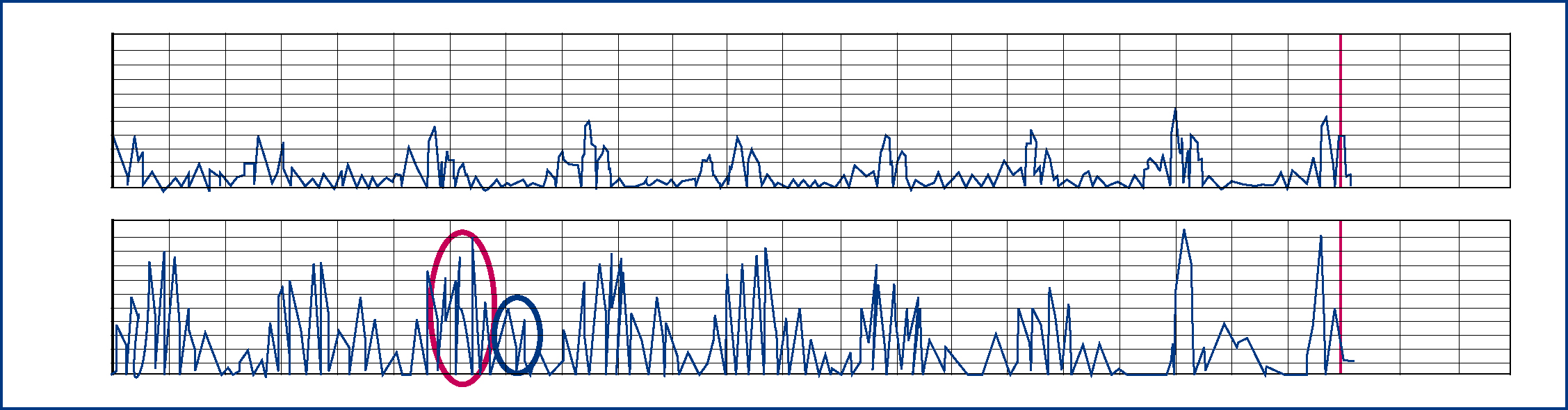
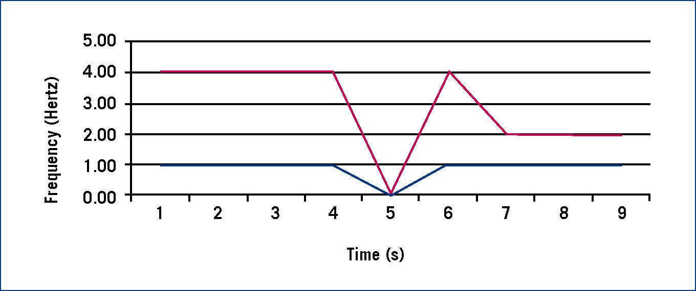
Data processing was performed using analysis software (Delsys EMG Works™ Version 4.13). In accordance with other EMG studies in the horse; data were rectified and a 5th order Butterworth low-pass filter with a cut-off frequency of 10 Hertz was applied prior to analysis (Licka et al, 2009; Zsoldos et al, 2010). Visual assessment of the sEMG trace identified onset and offset in the Gluteal superficialis within the defined study period. Zsoldos et al (2010) have successfully demonstrated that direct comparison of related EMG events is a valid approach when evaluation of overall muscle activity is the defined objective. In this work, evaluation of repeated sEMG profiles for defined bouts of activity within subjects was one of the core objectives of the study therefore this approach rather than standardisation to limb phasing was deemed appropriate. For each run mean MUAP (mMUAP) for the left and right Gluteal superficialis, mean amplitude frequency (mEMGF) and median amplitude frequency (MeEMGF) were calculated and exported to Microsoft Excel™ version 2010 to organise repeated runs for individual horses, and was subsequently analysed in Statistics Package for Social Scientists (SPSS; version 19). Repeated measures ANOVA tests assessed differences in mM-UAP and mEMGF between runs for the left and right Gluteal superficialis, across the cohort and between runs for individual horses. A Pearson's product moment correlation identified if mEMGF was related to perceived fitness level. mEMGF and MeEMGF for individual horses were plotted over time (0.25 second intervals) for each piece of canter work to visually assess fitness and fatigue levels for the duration of the training periods that were recorded.
Results
Visual assessment of the sEMG data
Examination of the sEMG data traces allowed the onset of muscle activity to be evaluated (Figure 4). The values observed were comparative to those obtained in a laboratory environment in the horse (Licka et al, 2009; Zsoldos et al, 2010) indicating that the field methodology was sound.
Muscle activity and lateralisation
Highly variable and individualised mMUAP readings were observed both intra and inter subjects for left and right Gluteal superficialis activity (Table 1); all horses except for horse 2 cantered with a right canter lead, foreleg. Similarly the amplitude of the maximal contractions per run across the population exhibited a wide degree of variance (mean maximal amplitude = 157.24±57.1mV). Across the cohort, no lateral difference in Gluteal superficialis activity was observed (p>0.05) however, highly significant differences were exposed in 58% of the population (horse 1: p=0.0001; horse 2: p=0.002; horse 3: p=0.0001; horse 5: p=0.0001).
| Horse | 1 | 2 | 3 | 4 | 5 | |||||||||||||
|---|---|---|---|---|---|---|---|---|---|---|---|---|---|---|---|---|---|---|
| Canter | 1 | 2 | 3 | 4 | 1 | 2 | 3 | 4 | 1 | 2 | 3 | 1 | 2 | 3 | 1 | 2 | ||
| RG.S. | Mean (mV) | 44.27 | 38.89 | 47.75 | 33.94 | 35.10 | 27.51 | 22.20 | 18.30 | 40.31 | 111.26 | 9.98 | 21.12 | 21.40 | 21.49 | 13.65 | 54.65 | |
| LG.S. | Mean (mV) | 11.93 | 14.49 | 12.24 | 5.84 | 34.38 | 27.94 | 18.75 | 18.53 | 13.01 | 15.11 | 13.85 | 13.49 | 15.29 | 12.47 | 2.75 | 474.93 | |
| Horse | 6 | 7 | 8 | |||||||||||||||
| Canter | 1 | 2 | 3 | 1 | 2 | 3 | 4 | 5 | 1 | 2 | 3 | 4 | 1 | 2 | 3 | 4 | 5 | |
| RG.S. | Mean (mV) | 9.32 | 15.08 | 14.50 | 80.14 | 88.49 | 30.73 | 61.08 | 32.76 | 27.71 | 33.72 | 19.52 | 105.12 | 55.76 | 54.58 | 47.09 | 51.80 | 31.66 |
| LG.S. | Mean (mV) | 20.94 | 27.00 | 21.71 | 42.35 | 41.25 | 13.98 | 35.54 | 24.46 | 449.19 | 240.64 | 53.09 | 106.81 | 22.97 | 27.78 | 9.20 | 564.36 | 6.70 |
Fitness and fatigue
No significant differences were found for mEMGF between runs within the group (p>0.05), however differences were seen at an individual level for 72% of the study sample (p<0.001). These were found between the first and all subsequent runs in three horses (p<0.001), and between run 2 and 3 (p<0.01) in two horses. mEMGF varied throughout runs suggesting increased and decreased muscle effort occurred during training, but interestingly, no relationship between fitness level (as predetermined by the trainer) and mEMGF was exposed (p>0.05). MeEMGF for seven horses remained consistent throughout runs indicative of a lack of fatigue, however in two horses partial fatigue was observed (Figure 5).
Discussion
This study has shown that sEMG exhibits the potential to be an objective tool to assess muscle activity levels and fitness, via visual assessment of sEMG traces and repetitive analysis of mMUAP and mEMGF in individual horses, although more extensive studies are warranted to confirm this. It was not possible to determine a group effect. The exercise speeds observed indicate submaximal exercise levels occurred during this study, yet the variation in EMG data obtained suggests individualised muscular responses occurred in the cohort of horses supporting Ferrari et al's (2009) suggestion that assessment of training on muscle conditioning is warranted and Ely et al's (2010) hypothesis that submaximal exercise conditions the musculoskeletal system. While this work highlights the potential applications of telemetric sEMG for analysis of musculoskeletal performance within the horse, it should be noted that the data collated represent ‘snap-shots’ of locomotory activity and not the entirety of the training period. Future work would benefit from continuous recording of muscle activity related to speed via a datalogger to establish complete fitness and fatigue profiles for assessment of efficacy of training for the complete duration of work.
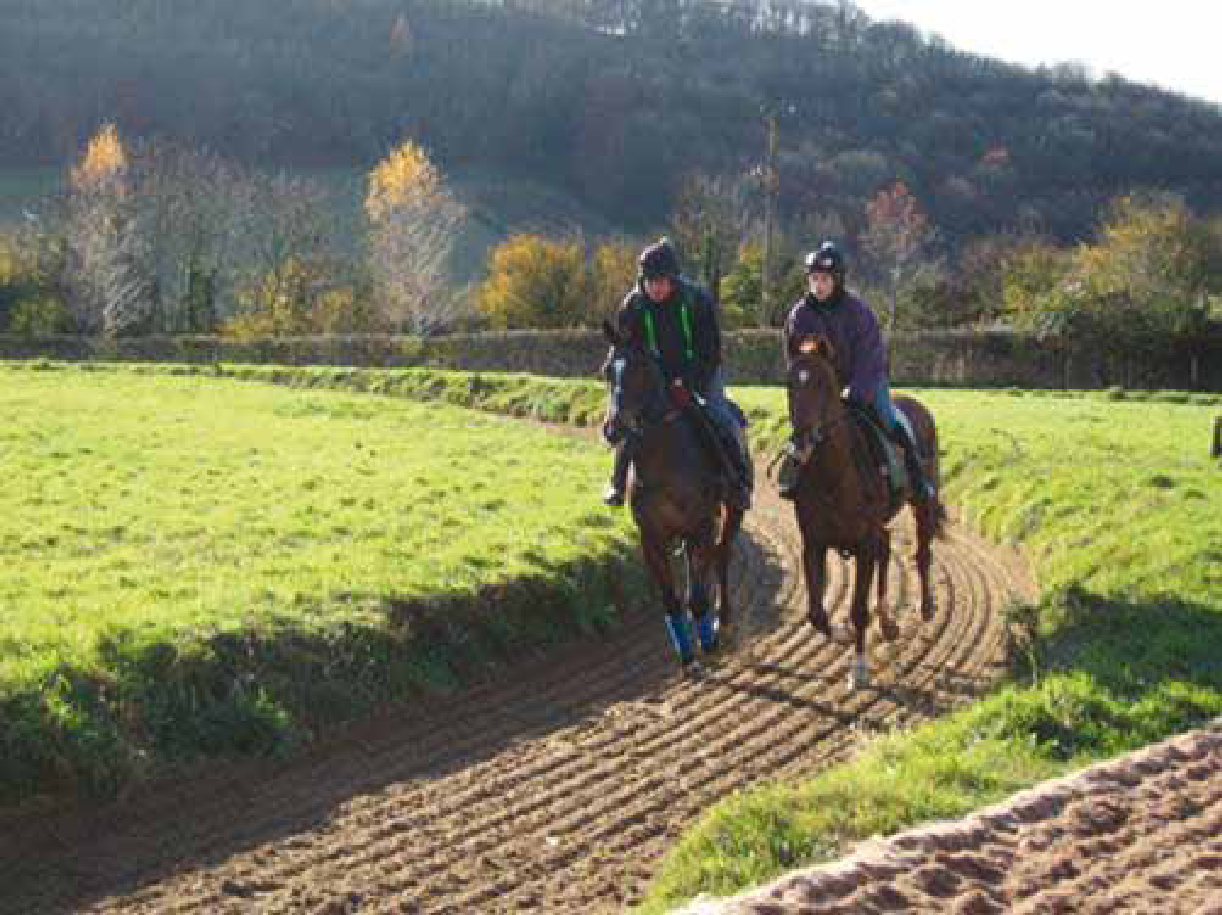
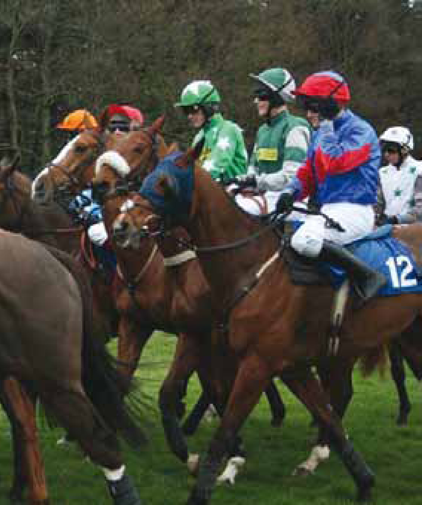
Some lateral variance in MUAP activity was expected to be observed due to increased muscle recruitment arising from the lead hind leg during canter; however the level of variance recorded within horses 1, 2, 3 and 5 was considered to exceed expected biomechanical lateralisation. Some individuals favour a specific hind limb to drive locomotion; this may be due to inherent or acquired laterality which could be associated with asymmetrical training or injury (McGreevy and Rogers, 2005). This study has shown that sEMG also exhibits potential as an objective tool to assess lateralisation of muscle activity in the individual horse and scope exists to relate this information to inform training practices or assess rehabilitation post injury to work towards a balanced athlete across equestrian disciplines. This is of particular interest in National Hunt racehorses (Figure 6) due to the high levels of musculoskeletal injury reported (Williams et al, 2001; Parkin et al, 2004) as training to promote symmetry could reduce uneven loading during higher intensity performance in races, galloping and jumping, on the tendons and ligaments of the distal limb predominately injured (Williams et al, 2001, Pinchbeck et al, 2004; Thorpe et al, 2010).
Within canter training mEMGF varied throughout runs suggesting increased and decreased muscle effort occurred; while MeEMGF for most horses remained consistent, evidence did present of partial fatigue. These results suggest that individual horses respond to interval training in different ways. This could be attributed to genetic variation, heritability of performance and/or muscle fibre profiles, or possibly could be related to the rider's technique as their riding style can obviously influence the effort from the racehorse, speed, and thus alter muscle activity levels. This technology exhibits potential to be used to design programmes to maximise performance and monitor fitness and fatigue within the equine athlete. However, it should be noted that the horse as a species has no defined and validated anatomical reference points for EMG assessment to date which could limit reliability between repeats to the experience of the experimenter. Muscles are also larger than their human counterparts, therefore as one sensor analyses fewer motor units, future work may wish to utilise multiple sensors to more fully assess muscle performance. Cross talk between muscle groups can also introduce errors in data collection, therefore accurate palpation and location of sensors is paramount in future work. The dynamic nature of canter and gallop exercise also presented its own challenges, movement and sweat production, and to retain the sensors in place, duct tape was utilised which although visually did not appear to influence gait may have exerted some residual effect on muscle activity. The main limitation of this study is that only one muscle was assessed for movement which comprises multiple muscle groups; however the success of data collection in this one area suggests a positive future for this emerging field.
Interpretation of sEMG data is acknowledged as challenging as variation in amplitude has been attributed to increased numbers of active motor units being recruited or the result of a change in the frequency of activation of motor units, i.e. firing rate has increased but the same numbers are being recruited (Stegeman et al, 2000; Konrad, 2005). Equally not all motor units fire at the same time and this is dependent on muscle fibre type; this will vary with the individual although Thoroughbreds will generally present with a greater ratio of fast twitch fibres compared with other horse breeds. An elevated frequency for amplitude of the sEMG signal can be due to an increase in fast twitch fibre recruitment or a higher firing rate in slow twitch fibres, both equalling more muscle effort, or could be the result of decreased synchronisation between motor units that are firing, muscle starting to fatigue. In contrast, a decreased frequency may be attributed to a reduced firing rate, fatigue, or increased motor unit synchronisation, coordinated muscle activity, no fatigue present.
Conclusion
sEMG adds a potentially useful tool to assess racehorse training methods within individual horses. Visual assessment of sEMG traces can indicate muscle recruitment during exercise while analysis of mM-UAP and mEMGF could provide comparative evaluation of performance and test efficacy of training systems to facilitate muscular conditioning. Further work evaluating MeEMGF recorded consistently over time may also prove useful to determine muscular fatigue.