Dermatophytosis in cats and dogs

01 July 2011
8 mins read
Volume 2 · Issue 6

Abstract

Dermatophytoses are a common dermatological disease. They can be challenging to manage as they are expensive to treat, difficult to control and zoonotic. Correct identification through good laboratory analysis is essential, with fungal culture being the gold standard. Knowledge of drugs available to treat dermatophytosis is helpful, although most animals with a competent immune system will resolve their dermatophyte infection spontaneously. Treatment of animals in multi-pet households or where there are young children or immuno compromised individuals is essential because of the highly contagious, zoonotic nature of this disease.

Dermatophytosis is one of the most common dermatological diseases seen in pets. It can be challenging to manage as it is expensive to treat, difficult to control and zoonotic. Correct identification through good laboratory analysis is essential and knowledge of drugs available to treat dermatophytosis is helpful.

Dermatophytosis is more common in cats than dogs and prevalence is higher in geographical areas with a warm and humid climate (Scott et al, 2001).

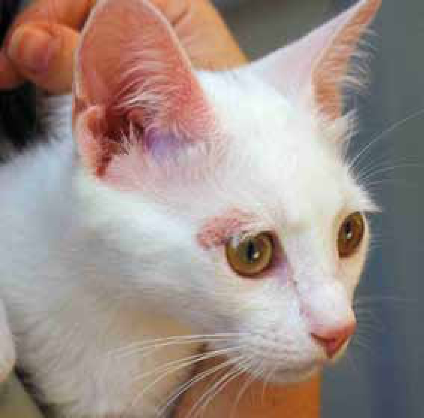
  • Microsporum canis — 90% of cats with ringworm have this type, 60% of dogs are likely to have caught it from a cat. Persians are genetically predisposed. (Gaudiano, 2005) (Figure 1)

    Figure 1. Presence of a focal area of hair loss caused by M. canis on the face of a domestic short hair kitten.

  • Microsporum gypseum — contracted through contact with contaminated soil, rare in the UK. Causes kerions (a nodule accompanied by a deep suppurating inflammatory response)

  • Trichophyton mentagrophytes — carried by wild rodents. Only 30% of dogs with dermatophytosis have this type, mostly Jack Russell terriers as they tend to attack these rodents (Gaudiano, 2005) (Figure 2)

  • Figure 2. Focal crusting of the bridge of the nose in a Yorkshire terrier with a Trichophyton mentagrophytes infection.

    Most human infections are caused by various Trichophyton (mentagrophytes, rubrum, verrucosum, tonsurans etc.) or Epidermophyton species (floccosum etc) (Outerbridge, 2006; Shaw, 2010).

    Transmission

    Transmission occurs through direct contact with infected animals or indirectly from contaminated fomites (such as bedding). Infection occurs when arthrospores attach to the upper layer of the epidermis, the stratum corneum. Spores germinate into fungal tubes known as ‘hyphae’, these are aided by enzymes produced by the fungal spores. This causes an inflammatory reaction, damaging the hair shaft and causing hair to fall out easily (Figure 3)

    Figure 3. Hyphae and arthospores can be seen along the hair shaft.

    Factors involved in the development of fungal infection

  • Host status — youth (kittens), debilitating disease, compromised immune status (from disease or drugs), poor nutrition, stress
  • Host immunity to dermatophytosis (both natural immunity and specific acquired immunity following infection)
  • Infective dose of the organism.
  • Clinical signs of dermatophytosis

    The typical lesions are characterized by areas of alopecia with peripheral erythema, scaling and crust. Hairs are broken and have a frayed aspect.

  • Moth eaten alopecia
  • Diffuse truncal alopecia
  • Scaling
  • Hyperpigmentation
  • Alopecic, papulo-pustular, crusting lesions on the face and nose or limb
  • Pruritus
  • Claw infection
  • Chin furunculosis (Figure 4 and Figure 5).
  • Figure 4. Multifocal areas of hair loss on the face of a Boxer puppy with M. canis infection.
    Figure 5. Generalized dermatophytosis in a Yorkshire terrier.

    The lesions may affect the whole body or can be localized to the head, ears, tail and front paws. In cats there could be a severe inflammatory response (Scott et al, 2001).

    Diagnosis

    There are a number of ways that dermatophytosis can be diagnosed; diagnosis should be based on clinical history, appearance and diagnostic procedures. With training and practice a qualified veterinary nurse can perform all of the following diagnostic procedures.

    Wood's lamp

    A Wood's lamp is an ultravoilet hand held lamp that causes infected hair to fluoresce apple green. This must be done in complete darkness with a good quality lamp, which has been properly warmed up.

    The benefits of this test are that it is fast, inexpensive, and can enable the screening of a large number of animals from the household at the same time. It also helps in the selection of infected hair for culture/trichogram.

    On the negative side only 50% of Microsporum canis strains fluoresce, and false positives may be caused by scales, crusts, topical medications.

    Direct hair examinations (Trichogram)

  • Place hairs in 10% potassium hydroxide for 5–10 minutes for clearing
  • Place 1–2 drops of lactophenol cotton blue on the slide
  • Apply sample to the slide
  • Examine at 10X–40X
  • Identify arthrospores/hyphae.
  • The benefits of this procedures are that it is possible to get fast results and it is inexpensive. On the negative side the process is time consuming and results are difficult to interpret due to the presence of debris and the complex structure of normal hair. False negative results can occur if unaffected hairs are examined.

    Fungal culture

  • Collect hair and scale from periphery of lesion, or hair that fluoresces by using a sterile brush, brushing all over the pet if it has no lesions. Apply sample to dermatophyte test media (DTM) or Sabouraud dextrose agar (SDA)(Figure 6). DTM is SDA with the addition of cycloheximide, gentomycin and chlortetracycline to prevent growth of fungal and bacterial contaminants. Some contain a colour indicator to help demonstrate the presence of dermatophyte

    Figure 6. Fungal plates with dermatophyte test medium and a round brush (Mackenzie brush) for sample collection.

  • Incubate for 3 weeks at 27°C (most dermatophytes grow within 7 to 14 days but keep for 3 weeks to be certain)

  • Check daily for white, fluffy growth (Figure 7)

    Figure 7. Fungal plate with a few colonies of M. canis

  • Use acetate tape to sample colony, place on a slide with 1–2 drops of lactophenol cotton blue (Figure 8)

    Figure 8. Presence of macroconidia in a sample collected from a colony of M. canis and stained with cotton blue lactophenol.

  • Examine at 10x–40x. Generally, many long strands of fungal hyphae, and at least a few macroconidia (spores) will be seen.

  • On the negative side results may take up to 4 weeks to obtain and false negative results may be seen if samples are incubated at the incorrect temperature.

    Skin biopsy

    Histopathological examination can confirm diagnosis in about 75% of cases (Mignon, 2008), but is no more sensitive than microscopical examination of hair and scale and is rarely necessary.

    Treatment

    Before initiating treatment it is worth considering that most animals with competent immune systems will resolve their dermatophyte infection spontaneously without treatment.

    Dermatophytosis is highly contagious and zoonotic in nature. For this reason animals in multi-pet households or in homes with young children or immune compromised individuals should always be treated.

    As cats may appear clinically normal, but may be chronic carriers, it is important to examine and culture all in-contact animals, systemically treat all animals with positive cultures, and decontaminate the environment simultaneously.

    During treatment fungal culture should be repeated every 2 weeks until 2–3 successive negative cultures are obtained.

    Clipping

    The aim of this simple treatment is to prevent further environmental contamination and facilitate topical therapy. The benefits of this treatment are that removing infected hairs minimizes contamination to the patient's environment but contaminates only the area where clipping is performed. Clipping encourages new hair growth, pushing out old diseased hair, and makes shampoo therapy more effective.

    On the negative side disease may worsen at 7–10 days post clipping if lesions are spread due to microtrauma caused by clippers.

    Environmental treatment

  • Use sporocidal disinfectant such as bleach or Virkon.
  • ○ Chlorine laundry bleach solution, 0.05% final concentration (most solutions as purchased are 5%, thus use a dilution of 1/100 = 10 ml/litre)
  • Vacuum thoroughly all areas where the animal has been and then dispose of vacuum bags (if used)
  • Discard bedding, brushes and toys, which may form fomites
  • Enilconazole environmental spray or smoke generator is licensed for cattery use in some parts of Europe.
  • Topical therapy (rinses or creams)

    The main aim of topical therapy is to destroy fungal elements on hairs and reduce further contamination (Morriello, 2003a

  • Enilconazole 0.2% (Imaverol®, Janssen-Cilag) topical dip licensed for dogs. Apply every 3 days.
  • Unpredictable toxicity was initially reported with enilconazole use in cats, caution should therefore be advised. It is theorized that the toxicity is related to oral ingestion by grooming after the rinse. In one study of 22 Persian cats treated with enilconazole, 6 developed elevated serum alanine aminotransferas and one developed transient muscle weakness (Hnilica, 2002).

  • Lime sulphur (LimePlus Dip®, DermaPet) has been found to be effective and safe (Newbury et al, 2007). It should be used at 5–7 day intervals. Licensed for use in dogs, cats and horses.
  • Antifungal creams are not recommended as topical therapy should be generalized not localized.
  • The benefits of topical therapy are that it limits spread and reduces contagion. Lime sulphur is very safe and can be used in pregnant, and nursing cats and kittens/dogs and puppies as young as 2–3 weeks. Enilconazole is very effective (Hnilica, 2002).

    On the negative side topical therapy may worsen disease initially, and it is important to be gentle and dry the patient thoroughly, keeping them warm until dry. Dips/rinses are time consuming. When Enilconazole is used liver enzymes should be monitored (see above).

    Shampoo therapy

    Used as an adjunctive therapy miconazole/chlorohexidine shampoo (Malaseb®, Dechra) can alleviate inflammatory response and reduce environmental contamination. Miconazole plus chlorhexidine (Malaseb®) shampoo was recently studied in cats as an adjunct treatment to oral griseofulvin (Paterson, 1999). Cats treated with shampoo+griseofulvin recovered visually at about the same rate as cats treated with griseofulvin alone. However, shampoo+griseofulvin cats achieved negative fungal cultures much more quickly than those treated with griseofulvin alone. In vitro data clearly show that there is synergism between these two ingredients for some strains of dermatophyte fungi.

    Systemic treatment

    Systemic treatment is the treatment of choice as it has been shown to decrease the duration and severity of feline dermatophytosis. There are a number of available systemic treatments.

    Griseofulvin

    Griseofulvin is a fungistatic antifungal agent. It should be given orally for 6–8 weeks. It is difficult to obtain in many countries. Not licensed in the UK.

  • Cat dose. Microsized = 25mg/kg SID or divided into twice daily. Ultramicrosized = 5–10 mg/kg/day
  • Dog dose. Microsized = 50–100 mg/kg/day. Ultramicrosized = 10–30 mg/kg/day
  • Do not use in cats that are FIV positive, pregnant animals or those under 6 weeks of age
  • Give with a fatty meal (e.g. Hills a/d) to enhance absorption
  • Side effects include vomiting and/or diarrohea, anorexia, bone marrow suppression, neurological signs
  • Advise owners to wear gloves when administering the drug.
  • Itraconazole

    At low doses itraconazole it is fungistatic, at high doses it is fungicidal (Itrafungal®

  • For cats use 10 mg/ml oral solution
  • 5–10 mg/kg once daily on alternate weeks, some cats may require 10 mg/kg once daily, every day, if they are not responding to treatment
  • Toxicity problems seem rare
  • For dogs a dose of 5 mg/kg once daily may be used off licence.
  • Terbinafine

    Terbinafine (Lamisil®

  • 10–40 mg/kg given orally once daily
  • Side effect — vomiting
  • Monitor liver enzymes as terbinafine may elevate serum alanine aminotransferas (ALT).
  • Lufenuron

    Lufenuron (Program® Novartis) is no longer recommended for treatment or prophylaxis as in recent studies it failed to prevent or alter the course of infection (Zur and Elad, 2006).

    Vaccination

    Effective prophylactic vaccines have been developed for cattle and for fur-bearing animals. However, to date, vaccines developed for use in cats and dogs (and probably for horses) have shown no prophylactic effect (Scott et al, 2001), rather their effect has been to hasten resolution of clinical signs in already infected animals. They are a treatment, not a preventative; such treatment may result in clinical improvement but not necessarily mycologic cure. Some authorities question their usefulness because they may cause premature cessation of treatments, such as topicals etc, that are important for total infection control (Westhoff et al, 2010).

    Monitoring treatment

    When treating M.canis it is important to monitor to make sure it is working and to know when the patient is cured. This is done via weekly fungal cultures. Previously it was recommended that monitoring started after 4 weeks of treatment now the recommendation is to start monitoring treatment once weekly throughout the treatment (Moriello, 2003b).

    If using the itraconazole and lime-sulphur protocol, treatment should be continued until two consecutive negative cultures have been obtained. Remember, cultures reveal what was happening on the patient's skin on the day of culture. Hold all cultures for 21 days. The patient is cured if it has two consecutive negative weekly cultures. In most cases, expect to treat the patient for at least 30 days if the itraconazole and lime sulphur are used twice weekly. If using any other protocol, do not release the patient until there have been three negative cultures a week apart.

    Causes of treatment failure

    There a number of possible causes of treatment failure. Incorrect diagnosis, reinfection through exposure to a contaminated environment or other infected animals, resistant organisms, concurrent illness such as hyperthyroidism, diabetes mellitus, neoplasia or refusal to clip hair in long-haired cats may all cause treatment to fail. It is therefore important to monitor treatment. Improvement should be seen within 2 to 4 weeks of starting therapy.

    Prevent reinfection

    In order to prevent reinfection culture all new pets added to the household, and keep cats inside.

    The role of the veterinary nurse

    Veterinary nurses can take an active role in the diagnosis and treatment of dermatophytosis and through training and practice enjoy a challenging and rewarding role as a dermatology nurse. By understanding the subject they can provide clients with invaluable advice and thus aid in reducing the number of animals affected by this zoonotic dermatological disease.

    Conclusion

    By understanding the varied clinical signs of dermatophytosis and by helping to process diagnostic tests it is possible for the veterinary nurse to be of great help to the veterinary surgeon in making a diagnosis.

    Fungal culture remains the gold standard of diagnostic tests despite results not being available immediately. Faster results may be gained from trichogram although training and practice are required to ensure accuracy.

    Multimodal treatment of both patient and environment are essential to ensure treatment is successful and to prevent reinfection.

    Hopefully in the future a prophylactic vaccine may be developed to significantly reduce the number of cats and dogs diagnosed with this difficult to treat, zoonotic dermatological disease.

    Key Points

  • Dermatophytosis is a common dermatological disease seen in pets.
  • Most animals with competent immune systems will resolve their dermatophyte infection spontaneously without treatment.
  • Dermatophytosis is highly contagious and zoonotic in nature. For this reason animals in multi-pet household or in homes with young children or immune compromised individuals should always be treated.