
Feline hyperthyroidism (FH) is a disease affecting the thyroid glands whereby an increase in the thyroid hormones, thyroxine (T4) and triiodothyronine (T3), speed up the body's metabolism. As a result, clinical signs such as weight loss, poor body condition, polyphagia, polydipsia and a poor coat may be seen in uncontrolled hyperthyroidism. In addition, a goitre is usually palpable in the ventral neck region on physical examination (Mooney and Peterson, 2012).
FH can be a difficult disease to control however there are various treatment options available. Management with medication (methimazole/carbimazole) can be in the form of oral tablets, oral liquid or a transdermal gel. These methods rely on the owner's ability to give the medication daily as well as the cooperation of the cat. Unwanted side effects of the drugs such as vomiting, anorexia and pruritus around the ears and eyes can also become a problem (Gunn-Moore, 2005).
An iodine restricted diet (Hills Y/D) can be used solely in an attempt to control hyperthyroidism, but this may be hard, particularly if the cat goes outdoors or is fussy with food. A surgical option is a thyroidectomy however there are complications that can be associated with this technique, such as, damage to or accidental removal of the parathyroid gland resulting in a post-operative hypocalcaemia and incomplete removal of the affected thyroid tissue (Birchard, 2006).
Many specialist veterinary hospitals are able to provide radioactive iodine (RAI) therapy. Due to the nature of the radioactive material used, special precautions must be strictly adhered to when working with RAI and cats that receive treatment must be isolated for a designated amount of time. Isolation can range anywhere from 3 days to 4 weeks (Carney et al, 2016) dependent on the protocol of the hospital. If treatment is successful, no further medication is required which is hugely beneficial for both the cat and the owner.
Background on radioactive iodine
Radioactive iodine (I-131) treatment is a form of nuclear medicine. I-131 is a high-energy radioisotope of iodine and begins in an unstable form. Its primary uptake is by the thyroid gland where iodine is used by the cells to make thyroid hormones. After administration, it quickly begins a process of degeneration in order to reach a more stable state. As it does so, it emits radiation (gamma rays and beta particles) known as ionising radiation, which destroy the hyperactive cells (Reid, 1963). The naked eye cannot see ionising radiation and healthy tissues can become damaged (Langley-Hobbs, 2014) if it is not carefully controlled.
RAI can be administered to the hyperthyroid patient in several ways: intravenously, orally or via the subcutaneous route. To administer it in the oral form (via a tablet), there is a greater risk to the personnel involved, such as the risk of being bitten by the patient during administration and the risk of the patient spitting the pill back out or vomiting after, thus making this method of RAI administration time consuming and ineffective. The intravenous route can also be time consuming and stressful for the cat, making this method of administration unsafe for personnel. Therefore, the subcutaneous route is preferred as the safest option (Scott-Moncrieff, 2015).
I-131 has a half-life of around 8 days (Scott-Moncrieff, 2015) and is primarily eliminated from the body via the urine. A small amount may also be found within the faeces and saliva. For this reason, patients treated with RAI are kept isolated so that the radiation emitted can be contained and monitored as best as possible and the risk of contamination from radioactive waste can be kept low. Isolation is usually within a referral veterinary hospital or specialist centre which must follow specific guidelines and legislation to protect personnel from radiation exposure.
Setting up the RAI unit and legislation
To be able to administer RAI as a treatment, registration and authorisation from The Environment Agency is needed. This allows I-131 to be kept and used on the premises and allows radioactive waste to be stored and disposed of in accordance with the Radioactive Substances Act (RSA) 1993. Once the permit is obtained, the employer is responsible for providing a set of local rules to ensure the risks of working with radioactive materials are clear to those who may encounter them.
The Ionising Radiations Regulations 1999 (IRR99) is a specific piece of legislation that is in place to protect the health and safety of those working with ionising radiation. A Radiation Protection Adviser (RPA) will be appointed to help the employer adhere to the IRR99 and a suitably trained Radiation Protection Supervisor (RPS) may be appointed to oversee the measures applied to protect personnel in the workplace. Both practical and theoretical training will need to be given to the assigned veterinary surgeons and veterinary nurses who will be administering the RAI and caring for the treated patients.
Within the veterinary hospital or specialist centre there must be a designated controlled area, i.e. ideally, a prep room and isolation ward, which will be specific for the use of RAI (IRR99, 2017).
At the author's place of work, RAI can be safely administered and the patient can be isolated in suitable accommodation post injection. Lead lined doors (or kennels) should be used to maximise protection of personnel and other animals from ionising radiation as the lead absorbs the gamma rays emitted from the RAI and therefore provides an extra barrier between the patient and the veterinary staff. Access to the controlled area must be restricted and it should be clearly labelled as a restricted area. Any personnel entering must be appropriately trained; they are then responsible for following the protocols suggested to restrict their exposure. The kennel management and feeding of radioactive patients can be organised on a rota basis which allows individual staff exposure to radiation to be kept as low as reasonably practical.
The veterinary premises will need adequate space to hold decaying radioactive waste from the patient post RAI injection. The waste is collected daily from the patient and must be stored appropriately in a sheltered area to allow sufficient time for it to decay, before being collected for disposal (RSA93, 2017). The radioactivity level from the waste will be at its highest in the first week following injection and can be measured using a Geiger counter.
A Geiger counter is a hand-held device which monitors radiation and can be used to detect any radioactive contamination. Personnel can use it when exiting the RAI unit to check for contamination on themselves and also to check specific areas which may have been exposed to radioactive material such as the doors, walls or floor of the unit.
In addition, to monitor personal exposure to ionising radiation, all personnel are required to wear a dosimeter on entering the RAI unit as well as during the administration of the injection. This is a badge which should be worn at hip height, on the front of the body. It should be stored correctly outside of the unit when not in use and is sent away externally each month for monitoring. In the unlikely event that a member of staff has been overexposed to radiation, or if a spill or contamination of the radioactive material occurs, the RPA and Health and Safety Executive (HSE) are to be notified.
Admission to discharge
During the admission process for RAI, the patient should receive a full physical examination from the veterinary surgeon (VS), blood samples (ideally a total T4 and biochemistry sample) should be taken, a full urinalysis and blood pressure. After procedures, the patient can be admitted to the RAI unit to start settling into its new environment prior to the RAI injection. The dose of RAI administered is tailored to each patient based on the T4 results, along with the severity of the patient's clinical signs (Scott-Moncrieff, 2015).
The majority of cats seen with FH are within the senior to elderly age range (Caney, 2015), between 11–15 years old, and so it is important to attempt to rule out any concurrent health issues. Patients that potentially have signs of congestive heart failure (CHF) on physical examination, or those that are azotaemic on bloodwork, may not be considered as good candidates for RAI therapy until further investigations have been carried out to ensure that they are stable enough for treatment.
Each hospital offering RAI treatment will have its own way of running a radioactive unit as well as its own set of local rules. Once the RAI injection has been given, the patient will usually stay hospitalised up to 4 weeks to allow radioactivity levels to decrease within the body prior to discharge (it should be noted that hospitalisation time post injection will depend on the local rules of each individual veterinary hospital).
The patient can be discharged at 2 weeks post injection although the veterinary hospital will advise the client to follow specific health and safety precautions with their cat post treatment for a further 10 to 14 days. This can include limited contact time with the cat as well as keeping the cat indoors and away from pregnant women and small children.
Alternatively, there may be options for the patient to stay hospitalised in the RAI unit for this extended time period. Both options allow radioactivity levels to decrease further with the latter meaning that extra precautions are then not needed once the patient is allowed home.
The veterinary surgeon and registered veterinary nurse (RVN) play important roles in assessing the temperament and behaviour of the patient from admission through to discharge. The hyperthyroidism is uncontrolled at the time of admission and so this, combined with the added stress of travelling to the hospital as well as possible anxiety from the owner, can make the patient slightly more difficult to examine and handle than they might otherwise be. Flattened ears, hissing or growling, a crouched body position or tail twitching can all be warning signs that the patient is feeling fearful, agitated or may become aggressive (Bowen and Heath, 2005).
The veterinary surgeon and RVN will assess for such signs and can make plans if necessary, to make blood sampling and the administration of the RAI injection less stressful for both the patient and the staff. Such methods are:
Accommodating the radioactive patient
Some feline patients will adapt more quickly to a veterinary environment than others and so not all patients coming in for RAI treatment are stressed. However, the feline patient can hide stress well, and to help them settle in clients are often encouraged to bring in something that smells familiar from home (Ellis et al, 2013). This can be a favourite blanket, toy or treats. In the iodine unit, the use of a Feliway Plug-in Diffuser (Ceva) in the kennel area may help the patient feel more secure in their new surroundings (Figure 1).
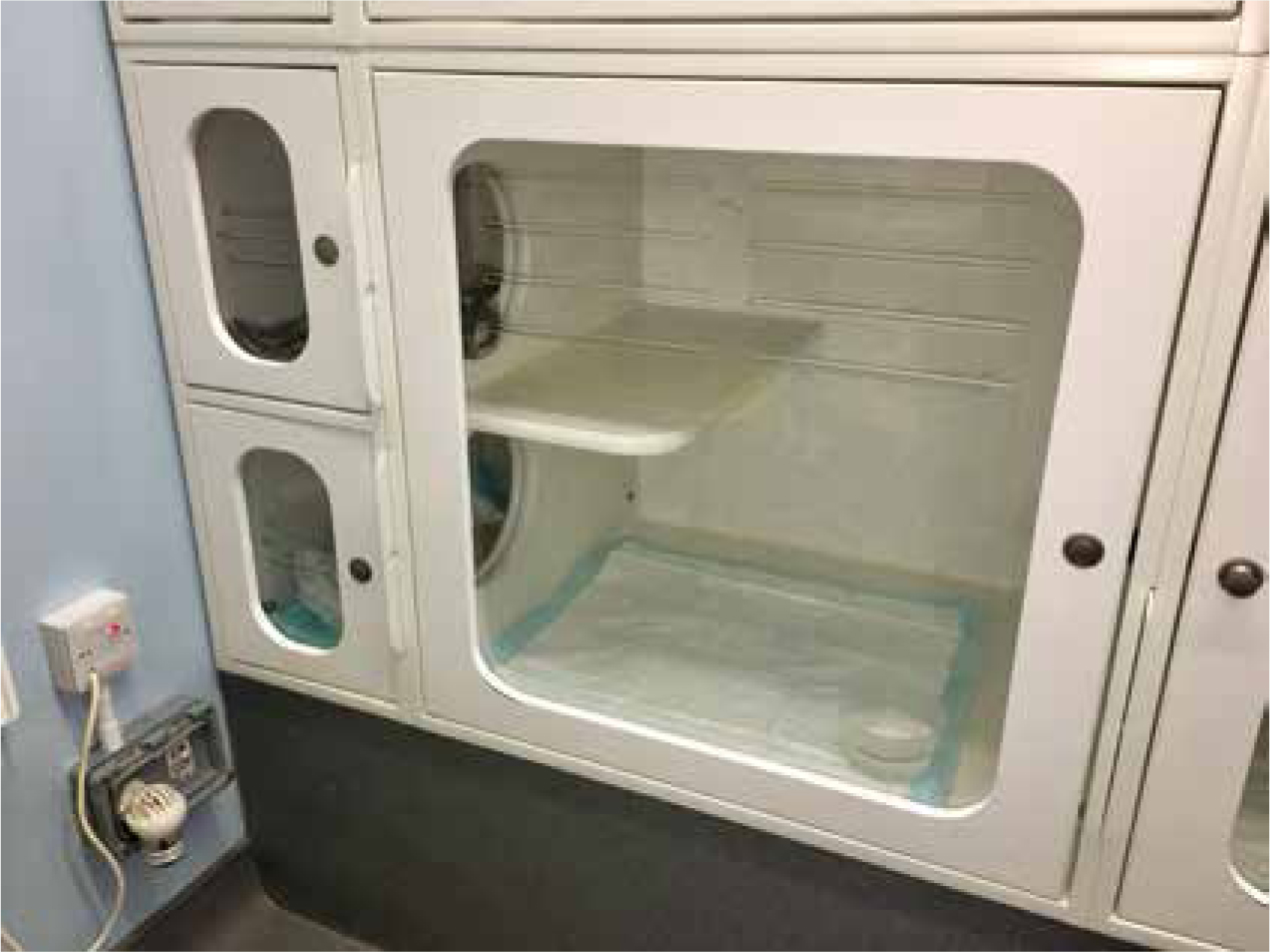
As the patient may be hospitalised for up to 4 weeks, the iodine unit kennels should be a reasonable size to allow space for the patient to be active and feel as comfortable as possible during their stay. Ideally a kennel of 70 x 90 x 55 cm should be used — this is the ‘gold standard’ size for feline patients staying over 24 hours in a veterinary practice as recommended by the International Society of Feline Medicine (International Cat Care, 2017). Large kennels are beneficial as there can be room to create a separate bedding and toilet area. Some kennels may have this built in, but cardboard boxes can also be used and disposed of after discharge. Older patients may spend longer periods of time sleeping and so bedding should be thick and warm to provide comfort (Carney et al, 2012). As the patient will be restricted with limited human interaction and away from their home environment, it is important to create a space where the patient can feel safe and comfortable while carrying out its normal behaviours and routine where possible (Ellis, 2009), therefore keeping stress levels minimal.
Many cats enjoy climbing and jumping as it allows them to reach places high off the ground where they can keep a look-out and may feel more secure (Ellis, 2009). The addition of a shelf in the kennel gives the patient this opportunity. Some wards may even have windows, which can provide a source of visual stimulation for the isolated patient (Halls, 2015). Cardboard scratch pads infused with catnip can also be provided in the kennel to stimulate the patient's senses and encourage scratching — a natural method of marking territory and claw sharpening that is often used in the wild or outdoors.
Cats are known to be very clean animals, often covering up their urine or faeces with soil/dirt either outside or in a litter tray. Litter trays should be kept away from the food and water bowls to prevent the patient from avoiding these if there are strong odours nearby. Use of a grit-style cat litter is ideal along with an incontinence pad to line the litter tray. This soaks up the radioactive urine and the grit litter minimises dust inhalation. The incontinence pad provides extra absorption for any toileting accidents (this can happen when a patient is becoming familiar with its new surroundings, if stressed or if the patient usually toilets outside). All waste from the radioactive patient must go into the designated clinical waste bin, including bedding if soiled. Disposable water bowls/food bowls should be used where possible in case of surface contamination. Waste bins are emptied daily and the bag is kept in the designated area on site for decaying radioactive waste.
Personal protective equipment
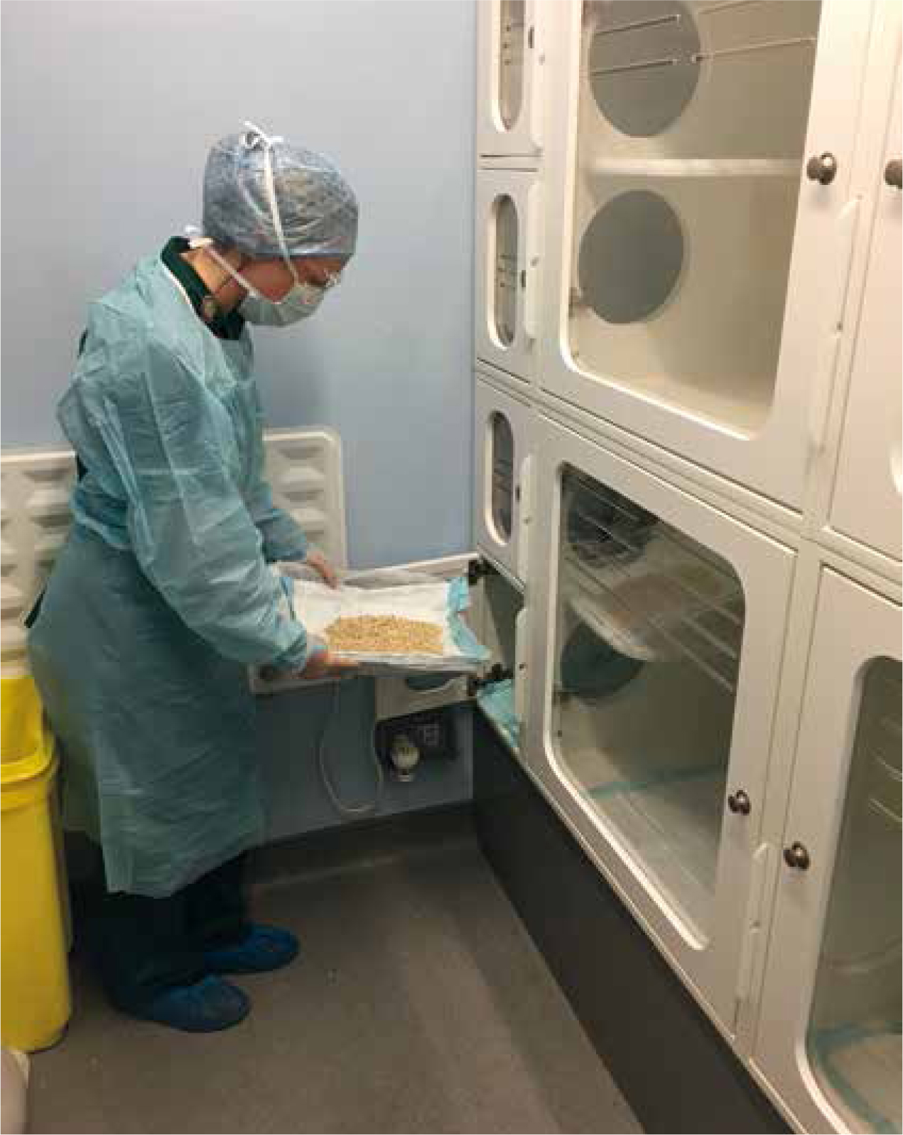
To protect personnel from contamination with radioactive urine and faeces, personal protective equipment (PPE) should always be worn within the ward area during the feeding/cleaning out time (Figure 2) and when handling the radioactive patient. PPE includes a full body gown, mask, surgical cap, shoe covers, protective goggles and two pairs of gloves.
Nursing care
During RAI treatment, daily hospitalisation sheets help keep track of the clinical status of the patient. Urination/defecation should be recorded to monitor for signs of stress cystitis, diarrhoea or constipation for example, as this may need to be treated. Constipation can be something to look out for in the older cat. Food and water intake should be closely monitored as a reduction in this could lead to dehydration and anorexia. Attention to the patient's demeanour and general wellbeing should also be considered to ensure the patient is bright, alert and comfortable.
Throughout hospitalisation of the patient, it is often the responsibility of the RVN to give the client regular updates. The client may be stressed or upset about leaving their pet for a long period of time and many worry about the lack of human contact their cat will be allowed during its treatment. The veterinary surgeon and RVN ensure clients are supported throughout this potentially stressful period of separation (from their pet) by giving regular updates via a telephone call or email. Photographs of the patient throughout its stay may also help to put the client at ease.
Conclusion
Setting up and running a radioactive unit can be complicated. There are many health and safety regulations that must legally be adhered to in order to keep the patient, veterinary personnel and client safe from radiation exposure. Providing stimulation for a feline patient in confinement can be hard to achieve particularly where human interaction is limited. The RVN has a key-role in helping the RAI unit run smoothly and in creating a stress-free environment for the feline patient throughout their stay. If the patient is discharged with the desired euthyroid result then this can be extremely rewarding for all involved.