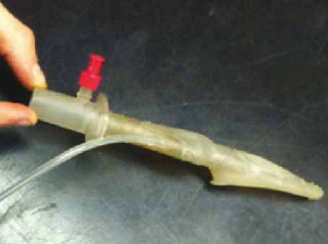
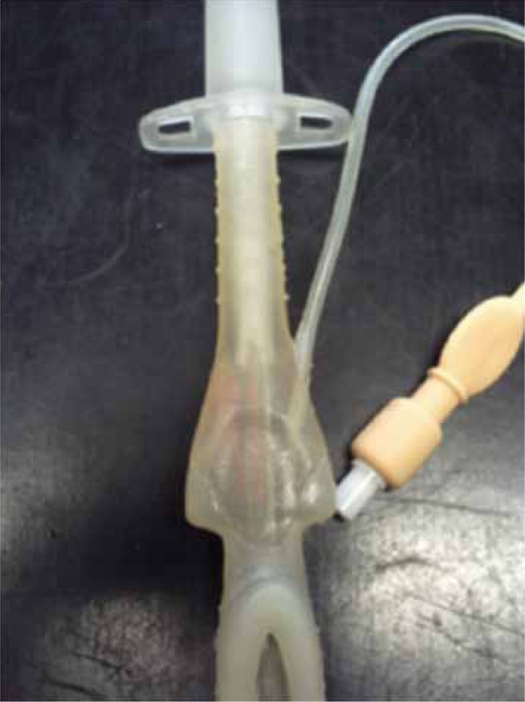
This article reports on the use of a novel supra-glottic airway device that has been designed for use in veterinary anaesthesia (v-gel, Docsinnovent Ltd). This device offers an alternative to endotracheal intubation. The development of the device has consisted of cadaveric studies to define the optimal shape and sizes of the device (Crotaz, 2010), and the sealing feature has been made from ultra-soft rubber materials to ft anatomical structures, to avoid causing trauma to the airway tissues and blood vessels during insertion and when in situ.
The use of endotracheal tubes is associated with coughing on recovery, tracheal irritation, endobron-chial intubation and in extreme cases necrosis due to over inflation of the cuff (Watney, 2003). Cats are also prone to laryngeal spasm (Evans, 2005). In addition in more serious circumstances, trauma can be caused to the trachea if a cuffed endotracheal tube is over in-fated (Hardie et al, 1999).
Clinical trials have been conducted to ensure and maximize design reliability and robustness of the novel supraglottic airway device (submitted for publication). This article describes the use of this novel device in three cats undergoing routine ovariohyster-ectomy.
Development of supraglottic airway devices
Supraglottic airway devices were developed for use in human anaesthesia to offer a simple and effective alternative to endotracheal intubation. They are de-fined as devices that ventilate patients by delivering anaesthetic gases/oxygen to a patient above the level of the vocal cords (Vaida, 2004).
There are many variations used in human anaesthesia but they are all designed to overcome the disadvantages of endotracheal intubation including damage to soft tissues and the trachea. Similarly they all have the same advantages including less coughing, a reduced occurrence of sore throats and ease of placement (Vaida, 2004).
The use of human paediatric devices has been studied by Kazakos et al (2007), who looked at the use of the laryngeal mask airway in 50 rabbits. The study concluded that the use of laryngeal mask airways provides an attractive alternative to endotracheal intubation, as the mask can be inserted easily and rapidly and its correct placement is easily confirmed (Kazakos et al, 2007). The v-gel, a novel supraglottic airway device which has been in development in the author's practice for 3 years, has been developed specifically for each species (cat, dog, rabbit and horses) so that it conforms to the anatomy of that species for an ideal ft to deliver an ideal seal (Crotaz, 2010).
There are currently three sizes of cat v-gel available: C1, C2 and C3. C1 and C2 are designed for use in cats under 4 kg. The C1 device is shorter than C2 to reduce the anatomical dead space and also to prevent a leveraging effect when the device length sticks out of the nose too much — the connector of the v-gel should be just in front of the patient's nose to minimize dead space. It has been noticed in the clinical trials that should the device be any longer, the weight of the anaesthetic circuit pulls the v-gel out of position. The C1 is therefore more appropriate for use in brachycephalic breeds and young cats. The C3 is designed for cats over 4 kg such as larger breeds and tomcats. It should be noted that overweight or obese cats may require a smaller device as excess weight does not affect the size of the upper airway. In addition to these there are three more cat sizes in development: two small-er than the C1, the C0.8 and C0.9; and the C4 which is a shorter version of the C3.
The v-gel has been developed by the same core people who invented and developed the i-gel (www.i-gel.com — Intersurgical Ltd, Wokingham, Berkshire), a similar device which is used in human anaesthesia. The device is designed to ft over the anatomical structures of the pharyngeal, laryngeal and perilaryngeal structures while avoiding compression trauma (Intersurgical, 2010). One of the important factors with the use of supraglottic airway devices is to allow easy identification of malposition of the device (which could allow gas leakage or reduce the airway diameter). For this reason, the v-gel has been designed with an integral capnometry monitoring port.
Continuous capnometry monitoring is designed to alert the anaesthetist if the v-gel becomes dislodged and is not forming a seal over the trachea. Capnometry also provides information about the patient's respiratory system and can alert the anaesthetist to any problems that may arise thus allowing the anaesthetic to correct any such problems. Pulse oximetry is also used routinely during the procedures at the author's practice. This is to provide information on arterial oxygen saturation and can alert the anaesthetist to falling atrial oxygen saturation in situations such as obstruction or hypoxia (McKelvey and Holloingshead, 2003). The v-gel device is designed to be fully autoclavable to prevent cross contamination.
Case studies
The novel supraglottic airway device (Figure 1) was to be trialled in a number of cats brought into the practice for ovariohysterectomy. Full owner consent was obtained after detailed discussion with the owners. Maintenance of anaesthesia was to be achieved by using a novel supraglottic airway device (v-gel, Docsin-novent Ltd, UK) in place of an endotracheal tube in all cases.
To place a v-gel the animal has to be at a surgical plane of anaesthesia otherwise, in the author's experience, the animal may gag, which can increase the risk of laryngeal spasm. In addition the patient should be placed on oxygen for a few minutes before device insertion. This is a pre-emptive precaution that Ivan Crotaz, the lead veterinary consultant to Docsinnovent, has put in place in case of intubation problems. Pre-oxygenation is part of the initial risk assessment and is considered good practice. Room air contains 21% oxygen, which is not adequate for oxygenation in the presence of respiratory depression which can accompany most anaesthetic agents (Hughes, 2008).
A surgical plane of anaesthesia can be achieved by a short period of inhalation anaesthesia via a facemask or induction with propofol (Abbott, Maidenhead, Berkshire) for placement of the device. The application of a facemask did not appear to cause stress to any of the cats included in these case studies. The cats were sedated from the premedication but not at the correct surgical plane for device insertion. It is the preference of Ivan Crotaz that inhalation anaesthetic is given. It should be noted however that while using a mask there is the potential for environmental pollution with inhalation anaesthetics.
Case 1
A 2.95 kg, 6-month-old female domestic short haired cat was anaesthetized for routine ovariohysterectomy using a premedication of 0.14 mls medetomidine, 0.11 mls butorphanol, 0.17 mls mg meloxicam (given sub-cutaneously). The medetomidine was reversed after surgery with 0.07 mls of atipamezole injected into the quadriceps muscle.
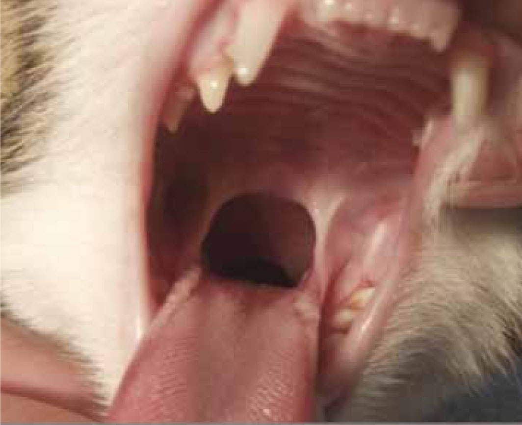
The cat was placed on 2% isoflurane via a face-mask for 3 minutes to achieve the required depth. This was assessed by checking that the blink and pedal reflex were not present, and from the position of the eye, which was in the downwards position. A C2 v-gel was attached to the capnometer and the v-gel was lubricated with KY Jelly to aid placement; each side of the device was lubricated with care taken not to get any lubrication gel in the lumen of the device that could be aspirated by the patient. The cat was held in right lateral for normal intubation as performed in the author's practice and the larynx desensitized with lignocaine (Intubeaze, CEVA, Amersham, Buckinghamshire). After checking there was no foreign material in the mouth that could be pushed into the larynx by the device, the v-gel was inserted. The end point of insertion was identified by the device dropping slightly over the base of the tongue, and by the shoulders of the device as they gently touched the pharyngeal arch (Figure 2 and 3). The device was secured using a wide open weave (WOW) bandage.
During the early phase of the anaesthetic it was found that oxygen saturation dropped from 98% to 88% as seen from the pulse oximetry readings. This might have been due to lingual compression (Kazakos et al, 2007). Continued compression of the lingual artery would have decreased the oxygen supply to the tongue.
The v-gel was removed and the cat was maintained on a facemask for 2 minutes. This was to provide the patient with oxygen and isoflurane to allow the veterinary surgeon to continue operating. When the SPO2 reading was normal the v-gel was then re-inserted quickly and easily. No further signs of lingual compression or low oxygen saturation occurred and the anaesthetic proceeded smoothly. Device removal and reinsertion was achieved by the author (an RVN), without the assistance of a second person, and without having to reposition the cat during the surgical procedure. Due to the way the v-gel has been designed, it self-positions over the larynx and it is not necessary to visualize the upper airway before insertion, making insertions much easier than with an en-dotracheal tube. Recovery following use of the v-gel is smooth and free from cough.
Case 2
A 2 kg cat was given a premedication of 0.1 mls me-detomidine, 0.08 mls butorphanol, 0.12 mls meloxi-cam (given subcutaneously). The medetomidine was reversed after surgery with 0.05 mls atipamezole (given into the quadriceps muscle).
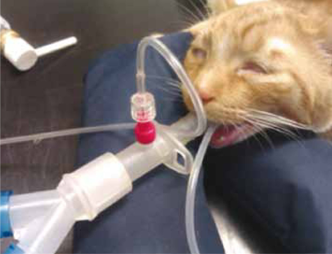
The cat was placed on oxygen for 2 minutes via a facemask to pre-oxygenate the patient before intubation. The cat was anaesthetized with propofol given to affect. A size C2 v-gel was selected and thoroughly lubricated with KY Jelly on both sides of the cuff area and attached to the capnometer (Figure 4) via a monitoring line. The patient was then held as for normal intubation. After checking there was no foreign material in the mouth the v-gel was inserted in one single fast smooth movement (on average placement of the device takes 5 seconds as recorded in the author's practice.). The capnometer was observed for a trace and when this was present the v-gel was tied in place using WOW bandage and the cat maintained on oxygen and 2% isoflurane. Tying the v-gel helped prevent the device from rotating and becoming dislodged as the patient was moved from the prep area to the operating theatre. As with endotracheal tubes v-gels should always be secured when in situ.
During the procedure the patient remained stable. The pulse oximeter had a constant reading of 98–99% oxygen saturation; heart rate, respiration rate, capillary refill time, mucous membranes remained within normal ranges (Muir et al, 2000). During the recovery, parameters continued to remain with normal ranges. The v-gel was removed just before the cat started to swallow. It has been observed by the author that after removal of the v-gel the cat shows no sign of coughing and regained consciousness with a smooth transition from anaesthesia to lifting their head.
Case 3
A 2.92 kg cat was given a premedication of 0.14 mls medetomidine, 0.13 mls butorphanol, 0.17 mls mel-oxicam (given subcutaneously) and 0.17 mls atipam-ezole (given into the quadriceps muscle) to reverse the medetomidine after the procedure. The cat was anaesthetized with propofol given intravenously to effect after pre-oxygenation.
A size C2 v-gel was selected (Figure 5), attached to the capnometer and lubricated. The cat was held as for normal intubation and after checking the oral cavity for foreign material the v-gel was inserted with ease until situated over the larynx. When a trace was observed on the capongraph the v-gel was tied in place and the cat maintained on oxygen and 2% isoflurane.
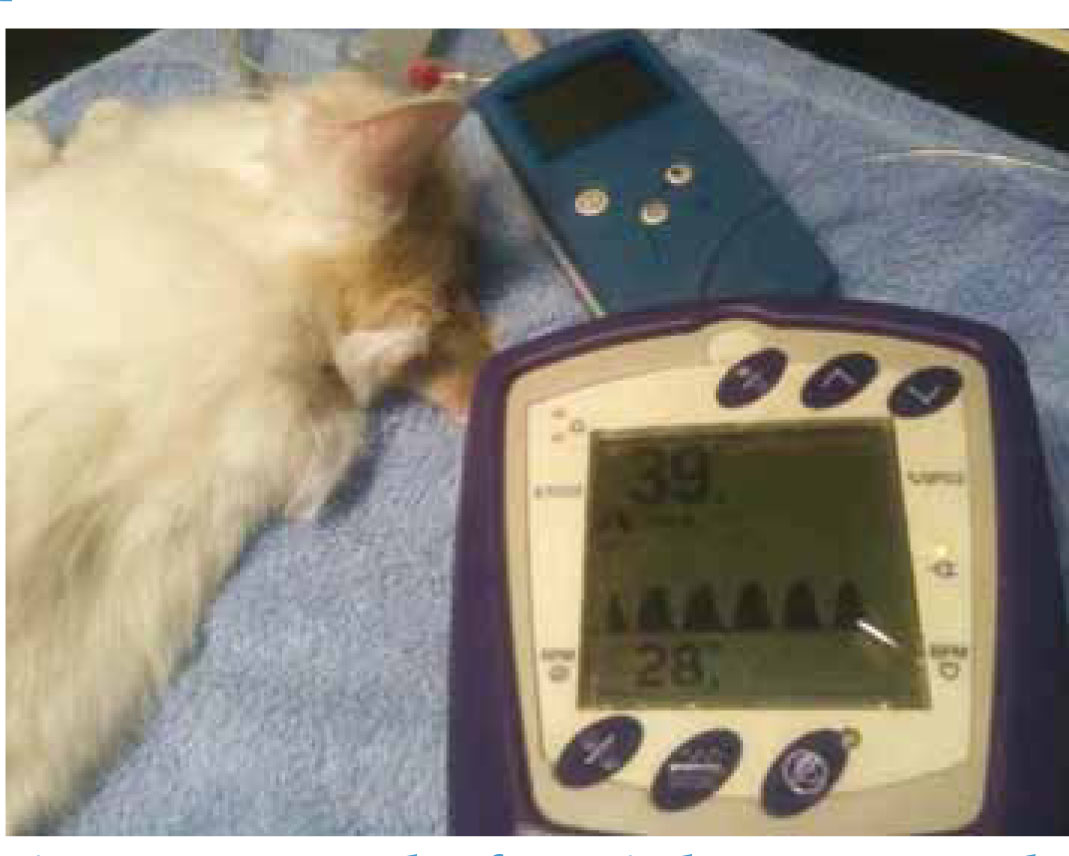
During the procedure the cat's parameters were all within normal ranges and remained stable. The average readings were: heart rate, 100 beats per minute; respiration rate, 20 breaths per minute; capillary re-fill time, 2 seconds; mucous membrane, salmon pink. As with the other case studies, this cat had a smooth transition from anaesthesia to waking. No coughing was observed and all clinical parameters stayed within normal ranges.
Discussion
The use of the v-gel speeds up the process of intubation (the author has found that on average it takes 5 seconds). Human studies have shown that intubation of the i-gel can be achieved in less than 5 seconds in most cases (Bamgbade, 2008); as the v-gel is based on i-gel technology, it is reasonable to conclude that similar intubation times would be expected. Anecdotal evidence would suggest that it also appears to reduce the risk of laryngeal spasm as the device is not passed through the larynx, but sits over the top of it. This is an advantage with cats as they have a high risk of laryngeal spasm (Evans, 2005). This makes the anaesthetic safer for the animal. In addition none of these cats showed any coughing on recovery following the use of these devices. In the author's experience the quality of recovery is subjectively better when comparing v-gel and endotracheal tube recoveries. Coughing is rarely observed in the immediate or later post-operative periods. Further double blind research work is planned to quantify and test this further.
It was likely in case 1 that the v-gel was slightly over-inserted, producing a greater pressure on the tongue and the lingual artery. This appears to subject the patient to no immediate danger, however, it is recommended that if this occurs it is corrected as soon as possible. The device has been designed with shoulders which prevent rotation and also help prevent over insertion, however the position of the device should always be checked on insertion. Compres-sion of the vagus nerve has not be noted in any of the trials. In addition, case 1 was discussed with Peter Jassell (the Innovation and Operations Director of Docsinnovent and ex Research and Development Director of Intersurgical Ltd who led the development of the i-gel from concept through to finished commercial product), who advised that the device being too large and therefore an inappropriate size for the patient could result in the lingual artery suffering excessive pressure. The v-gel is selected by using the patient's weight and head shape as a guide. In addition, the device not being lubricated sufficiently and getting stuck in an inappropriate position could also result in excessive pressure being placed on the back of the tongue. The device needs to slide into the correct place over the larynx and the tongue on insertion. When the shoulders on the device come into contact with the pharyngeal arches, however, if it has not been sufficiently lubricated then friction between the device and the immediate anatomical structures can cause sticking, preventing the v-gel from being correctly positioned. It is a prerequisite of the human i-gel that insertion success requires thorough lubrication of both sides of the device and is part of their user guide in the literature and on the dedicated i-gel website (www.i-gel.com). This may also be the case in veterinary patients as well. As this has been observed as a one of incident which was completely rectified on redoing the insertion process, a lack of lubrication can be considered the cause of the problem; incorrect size of the device used can be eliminated as being the cause, and a smaller size would not have formed a seal over the larynx. It is therefore crucial when using the v-gel to always ensure the v-gel device is thoroughly lubricated on both sides of cuff and shoulders to avoid lingual compression as seen in case 1. At the author's practice 57 cats have had the supraglottic airway device inserted without complication, and further trials are being carried out at other veterinary surgeries.
The v-gel includes an integral capnometry port which helps the user ensure the device is in the correct position and facilitates anaesthetic monitoring. It is possible to see if the device is positioned correctly as a trace will be present on the screen as the animal breathes. In addition, movement of the reservoir bag on the anaesthetic machine should also be observed when the device is positioned correctly.
Conclusion
In conclusion the v-gel device provides an alternative to using an endotracheal tube. It is essential that a capnograph is used with the device to ensure the airway is patent. Observations to date have shown there to be less coughing on recovery than is seen with an endotracheal tube, and that the recovery is smooth and uneventful, although further double blind studies are planned to test and evaluate this.