Blood banking offers clear advantages to the veterinary surgeon, patient and donor in terms of both convenience and the ability to tailor blood products to an individual animal's needs, maximizing the benefits and minimizing the risk of complications associated with transfusion.
Blood banking systems ensure there is appropriate donor screening and typing and increase the products available for an individual patient. Many veterinary hospitals have permanent canine and/or feline blood donors to cover their transfusion requirements and some have their own blood bank.
Component therapy involves splitting a unit of whole blood into its parts, most commonly packed red blood cells (RBCs) and plasma. The development of component therapy allows tailoring of transfusion therapy to the patient's needs, allows a single unit to help more than one patient.
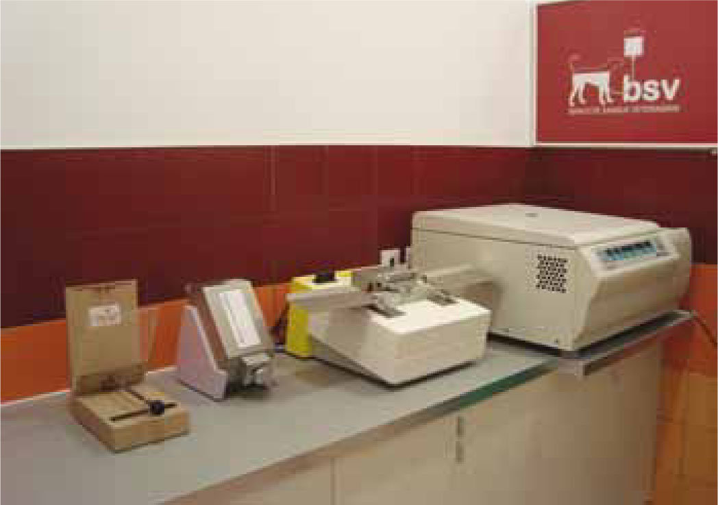
Blood components are prepared from a single donation of blood by simple physical separation methods, such as centrifugation, generally within 8 hours from collection (Giger, 2000). Blood component preparation is best accomplished by using plastic blood bags with satellite transfer containers in order to assure sterility (Knottenbelt and Helm, 2010).
Collected blood may be given as a fresh transfusion, stored and transfused at a later date, or separated into various components for fresh transfusion or storage (Knottenbelt and Helm, 2010). This article will look at the products, and the collection and administration techniques currently used in transfusion medicine.
Blood products
RBC products
Whole blood
Whole blood is indicated to replace erythrocytes capable of transporting oxygen. Whole blood contains erythrocytes, leukocytes, platelets and coagulation factors (Prittie, 2003). Platelets do not survive cooling, thus the whole blood is not a good choice for patients with thrombocytopenia (Chiaramonte, 2004).
Packed RBC
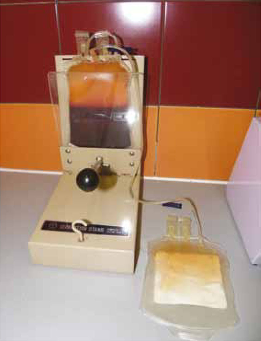
Fresh whole blood is separated into packed RBCs and plasma by centrifugation (Figure 1) or sedimentation (Littlewood et al, 2000). The infusion of packed erythrocytes will increase the oxygen capacity of the patient's blood by increasing the number of circulating erythrocytes (Chiaramonte, 2004).
The advantage over whole blood is to avoid volume overload in patients who do not require proteins or clotting factors (for example, those with non-regenerative annaemia) (Chiaramonte, 2004).
RBC substitutes
Oxyglobin, a polymerized bovine haemoglobin solution, is an example of a RBC substitute (Feldman, 2000). The principal advantage of Oxyglobin is to avoid incompatibility reactions and it is superior to RBC in delivering oxygen to the tissues. The main disadvantage is its short duration of effect (11–82 hours), compared with RBC (4–6 weeks, unless there is accelerated RBC destruction) (Feldman, 2000; Chiaramonte, 2004; Haldane et al, 2004). Oxyglobin acts as a plasma volume expander and causes vasoconstriction; this increases systemic and pulmonary arterial blood pressure (Chiaramonte, 2004; Haldane et al, 2004).
Plasma products
After RBCs are separated from plasma (Figure 2), the plasma may be processed into various products. All plasma products are poor in platelets, which remain in the packed RBC fraction (Littlewood, 2000).
Fresh plasma and fresh frozen plasma
Fresh plasma consists of plasma separated from RBCs within 8 hours of collection and immediately transfused. Fresh frozen plasma consists of plasma separated and placed at −18°C or colder, within 8 hours of collection (Kirstensen, 1995; Littlewood, 2000; Chiaramonte, 2004).
Liquid plasma
Liquid plasma is plasma stored at 1–6°C, for more than 8 hours after blood collection. The maximum storage time is 6 weeks; at this point the plasma should be placed at −18°C and labelled as frozen plasma (Littlewood, 2000).
Coagulation factors
Liquid and frozen plasma typically do not contain clinically adequate levels of factor VIII antigen (FVIII) or von Willebrand factor (vWf), especially if prepared from outdated whole blood. The vitamin K dependent coagulation factors (II, VII, IX and X) are stable in liquid and frozen plasma, so these products may be used to treat bleeding due to anticoagulant rodenticide poisoning, vitamin K deficiency and haemophilia B (Littlewood, 2000).
Cryoprecipitate (Cryo-P)
Cryp-P is a precipitate of fresh frozen plasma, also known as cryoprecipitated antihaemophilic factor, stored at −18°C or colder for up to 1 year from the unit's collection date. Cryoprecipitate is enriched in FVIII and vWf and is the product of choice for treating haemorrhage due to haemophilia A, and von Willebrand disease (LIttlewood, 2000; Chiaramonte, 2004; Haldane et al, 2004).
Cryosupernatant (Cryo-S)
Cryo-S is a plasma fraction which is separated from fresh frozen plasma by a process of controlled thawing and centrifugation. It is the remaining fraction after cryoprecipitate production. It contains plasma proteins including albumin and vitamin K dependent clotting factors II, VII, IX and X (Littlewood, 2000).
Platelet products
Platelet concentrate
Platelet concentrate is used to for treating patients that have suffered haemorrhage due to thrombocytopenia and thrombocytopathy (Kirstensen and Feldman, 1995; Littlewood et al, 2000). However, this product is not available in the UK.
Donor criteria
Donors should be considered to be clinically normal on the basis of standard history and physical examination findings. These should be rechecked before each donation and the donation cancelled if there is any suspicion of a relevant disorder. Typical reasons for cancelling a donation include bite wounds or acute vomiting and diarrhoea. Similarly, blood should be discarded from a dog that develops a fever, or other signs of potentially infectious disorders, within a week following blood donation. Annual laboratory evaluation, consisting of a complete blood count, serum biochemistry profile, urinalysis and faecal examination for parasites, is often recommended (Littlewood, 2000). Many of the recommendations are based on current human blood banking and transfusion standards. The donor's packed cell volume (PCV) and total proteins should be measured before each donation (Littlewood, 2000).
The characteristics of an ideal donor (dog or cat) are summarized in Table 1.
| Dogs | Cats |
|---|---|
| >25 kg | >4 kg |
| 1 to 8 years old | 1 to 8 years old |
| Nulliparous and spayed females | Spayed females |
| Healthy, not medicated beyond deworming | Healthy, not medicated beyond deworming |
| No history of serious illness or anomalous auscultatory findings | No history of serious illness or anomalous auscultatory findings |
| Without potential to develop bacteraemia (periodontal disease, skin lesions, abscesses) | Without potential to develop bacteraemia (periodontal disease, skin lesions, abscesses) |
| Normal temperament and good body condition Hematocrit >40% | Normal temperament and good body condition Hematocrit >35% |
| Without prior transfusion | Always indoors and fed only a commercial diet |
| Blood typed for D.E.A. 1.1 positive/negative | There is no universal donor (blood group of donors should be known) |
| Up to date vaccination and deworming | Up to date vaccination and deworming |
| Complete blood count, complete biochemical | Complete blood count, complete biochemical |
| panel and platelet count (annually) | panel and platelet count (annually) |
| Free of infectious diseases (agents to test vary | Free of infectious diseases (agents to test vary |
| with geographic location) | with geographic location) |
| Has not received a transfusion himself | Has not received a transfusion himself |
Blood collection: volume, frequency and storage
The main aspects of blood collection technique are summarized in Table 2.
| Collection systems (consisting of a 16G thin-walled needle, tubing and plastic collecting bag, containing an anticoagulant preservative solution, available with satellite bags) | Open systems — increases the risk of microbial contamination |
| Closed systems — no potential for environmental contact of the blood | |
| Syringes | 60 ml and a winged infusion set |
| Blood collection | Jugular vein (cephalic vein may be used in large dogs) |
| Sedation or anaesthesia may be required | |
| The donor should be fasted; if not, postprandial lipaemia does not seem to affect the quality of fresh or stored blood products. However, lipaemia may increase rouleaux formation, which complicates cross matching |
Canine blood donation
In dogs, 15 to 20% of estimated blood volume can be safely donated. Using the formula, the maximum acceptable donation is about 16–18 ml/kg. A standard blood donation in the dog is approximately 450 ml, which is referred to as 1 canine unit (Littlewood, 2000).
Estimated blood volume (l) = 0.08 – 0.09 × body weight (kg)
As a guideance:
Feline blood donation
In cats, 15 to 20% of estimated blood volume can be safely donated. Using the formula, the maximum acceptable donation volume is approximately 11–13 ml/kg (1 feline unit), which equates to 50 ml from a 4–5 kg cat (Littlewood, 2000).
Estimated blood volume (l) = 0.055 – 0.065 × body weight (kg)
As a guideance:
Blood is commonly collected from cats with a 19G winged infusion set attached to a 60 ml syringe into which the anticoagulant/preservative solution has been drawn. For blood banking purposes, the blood is then transferred to a 100–150 ml storage bag (Littlewood, 2000).
Storage
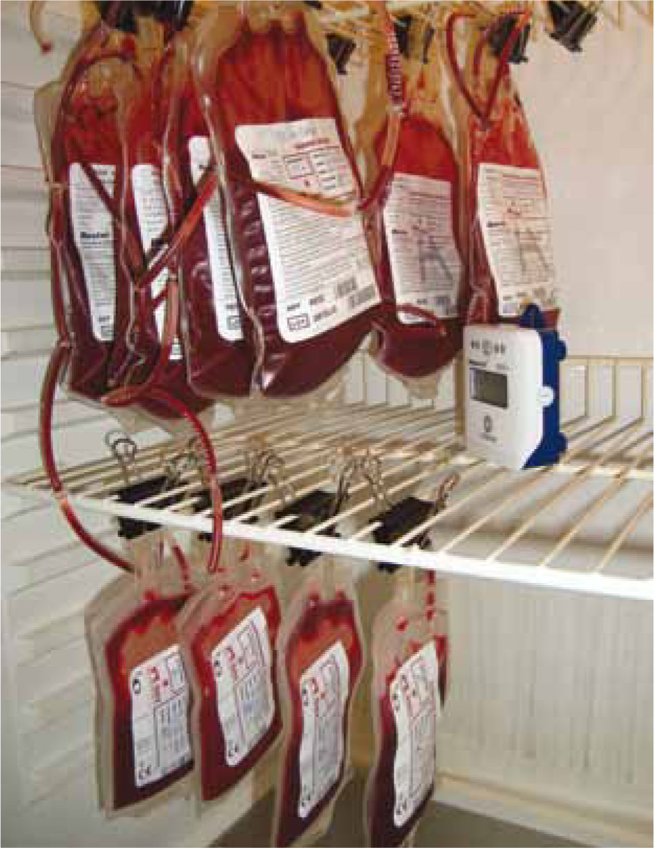
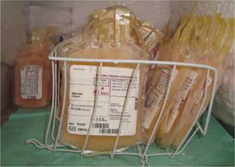
After the donation, the storage bag should be clearly labelled with the type of product, collection date, expiration date, donor, blood type, PCV (for whole blood and packed RBCs) and total solid protein (for whole blood, packed RBCs and plasma products) (Wardrop, 1995; Littlewood, 2000), and storage be performed in the correct way (Table 3; Figures 3 and 4).
| Component | Storage | Validity |
|---|---|---|
| Whole blood | Refrigerator (4μC) | 5 weeks |
| Packed red blood cells + solution of nutrients | Refrigerator (4μC) | 6 weeks |
| Fresh frozen plasma | Freezer (−20μC) | 1 year (after this period is considered Frozen Plasma) |
| Frozen plasma | Freezer (−20μC) | 5 years |
Blood collection technique
The canine donor
The feline donor
Blood administration
Warming and mixing
Refrigerated whole blood and blood components do not need to be routinely warmed before administration. The products may be gradually warmed to room temperature, during administration or may be warmed to body temperature (for recipients at risk of hypothermia and when large volume and/or rapid transfusions are planned) (Littlewood, 2000).
To warm to room temperature, the blood product may be at room temperature 30–60 minutes before beginning the transfusion. To warm to body temperature, the intravenous line may be passed through a bowl of water or sandwiched, between oat bags warmed at 37–38°C. Electrical infusion warmers or Bairhugger warming systems are also available. The blood bag may also be warmed by immersion in a 37–38°C water bath, although this increases the risks of contamination and decreased RBC viability, compared with warming the infusion line. Microwave warming is not recommended because of the risks of haemolysis (Knottenbelt and Mackin, 1998; Littlewood, 2000).
Stored whole blood should be mixed by gentle inversion before transfusion. With canine packed RBC stored in citrate phosphate dextrose adenine (CPDA), the PCV is often 70–80% and the product may be too viscous to transfuse easily. It is possible to decrease viscosity by adding 100 ml of 37°C saline to the unit and resuspending the RBCs by gentle manual agitation and kneading (although feline packed RBCs have a lower PCV, dilution with 20–30 ml of saline facilitates passage through the filters recommended for blood administration) (Knottenbelt and Mackin, 1998; Littlewood, 2000).
Calcium-containing fluids such as lactated Ringers should not be used for dilution because the calcium may initiate coagulation (Knottenbelt and Mackin, 1998; Littlewood, 2000).
Citrate toxicity is an uncommon complication, resulting from chelation of recipient blood calcium by the anticoagulant in a transfused product. Citrate toxicity might occur after rapid, large volume replacement transfusion, plasmapheresis and severe liver failure with impaired citrate metabolism. The resultant hypocalcaemia causes tetany, tremors and cardiac arrhythmia (Hale, 1995; Harrell, 1995; Lanevschi and Wardrop, 2001).
Frozen plasma products may be more rapidly thawed in a 37–38°C water bath or incubator. Thawing time is 30 minutes or more for a unit of canine plasma. Agitating and manually kneading the bag, to break up ice crystals, speeds up thawing. Higher temperatures may result in protein denaturation (Knottenbelt and Mackin, 1998; Littlewood, 2000).
Venous access
A transfusion may be given using any vein. If venous access is not possible, the intraosseous route is the next choice (Littlewood, 2000) (Table 4). Neonates can also be transfused by intraperitoneal injection. About 50% of transfused RBCs will be absorbed into the circulation, from the peritoneal space, in 24 hours, and 70% within 48–72 hours, but they will have a shorter life time (Knottenbelt and Mackin, 1998; Littlewood, 2000).
| Needles | ||
|---|---|---|
| Access | Dogs | Cats |
| Venous access and | 16–19G jugular catheter (preferred for packed RBC transfusions) | 22G peripheral vein catheter (lower PVC smaller RBC size) |
| 18–20G peripheral vein catheter | ||
| Intraosseous | 18–20G or bone marrow aspiration needle placed into the throchanteric fossa | 22G |
| 22G (in neonatal transfusion can also be placed into the tibial crest) | ||
Transfusion rate
Whole blood and its components may be transfused at a rate of 5–10 ml/kg/hour. The initial rate should be 0.25 ml/kg/hour, for the first 15–30 minutes, to allow for early detection of potentially severe transfusion reactions. The maximum rate of transfusion is 22 ml/kg/h, which is usually only used in an emergency situation (Turnwald and Pichler, 1985; Littlewood, 2000). To minimize the risks of bacterial proliferation in RBC and plasma products, a transfusion should be completed within 4 hours (Turnwald and Pichler, 1985; Littlewood, 2000).
Transfusion reactions
Transfusion reactions are adverse events occurring following administration of blood or blood products. Their effects can be fatal in some cases or may merely result in the transfusion providing limited benefits to the recipient (Weingart et al, 2004). Reactions that occur within 24 hours of administration are termed acute reactions while those occurring more than 24 hours following transfusion are referred to as delayed reactions. Reactions can be caused by immunological or non-immunological mechanisms, both of which should be rare when a matched transfusion is given and blood has been stored and administered appropriately. Despite this, all patients should be monitored closely during the transfusion period in order to detect unexpected adverse reactions.
If signs associated with a potential transfusion reaction (e.g. anaphylactic reaction, pyrexia, vomiting, hypothermia, circulatory overload) are seen the transfusion should be stopped immediately (Turnwald and Pichler, 1985). The type of reaction encountered should then be determined and the appropriate treatment given. Although transfusion reactions can be due to poor storage or administration techniques, most significant reactions are associated with the administration of mismatched transfusions. Some clinicians recommend slow flow rates (0.25 ml/kg/hour) during the first 30 minutes of transfusion in order to detect unexpected transfusion reactions (Turnwald and Pichler, 1985).
Filter
Special administration sets for blood transfusion contain in-line filters to remove clots, platelet aggregates and some fat. Filters should always be used with stored whole blood and blood components (Turnwald and Pichler, 1985; Littlewood, 2000).
Transfusion amount
A number of calculations have been suggested to evaluate the amount of red cells needed:
The volume of blood to be transfused to a patient can be calculated with the following formula, which assumes a total blood volume of 66 ml/kg in cats and 88 ml/kg in dogs (Littlewood, 2000):
Volume to be transfused = 66 or 88 × weight of patient (kg) × (desired PCV% – patient PCV %) ÷ PCV% of donor
The desired PCV following transfusion is usually around 20% and 30% in cats and dogs respectively (which equates to 50 ml for a 4–5 kg cat and up to 500 ml for a 30 kg dog), which allows clinical improvement, without suppressing the regenerative response too much.
These equations should be used as a guide only when considering how much blood to obtain or administer (Littlewood, 2000).
The volume of plasma to transfuse is calculated by using the following formula:
Volume of donor plasma to be transfused (ml) = recipient weight (kg) × 4.5 × (desired – current recipient plasma albumin level (g/l))
This formula assumes that normal canine plasma volume is 4.5 % kg bodyweight, albumin level in the donor plasma bags averages 25 g/l and the distribution of albumin is 40% in the plasma (Littlewood, 2000).
The role of the veterinary nurse
It is the responsibility of the veterinary nurse to monitor patients and recognize any transfusion reaction. She/he should also assess the value of haematocrit before and after the transfusion, as well as the presence or absence of haemoglobin in plasma or urine (Feldman, 2000).
Conclusion
A high number of blood transfusions are performed each year on dogs and cats and the demand for blood products continues to grow. Risks associated with transfusion reactions include the risk of disease transmission. Appropriate screening of blood donors for infectious disease should be performed to lessen this risk. In addition, factors involving general health care and management of blood donors should be employed to further ensure blood safety.
The ability to collect and administer blood safely is an important skill in veterinary practice. The availability of commercial blood products means that many more animals can benefit from transfusion therapy and provides essential support to many critical patients.