

In 2021 there were 900 000 pet rabbits living in the UK, and 2% of adults owned a rabbit (PDSA, 2021). According to the PAW report 2021, a higher proportion had been neutered and were insured in 2021, while the proportion that have received regular boosters had decreased when compared with pre-pandemic levels (PDSA, 2021) — 50% (450 000 rabbits) are not receiving regular boosters (higher than pre-pandemic levels — 42% in February 2020). There are a number of viral diseases against which rabbits should be vaccinated, including myxomatosis and rabbit viral haemorrhagic disease (RVHD).
Myxomatosis
Background
Myxomatosis is a disease caused by the myxoma virus that was first recognised in a laboratory in 1896 where it killed European rabbits (Oryctolagus cuniculus). It was originally introduced to wild rabbits in Australia in the 1950s as a means of managing the growing populations that were considered a pest species, and eventually spread across Europe and by 1953 Britain, rapidly killing tens of millions of animals. Many people in agricultural business and forestry, welcomed the disease as a pest control on rabbit populations. But others such as those that hunted the species for meat and fur deplored the virus as it resulted in loss of income for their business (Bartrip, 2008).
Transmission
Myxomatosis can be indirectly spread between rabbits by vectors, such as mosquitoes or fleas, that become carriers of the myxoma virus through contact with infected hosts. It can also be directly spread through contact with infected rabbits, where infectious secretions may be inhaled by an otherwise healthy rabbit. Therefore, it is important to stress to pet owners the risk of their rabbit contracting the virus, whether they are kept indoors or outdoors, as vectors are able to access indoor as well as outdoor housing. When introducing new stock with unknown health status this can also pose the same risk. If kept outdoors, rabbits can become infected by vectors (parasites) as well as throigh contact with a population of infected wild rabbits through grazing pastures or contact through housing (Rabbit Welfare Association & Fund (RWAF), n.d.).
Clinical presentation
Clinical presentation of rabbits with the virus depends on the strain of the virus and genus and species of the infected rabbit. It has been reported that in Sylvilagus species (American leporid (rabbit) species) depending on the severity of the virulent strain, myxomatosis can manifest at the site of viral entry as innocuous localised cutaneous fibroma (Kerr, 2012). However, in European rabbits (O. cuniculus) the same virus causes the lethal disseminated disease myxomatosis. The incubation period is 1–3 days — the most classic clinical signs are lethargy, fever, anorexia, skin haemorrhages and seizures with a high mortality rate (Hess and Tater, 2012). Those that survive the initial infection period may go on to develop purulent blepharoconjunctivitis (inflammation of eyelid and conjunctiva), and erythematous oedematous nodules around the face and perineum region (Hess, 2012). Death is usually within 1–2 weeks after infection, but occasionally animals survive, and signs will slowly regress over about 3 months (Hess and Axelson, n.d.).
Diagnosis and treatment
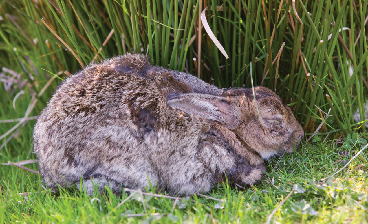
Diagnosis of myxomatosis is usually by clinical signs (Figure 1), histopathological analysis of affected tissue and viral isolation on cell culture testing. Treatment options are often unsuccessful as there are no medical interventions that specifically treat the virus and therefore supportive treatment should be given such as fluid therapy, antibiotics to prevent secondary infections occurring, pain management, ambient temperature environment, topical ointments which can help to soften and protect skin lesions and elimination of vectors carrying the virus. Corticosteroids should be avoided because of contraindication of their immunosup-pressive effects. Nursing care should be provided for effective supportive management such as ensuring provision of clean bedding to prevent sticking to lesions, tempting the patient with food and handfeeding to encourage nutritional benefit. Syringe feeding can be difficult because of blocked nostrils, and it is suggested that rabbits be euthanased for this reason if they are unable to eat on their own (Harcourt-Brown, 2022a).
Prevention
Myxomatosis can be prevented by keeping rabbits up to date with their vaccinations and educating pet owners of the risks. This will give rabbits the best chance should they be exposed to the virus. Although not an absolute guarantee that the rabbit will be 100% protected, it can somewhat dampen the effects should they catch the virus. According to the European Medicines Agency, Nobivac Myxo-RHD Plus vaccine protects against myxomatosis, RHD1 and RHD2, and is licenced to be given to rabbits from the age of 5 weeks, with an onset immunity of 3 weeks and a duration of immunity of 1 year (European Medicines Agency, 2021). The RWAF encourages pet owners with the following guidance on preventing myxomatosis spread along with vaccination:
- Owners should source hay and straw materials from producers where the farmer has not seen any rabbit with myxomatosis on the farm land
- Feeding dust-extracted hay or kiln-dried grass
- Fitting an insect screen to outdoor enclosures
- Eliminating standing water (where mosquitoes might breed) from the garden. If using a water butt, put a small amount of cooking oil into the water. This will form a film over the surface that will suffocate mosquito larvae. Or alternatively, have in place a sealed lid so mosquitoes are unable to make contact with the water supply
- Treating cats and dogs of households regularly for fleas to prevent the spread to rabbits
- Stopping wild rabbits from getting into gardens if possible. If this is not feasible, make it impossible for wild rabbits to have nose-to-nose contact with pet rabbits.
- Make sure there is nothing to attract vermin and wild birds to hutches/runs and use small-hole mesh on hutches/runs to keep unwelcome creatures out (RWAF, n.d.).
Rabbit viral haemorrhagic disease
Background
RVHD was first reported in China in 1984 and has since rapidly spread worldwide and is now considered endemic in several countries. In countries such as Australia and New Zealand rabbits were considered pests and in 1996 RVHD was introduced to control the numbers of introduced European rabbits (O. cuniculus) (Abrantes et al, 2012). RVHD is a type of calicivirus, and it consists of two strains known as RVHD1 and RVHD2. Both strains are lethal to rabbits; it is believed that RVHD2, which has been in the UK since 2013, has become more prevalent across Europe, after being first reported in France in 2010 — more cases of the RVHD2 strain are now being seen than the classic RVHD1 strain (RWAF, 2017). This disease has a high mortality in rabbit species with individuals dying from the disease between 48–72 hours post-infection.
Transmission
Various potential routes for transmission of the disease may include oral, nasal, conjunctival and parenteral contact. Insect vectors have been shown to be efficient in the transmission of the virus through biting and infecting the rabbit's bloodstream with the virus. RVHD is also transmitted by direct contact with an infected rabbit as they may shed viral particles in their secretions and excrement or through indirect contact through fomites such as bedding, water, food, accommodation and equipment that has been used, which have become contaminated with the viral spores (Hess and Axelson, n.d.). ‘Natural doors’ have been suggested as the viral entry, for example upper respiratory and digestive tract. The upper respiratory route includes breathing in viral spores and the digestive tract route, such as through coprophagia (re-ingestion of soft faeces) a natural behaviour in rabbits, is considered a preferential mode of transmission (Abrantes et al, 2012).
Clinical presentation
RVHD is characterised by acute necrotising hepatitis, but there may also be presence of haemorrhage in other organs, for example the lungs, heart and kidneys because of excessive blood clot formation in blood vessels known as disseminated intravascular coagulation (DIC) (Abrantes et al, 2021).
Often rabbits with the disease will die within 12–36 hours after onset of fever. The two types typically seen in rabbits are:
- Preacute form, whereby infected rabbit's display no symptoms and die suddenly
- Acute infections are seen through clinical signs of anorexia, apathy and congestion of the palpebral conjunctiva. As well as neurological symptoms which may be seen such as opisthotonos (muscle spasms causing backward arching of the head, neck and spine), excitement, paralysis and ataxia. Occasionally, some respiratory signs have been reported in the form of tracheitis (inflammation of the trachea), dyspnoea (difficult or laboured breathing) and poor circulation of oxygen presented with cyanosis (bluish discolouration of mucus membrane). There may also be a presence of foamy and bloody nasal discharge. Eye complaints include increased lacrimation, ocular haemorrhages, and epistaxis. (Abrantes et al, 2021).
The RVHD2 virus has some differences from the classic RVHD. In particular, it may affect rabbits of any age, as opposed to RVHD1, which is rarely if ever seen in rabbits under 8–10 weeks of age (RWAF, 2017). In addition, it appears that there are fewer symptoms with RVHD2 than the classic strain. Most rabbits that become infected die suddenly. The disease is highly transmissible through contact with other rabbits, surfaces, excrement, and food sources. Clinical presentation may include fever, lethargy, loss of appetite and muscular spasms, shaking or seizures, and bleeding from the nose or mouth. Some rabbits may be asymptomatic. Infected rabbits that are asymptomatic or recover from the disease may shed the virus for 2 to 4 months post infection (Justice, 2020).
Treatment
Unfortunately, there is no current treatment for RVHD1/RHD2. Those that have developed the disease require generalised supportive treatment, e.g. fluid therapy, syringe feeding and warmth. As these rabbits are highly infectious it is important to ensure barrier nursing is established to prevent risk of spreading the disease (Harcourt-Brown, 2022b).
Prevention
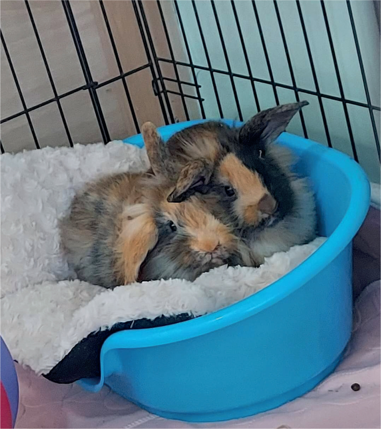
The best way to prevent rabbits from contracting the virus is vaccination. Until recently, only Filavac (CEVA Animal Health) and Eravac (HIPRA UK & Ireland Ltd) vaccines were licensed for use in the UK to prevent RHD2. In 2020, MSD launched their new vaccine ‘Nobivac Myxo-RHD PLUS’ which gives protection against myxomatosis, classic strain RHD1 and new strain RHD2 all in one single dose, giving protection for 12 months. It is important to have rabbits — both those kept outdoors and indoors — in for yearly boosters to maintain this protection (Figure 2) (MSD Animal Health, 2021).
Biosecurity steps can also be taken to prevent the spread of infection, for example barrier nursing, and if rabbits are housed outdoors for any length of time, owners should ensure that there has been no wild population of rabbits grazeing on the land, and they should take care when sourcing forage foods (Justice, 2020).
Additional rabbit diseases
Other diseases less commonly seen in rabbits include encephalitozoonosis (Encephalitozoon cuniculi infection) and pasteurellosis (Pasteurella multocida infection). E. cuniculi is a microscopic parasitic organism. It is an intracellular organism and is spread in rabbits from mother to offspring in the uterus and from rabbit to rabbit through infected urine. It usually causes a latent condition, and many infected rabbits therefore show no signs. When signs develop, the affected rabbit may have back leg weakness and paralysis as a result of the swelling surrounding the E. cuniculi causing destruction of the nervous tissue. Other signs commonly seen with this disease include a head tilt, fitting, loss of balance, tremors, kidney failure and bladder weakness. Unfortunately once the rabbit develops severe clinical signs this can lead to death. Signs of kidney failure may include increased thirst, increased urination, weight loss and decreased appetite. Treatment involves administration of anti-inflammatory and anti-parasitic drugs for several weeks, along with supportive care such as syringe feeding and motion sickness medication. There are currently no medications that are guaranteed to clear the infection, and many rabbits continue to show signs even after treatment (Hess and Axelson, n.d.).
P. multocida are carried by all rabbits, but only some will show signs of disease. Signs include abscesses, respiratory infections (in the upper airways known as snuffles), and chronic inflammatory disease in rabbits. It can infect the nasolacrimal (tear) ducts, eyes, ears, and nose, and can cause abscesses of tooth roots, bones (particularly the jaw), skin, tissues under the skin, and internal organs. Stresses, such as poor nutrition, change in diet, introduction of a new pet or person in the house, over-crowding, environmental stresses, immunosuppression, or the presence of other disease, may trigger clinical signs. Treatment usually involves either oral or injectable antibiotics, given for a minimum of 2–4 weeks (Hess and Axelson, n.d.).
Disease control
Disease control preventing further spread of infectious diseases, should be maintained through efficient cleaning and use of appropriate disinfection to animal accommodation to include fomites such as food bowls and litter trays. Infectious bodily fluids, urine and faeces should always be put into contaminated waste and not general household waste to prevent the spread of disease (Stull et al, 2015). Good hygiene practice in storage, handling, and preparation of food as well as regular sanitising of hands between patients can prevent certain diseases. Isolation or quarantine of animals is imperative if the animal poses a risk to infection particularly when introducing new stock of animals with unknown health status (Dawson and Cooper, 2020).
Barrier nursing should be used in the management of infected patients, and protective clothing should be worn when handling these animals and clothing changed if it becomes soiled. Providing good draught-free ventilation within the hospital environment and avoiding mixing animals in large groups in the same airspace is recommended, as well as reducing populations of stray animals with unknown health status particularly within the hospital environment where animals may be presented with depreciated immune systems as a result of illness (Dawson and Cooper, 2020). Accommodation should be rested between patients for as long as possible, and an effective disinfection protocol used to control diseases, particularly before reintroducing other animals, to help reduce the risk of residual environmental contamination. Optimum nutrition management, minimising stress through good husbandry and attending promptly to any ancillary health problems can help to speed recovery in most circumstances (Dawson and Cooper, 2020). Vectors of diseases, such as external parasites, should be controlled with preventative treatment, and patients should be shielded from the infected as they are more at risk of infection because of the presence of underlying disease. Encourage routine immunisation and preventative care at the earliest opportunity to prevent transmission of the disease. Routine veterinary visits for annual examinations and overall health assessments (which may include faecal examination for the detection of intestinal parasites depending on exposure risk) can play a vital role in ensuring pet owners are educated regarding disease risks, hygiene, and good husbandry (Stull et al, 2015).
KEY POINTS
- Myxomatosis can be indirectly spread between rabbits by vectors such as mosquitoes and fleas that carry the myxoma virus through contact with infected hosts.
- Corticosteroids should be avoided in the treatment of myxomatosis because of contraindication of the immunosuppressive effects.
- The Rabbit Welfare Association & Fund (RWAF) has set out guidance for pet owners on preventing the spread of myxomatosis alongside vaccination.
- Rabbit viral haemorrhagic disease (RVHD) can be spread by various routes such as oral, nasal, conjunctival and parenteral contact, as well as through insect vectors that can spread the virus by biting and infecting the rabbit's bloodstream with the virus.
- The best way to prevent rabbits from contracting these two viruses is by routine vaccination, for both indoor and outdoor kept rabbits as they are both at risk.
- Care should be taken when sourcing foraging food from unknown sources where infected wild populations may reside the viruses can be spread through ingestion of cross-contaminated foods.
- When nursing these patients, barrier nursing should be used to prevent the risk of spreading the infection to other patients. This includes resting accommodation between patients for as long as possible, and carrying out a thorough disinfection protocol to help reduce the risk of residual environmental contamination.
Conclusions
It is important for veterinary nurses to keep themselves and their clients up to date with rabbit health preventative measures, including vaccinations and education. If clients are proactive with biosecurity measures at home, keep up to date with vaccinations and boosters and take measure to prevent the spread of infection, this could lead to fewer cases of the common rabbit diseases, and potentially in the long run, lead to the eradication of disease.