
Canine atopic dermatitis (CAD) is a common genetically predisposed inflammatory and pruritic skin disease. It is associated most commonly with IgE antibodies to environmental allergens (Halliwell and DeBoer, 2001). The clinical presentations of CAD are protean and vary between breeds. The extent of the lesions, the chronicity of the disease, the type of secondary infection as well as the overlap of clinical signs with other non-atopic skin diseases can make diagnosis challenging (Hensel et al, 2015).
History and clinical signs
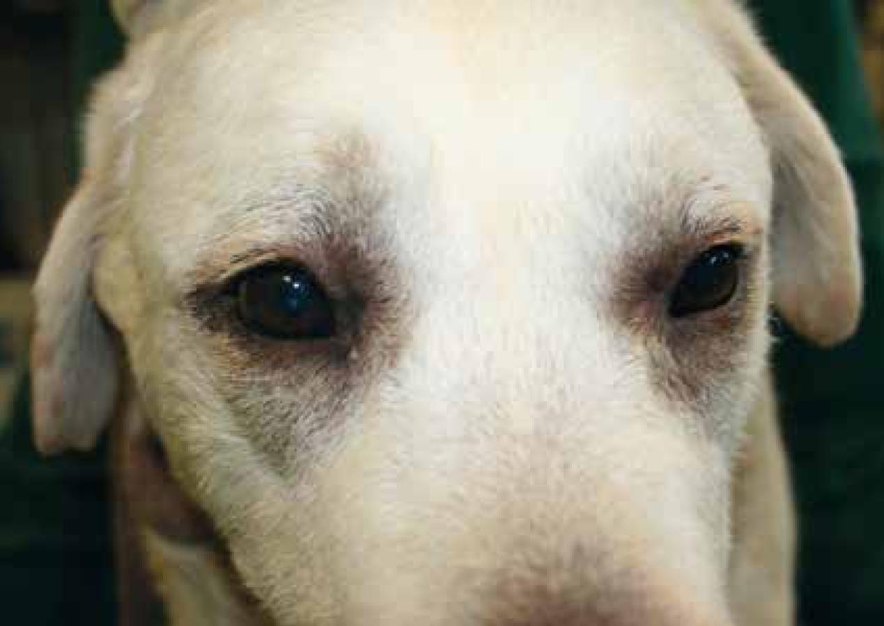
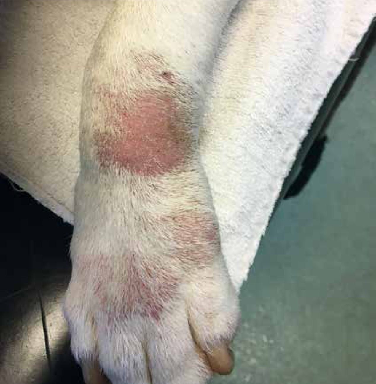
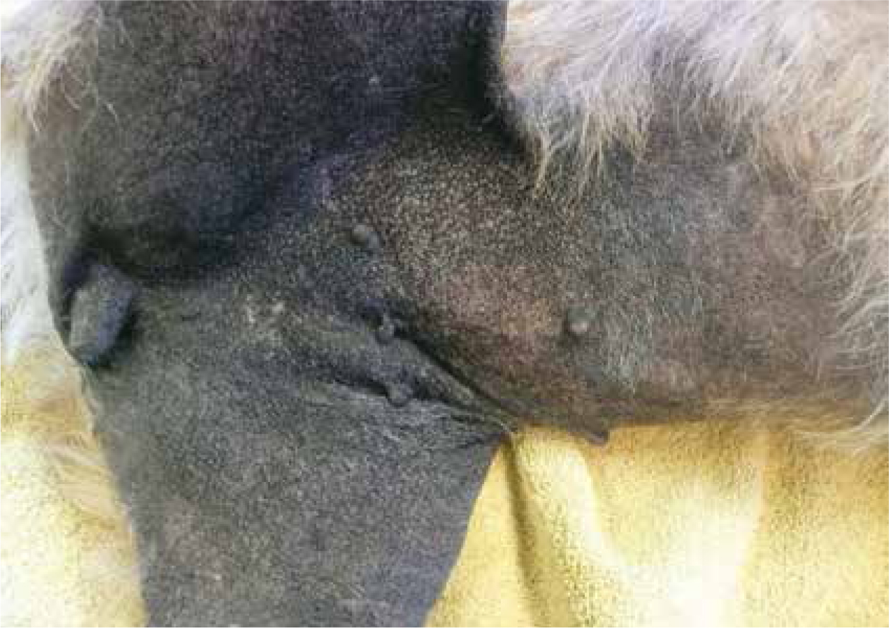
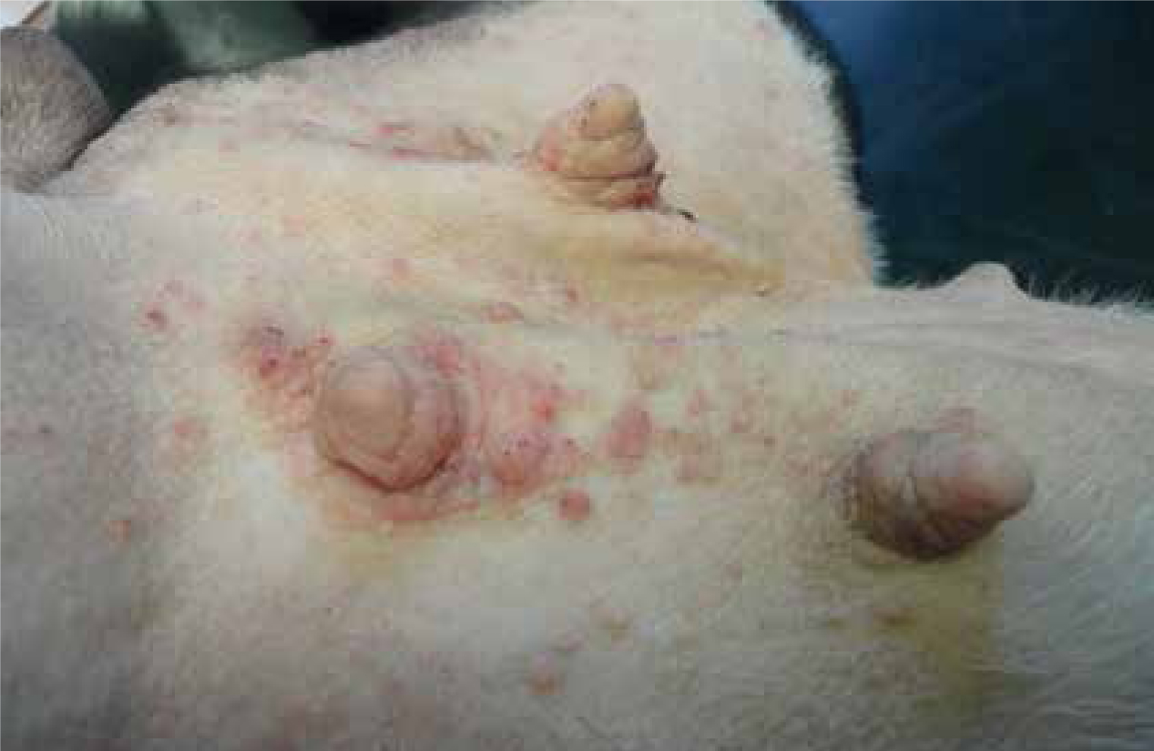
A consistent finding in CAD is the presence of pruritus (itching). Pruritus can manifest itself in a variety of ways including licking, chewing, scratching, rubbing or over grooming, all of which can lead to signs of self-inflicted trauma and chronic changes in the skin. Clinical signs in the early stages of the disease are often seasonal. In the UK dogs commonly present with pollen-induced disease in the early summer often going into remission at the end of the pollen season in the autumn. Some dogs will continue with a seasonal pattern, but many will go on to develop perennial disease with seasonal exacerbation. The areas of the dog's body that are most commonly affected are the face (Figure 1), concave aspect of the ear pinna, ventrum, axilla, inguinal area, perineal area and distal extremities (Figure 2) (Griffin and DeBoer, 2001). Primary lesions such as papules, which are common and a useful differentiator in such diseases as ectoparasites, are uncommon in CAD. Often dogs present with erythema, and lesions are created by the dogs as they self-traumatize due to the pruritus. In chronic disease lesions can become hyperpigmented, lichenified and excoriated and often become much more extensive to extend beyond the classical sites associated with CAD (Figure 3). A set of clinical criteria developed by Favrot (2010) are useful to try to differentiate cases of CAD from other pruritic skin disease (Favrot et al, 2010). These are best applied after ectoparasites, infection and other allergic diseases have been eliminated (Table 1).
| Age of onset <3 years |
| Mostly an indoor dog |
| Alesional pruritus at onset |
| Affected front feet |
| Affected ear pinnae |
| Non affected ear margins |
| Non affected dorso-lumbar area |
| Glucocorticoid responsive pruritus |
A combination of five satisfied criteria has a sensitivity of 85% and a specificity of 79% to differentiate dogs with CAD from dogs with chronic or recurrent pruritus without atopic dermatitis (AD) (Favrot et al, 2010).
Diagnosis of CAD
There are no specific diagnostic tests for CAD. CAD is a diagnosis of exclusion.
Guidelines recently published as an open access article by Hensel et al (2015) suggest four important steps should be followed to rule out other causes of pruritus:
Eliminate fleas
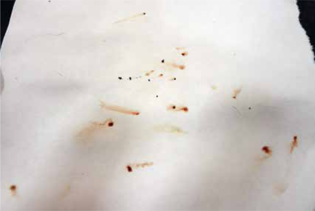
All pruritic dogs should be checked for fleas or flea faeces on direct examination or brushings of the hair coat. A wet paper test is a useful way to identify flea faeces and demonstrate (sometimes to a skeptical owner) the presence of fleas. The dog's coat should be brushed onto a piece of wet paper and any flea faeces will appear as red/brown streaks as their blood content seeps onto the paper (Figure 4). Where flea or faeces are not identified, it is still worthwhile instituting flea control. This acts as a safeguard where fleas have been groomed out of the coat by the dog or where the owner has bathed the dog prior to consultation. There is now a wide range of excellent flea control products on the market, the author's preference is for an oral preparation as these tend to be less affected by topical shampoos than spot on preparations.
Eliminate other ectoparasites
Other ectoparasitic diseases that need to be eliminated include sarcoptic mange, cheletiellosis, pediculosis, trombiculiasis, otoacariasis and demodicosis. A variety of diagnostic tests can be employed to eliminate these parasites (Table 2). All of them are quick and inexpensive and can easily be undertaken in a primary care practice. Microscopic examination can be performed using a low power objective of 4x or 10x and a low light intensity.
| Disease | Ectoparasite | Most important diagnostic tests |
|---|---|---|
| Sarcoptic mange (scabies) | Sarcoptes scabiei | Deep skin scrape, blood serum for serology testing, empirical therapy with a scabicide |
| Cheyletiellosis | Cheyletiella blakei | Coat brushing, acetate tape impressions and superficial skin scrapes |
| Cheyletiella yagsuri | ||
| Cheyletiella parasitovorax | ||
| Pediculosis (lice) | Trichodectes canis | |
| Trombiculiasis (harvest mites) | Trombicula autumnalis | |
| Otoacariasis (ear mites) | Otodectes cynotis | Examination of ear wax, acetate tape impression smears or superficial skin scrapes of coat |
| Demodicosis (demodectic mange) | Demodex canis | Deep skin scrapes, hair plucks, acetate tape impression smears of squeezed skin |
| Demodex injai | ||
| Demodex corneii |
Eliminate infection
Infection with both bacteria typically Staphylococcus spp. and yeast usually M. pachydermatis is common in cases of CAD (Figure 5). Both primary and secondary lesions can be identified in cases of bacterial infection (pyoderma). Pustular lesions (primary lesions) are less common that secondary lesions such as crusting and epidermal collarettes in pyoderma. Both types of lesions can be sampled cytologically. Pustules may be pricked so the contents can be absorbed onto a slide, to be stained and examined with a Diff Quik stain. Secondary lesions can be sampled by impression smears or acetate tape impressions and stained in the same way. Where bacterial infection is localised or superficial it can in most cases be treated with topical antimicrobial therapy; in such situations bacterial culture and microbial susceptibility is not necessary. However, where there has been a failure to respond to appropriate topical treatment or where the infection is severe, generalised antibiotic treatment is often needed and should be prescribed on the basis of appropriate culture results. M. pachydermatis is commonly identified in intertriginous areas (skin folds) where is may present as areas of localised erythema and seborrhoea. It can easily be identified as purple staining ‘peanut’ shaped organisms on Diff Quik stained acetate tape impression smears.
Eliminate a CAFR
Pruritic skin disease can be caused by a reaction to a food the dog is ingesting. The institution of a low allergy or hypoallergenic diet should therefore form an important part of any investigation of CAD. Selection of a diet can be challenging as a variety of different factors need to be considered. Where possible the diet should contain ingredients the dog has not been exposed to before; it should be palatable and reasonably priced so that it can be instituted for a prolonged period of time. A strict exclusion diet should be fed for a minimum of 8 weeks (Rosser, 1993). The most common food allergens recognised in dogs are beef, dairy, chicken, and wheat. Other proteins such as lamb, fish, soya and pork are less common, as is corn (Roudebush, 2013). Where a novel protein such as rabbit, kangaroo or venison is available these may be considered as possible hypoallergenic proteins. However recent work has suggested that cross reactions to proteins that are phylogenetically similar exist, meaning that venison may not be a sufficiently novel protein in a dog with an allergy to beef (Ayuso et al, 2000). Hydrolysed diets offer an excellent, convenient and cost-effective alternative to home cooked diets. While there are dogs with CAFR that may still react to these diets they never the less will identify a high percentage of dogs with food induced pruritus (Cave, 2006).
Where pruritus is still present after ectoparasites, infection and CAFR have been eliminated then a CAD remains the most likely diagnosis. All of the basic diagnostic tests such as wet paper tests, skin scrapes and hair plucks to identify ectoparasites and cytology to identify infection, can be undertaken by an experienced nurse. Nurses can also provide support and guidance to owners about flea control and about the institution of an appropriate food trial.
Allergy testing is useful after a diagnosis of CAD has been made. Allergy testing can be performed by assessment of skin reactivity by intradermal testing (IDT) or by detection of IgE by allergen-specific IgE serology (ASIS) testing. Both tests can be used to identify offending allergens so that allergen-specific immunotherapy (ASIT) can be formulated, neither test should be used as a diagnostic test for CAD. Intradermal injections for IDT are usually performed on a small clipped area on the lateral thorax in a sedated dog. Typically 0.05–0.1 ml of each allergen is injected intradermally into the skin. In all IDT, histamine is used as a positive control and sterile saline as a negative control. All other allergens are measured against these two intradermal injections. The author usually considers a positive reaction to be one that is larger than the mean value of the positive and negative controls. Reactions are usually read 15–20 minutes after the injections have been administered. After the allergy test has been read the results can be used to formulate ASIT, this is made up to include all allergens that have produced relevant positive reactions. Although IDT is generally considered by most dermatologists to be the test of choice in producing ASIT for a patient, ASIS does have several advantages to IDT. It is far more convenient for a client as it only involves the collection of a blood sample; it carries no significant risk as dogs do not generally need sedation; multiple intradermal injections are not needed and it is a much quicker test to perform. In most veterinary dermatology clinics, nurses with advanced training perform IDT and also ASIS.
Therapy for CAD
There is no one therapy that is suitable for every dog with CAD. The most successful protocols are usually multi-modal. All dogs with AD benefit from ongoing ectoparasite control. Many dogs also need some form of dietary manipulation. This is most important where the institution of a hypoallergenic diet may have produced some benefits in the dog's clinical condition but it has been incomplete. In such cases dietary challenge can be undertaken to establish which specific ingredients of the dog's diet induce pruritus, so ongoing dietary advice can be given. Topical therapy can also be used to great effect in cases of CAD. Antiseptic shampoo, sprays, mousses and wipes can help prevent recurrence of bacterial and yeast infection. Topical emollients and moisturisers can help restore skin barrier function which is known to be poor in these animals. A detailed discussion of the use of topical therapy is beyond the scope of this article and the reader is referred to other texts for more information (Olivry et al, 2015). Specific therapy for the pruritus can be divided into proactive and reactive regimens (Table 3). Proactive therapy aims to control the disease rather than responding to it after it has happened which is the mainstay of reactive therapy.
| Proactive therapy | Reactive therapy | |
|---|---|---|
| Allergen specific immunotherapy (ASIT) | Sublingual ASIT (SLIT) | Antihistamines |
| Glucocorticoids | ||
| Ciclosporine | ||
| Subcutaneous ASIT (SCIT) | Janus kinase (JAK) inhibitors | |
| Monoclonal antibodies IL 31 | ||
| Essential fatty acid | ||
Many different factors need to be considered when choosing therapy for cases of CAD. All treatment will be needed on a life time basis meaning that the convenience of the therapy for the owner and dog, the safety of the regimen (i.e. side effects versus benefits), and the ongoing expense all need to be discussed.
Allergen specific immunotherapy (ASIT) is a useful form of treatment in many animals. It can be given by subcutaneous injection or by sublingual administration. Where dogs are compliant and tolerate injections, owners can be coached to inject their own dogs. Once on maintenance therapy many animals only need injections every 4 weeks. Sublingual therapy often suits owners with small dogs, owners who have problems travelling to the veterinary practice, or where dogs are refractory to the administration of injections. Sublingual therapy, which involves giving drops onto the tongue, has the disadvantage that it is more labour intensive as most protocols involve giving the drops at least once and usually twice daily. The success of ASIT varies between sources, but is usually quoted at 60–65% (Griffin and Hillier, 2001).
Essential fatty acids do exhibit the potential to reduce inflammation through the modulation of prostaglandin and leukotriene production. However there is only a weak evidence base to recommend their use as there are few high quality clinical trials demonstrating a positive benefit. Many veterinary dermatologists use these as part of a multimodal treatment regimen, but rarely as the sole form of therapy (Olivry et al, 2001).
Antihistamines are often prescribed in cases of CAD, this is mostly on the basis that they have some benefit in human patients with AD. The quoted success rate in canine studies varies enormously from 0–70% depending on the study and the antihistamine. Many dermatologists will consider using several antihistamines evaluated in sequence for 7–14 days (DeBoer and Griffin, 2001).
Despite the development over the last 10 years of a range of new anti-allergy drugs for CAD, glucocorticoids can still be used in canine patients with enormous benefit. They represent a low cost option for therapy and there is considerable evidence demonstrating that oral low-dose glucocorticoid formulations can be used safely to control skin lesions and pruritus in dogs with CAD (Olivry and Sousa, 2001). The major disadvantages of glucocorticoid therapy is the polyphagia and associated weight increase, together with polydipsia and polyuria that is seen in many dogs.
Ciclosporine has been available as a licensed drug for CAD for more than 14 years (Forsythe and Paterson, 2014, DeBoer, 2014). There have been numerous studies demonstrating its efficacy in the therapy of AD (Olivry et al, 2010). It is available as both capsules and liquid formulations. Although it is a relatively higher cost option for therapy it has a high degree of efficacy. Disadvantages associated with ciclosporine therapy include gastro-intestinal disturbances, hirsuitism and gum hyperplasia (Radowicz and Power, 2005).
The discovery of the cytokine Interleukin 31 (IL31) as a major initiator of pruritus in CAD, has led, in the last few years, to the development and launch of two new drugs specifically targeting the action of this cytokine (Gonzales et al, 2014). IL31 released from activated T cells in CAD selectively binds to receptors on a wide range of cells including neurones. The binding of IL31 to Janus kinase (JAK) receptors on these neurons activates pruritogenic signals in peripheral nerves causing itch. Oclacitinib is a JAK inhibitor that selectively inhibits JAK1-dependent cytokines involved in allergy, inflammation, and pruritus (Gonzales et al, 2013). It has been shown to be a safe, effective, rapid acting drug to control pruritus in allergic skin disease in dogs (Cosgrove et al, 2013). Oclacitinib is given orally. The disadvantage of oclacitinib is its anti-inflammatory effect. This means it should be used with care in dogs with a history of infection, demodicosis or neoplasia. Lokivetmab is a caninised monoclonal antibody that specifically targets IL31. It is given as a single injection and provides long-lasting relief from pruritus for up to 4 weeks, and has a rapid onset of action. Although this is high cost option for therapy of CAD, initial use of this drug has shown it to be safe, well tolerated and useful in dogs with concomitant disease including neoplasia (Moyaert et al, 2017).
Conclusion
Canine atopic dermatitis is a common genetically predisposed inflammatory and pruritic disease. A diagnosis of CAD should be made on the basis of the exclusion of other pruritic skin diseases. Basic diagnostic tests that all nurses should be familiar with and be able to perform to help contribute to a diagnosis of CAD include coat brushing and wet paper tests to eliminate fleas; skin scrapes, hair plucks and tape strips to eliminate other ectoparasites as well as tape strips and skin cytology to assess for infection. Nurses should also be aware of the need to institute a hypoallergenic diet in all pruritic dogs and the principals behind the selection of an appropriate diet. Therapy can include systemic medication, such as essential fatty acids, antihistamines, glucocorticoids, ciclosporine, JAK inhibitors, monocloncal antibodies to IL31, and allergen specific immunotherapy.