
Atopic dermatitis or atopy is a common skin condition that can affect cats, dogs and even some exotics such as rats (Girling, 2013). It has been estimated that the incidence of the condition in dogs is around 10% (Scott et al, 2001), although it is acknowledged that this figure may be affected by geography and diagnostic criteria (Hillier and Griffin, 2001). Nevertheless, they also comment that it is commonly seen in general practice and dermatology referral clinics. It is a chronic condition that can impact on both the lives of the animal and their owner. The veterinary nurse can play an important role in the diagnosis and management of this condition providing support and guidance to clients.
Aetiology
Atopic dermatitis is an allergic skin condition that can come under the umbrella term of atopy. In human medicine atopy encompasses three conditions — allergic rhinitis, asthma and atopic dermatitis. While the other conditions are seen in animals the skin form is the most common presentation in dogs and can also affect cats or other mammals.
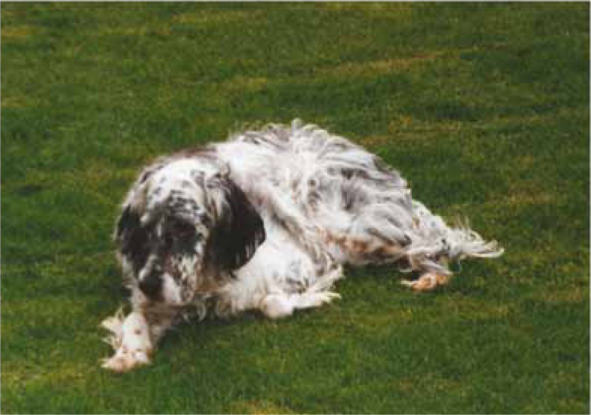
Many different theories have been put forward to explain the pathogenesis of the condition and it is likely that a combination of different factors are present in any one animal. The condition is more likely in particular breeds such as West Highland White Terriers, English Setters (Figure 1), English and Staffordshire Bulldogs, and Labrador Retrievers (Sousa and Marsella, 2001), but can be seen in any breed or cross breed. A genetic component to the condition has been shown (Bizikova et al, 2015). Work by Ravens et al (2014) examined the incidence of atopic dermatitis in feline patients in Australia. The most commonly seen breeds were domestic mixed (24/45), Abyssinian (6/45) and Devon Rex (3/45).
Affected animals can be allergic to a variety of different substances. Some of the more common allergens that dogs and cats can react to are shown in Box 1. However, it should also be remembered that dogs can be exposed to different allergens than those commonly associated with people (Fraser et al, 2001) as dogs at ground level are exposed to heavier pollens.
One of the main pathological processes in this condition is the activation of the Type 1 hypersensitivity reaction and the degranulation of mast cells resulting in clinical signs of pruritus. Historically it was thought that allergens came into contact with the immune system through inhalation (Wittich, 1941; Olivry and Hill, 2001). However, more recently it has been proposed (Day, 1996; Olivry and Hill, 2001) that percutaneous transmission of allergens takes place — this would certainly make much more sense when the anatomical distribution of lesions is considered — namely the paws, abdomen, axillae, ears and peri-ocular areas. In addition dendritic cells, keratinocytes and macrophages within the epidermis and dermis have been shown to contribute to the hypersensitivity reaction (Pucheu-Haston et al, 2015).
Alterations in the formation of the stratum corneum have been shown in atopic dogs where the barrier function of the lipid emulsion was abnormal (Marsella et al, 2011). This is another possible reason for percutaneous exposure to allergens, as removal of the outer protective later allows allergens to come into contact with dendritic cells and keratinocytes which will stimulate an immune response.
Other components of the immune system including T lymphocytes and inflammatory mediators such as interleukins and cytokines are also involved in the immune response. Abnormalities in the proportion of T helper or T suppressor cells and variations in the levels of chemical mediators also contribute to the development of the disease.
Once an animal develops pruritus they will start to scratch, chew or overgroom in response to the itch. It is this self trauma that results in more damage, inflammation or secondary infections that makes the condition even more difficult to treat. Chronic trauma can result in lichenification and hyperpigmentation of the skin.
In managing atopy, owners need to understand that there are a variety of different causes, clinical presentations and treatments, and that all of these can vary throughout the year in the one animal. If an individual animal has an allergy to pollens then it is more likely that they will have problems during the summer — if an owner can avoid allergens, pre-empt any problems or bring the animal to the practice as soon as they become pruritic then secondary problems are minimised, making it easier and quicker to treat.
Clinical signs
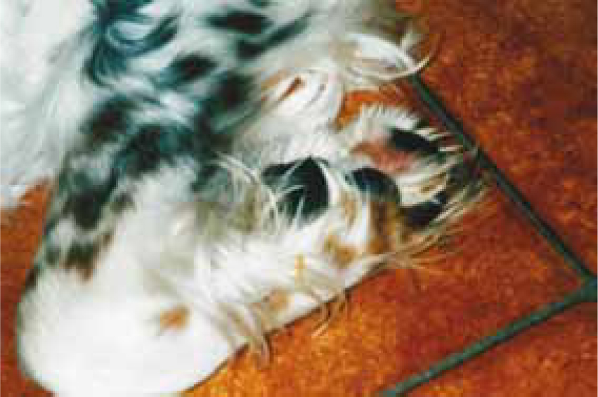
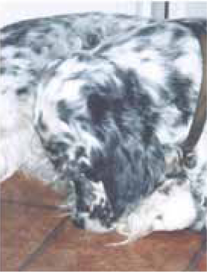
The underlying problem in atopic dermatitis is that of itch. The way in which an individual animal responds to this will vary. While cats can be seen to scratch when itchy, they are more likely to over-groom; this damages their coat and so they can present with areas of alopecia. In the author's experience dogs are more likely to be seen scratching or chewing at themselves, but they can focus just on one area such as one ear or foot, or they could scratch all over (Figures 2 and 3).
Animals with atopic dermatitis are generally young (between 1–3 years) (Olivry, 2010), but it is possible to see animals that are younger or older than this at first presentation. Depending on the underlying problem clinical signs may be seasonal (e.g. pollen allergen during the summer), or all year round (e.g. house dust mites).
Commonly affected areas are the peri-ocular skin, around the mouth, the external ear canal, feet, abdomen and base of the tail (Scott et al, 2001). Other clinical signs can include erythema, alopecia or saliva staining where reddish brown pigment deposits can be seen on white/light coated areas.
Diagnosis
As already explained atopic dermatitis is a complicated disease — due to this the diagnosis is also complicated. Different diagnostic criteria have been suggested over recent years (Willemse, 1986; Olivry, 2010), with a reliance on a combination of history, clinical signs, excluding other conditions and then carrying out allergy testing, either through intradermal skin testing or serology.
Serological tests have advanced over recent years and are now routinely used in practice. These assays can assess circulating IgE levels that are specific for individual allergens and use this as an indicator of the underlying problem (Table 1). However, non-atopic animals can sometimes show increased levels of allergen specific IgE and so these tests cannot be used in isolation to make a diagnosis. Medication is less likely to interfere with serological results compared to those of intradermal skin testing, and drugs such as corticosteroids may only need to be withheld for a short time before blood sampling.
| Result | Reference range >150 is considered a positive result |
|---|---|
| Flea saliva | 45 |
| Dermatophagoides farinae | 1674 |
| Dermatophagoides pteronyssinus | 203 |
| Tyrophagus putrescentia | 5036 |
| Lepidoglyphus | 1066 |
| Acarus siro | 3783 |
| Alternaria alternata | 29 |
| Aspergillus fumigatus | 288 |
Intradermal skin testing (IDST) is still carried out alongside or instead of serological testing. As the allergen is injected into the dermis it is evaluating a different component of the immune system to serology and so different results may be obtained. It is possible to obtain a negative result on serology and a positive on IDST and vice versa. Traditionally IDST has been regarded as the gold standard with serological tests being compared with IDST. However, the reasoning behind this has been questioned (DeBoer and Hillier, 2001) as IDST can give false positive results. Both diagnostic tests are evaluating different components of the immune response and may also be using different antigens. Therefore it cannot be expected that both tests will give 100% agreement.
IDST involves sedation, clipping and then injecting up to 40–60 different allergens into the skin and observing the response. Sedation is usually with an alpha-2 drug and an opioid. Acepromazine should be avoided as it is an antihistamine and will affect the response to the test. Patients who have been on corticosteroids or antihistamines need to be taken off this medication for a few weeks before the test — this can prove difficult for some patients and owners may need help in providing other methods of therapy. Although very rare, it is possible for a patient to have an anaphylactic reaction during a test and so a crash box, adrenaline, corticosteroids and oxygen should all be available. Owners need to be aware of the possible side effects when giving informed consent to the procedure. The patient should be monitored throughout the procedure.
As can be seen from Figure 4, a postcard sized area needs to be clipped on the flank, depending on the size of the patient. Again owners need to know that their animal will go home with a large clipped area. Where the patient has a long coat (e.g. in Golden Retrievers), then it is possible to pull back the fur and then clip. Following the test the fur above the clip site can then be pulled down over the test area, making it much less obvious.
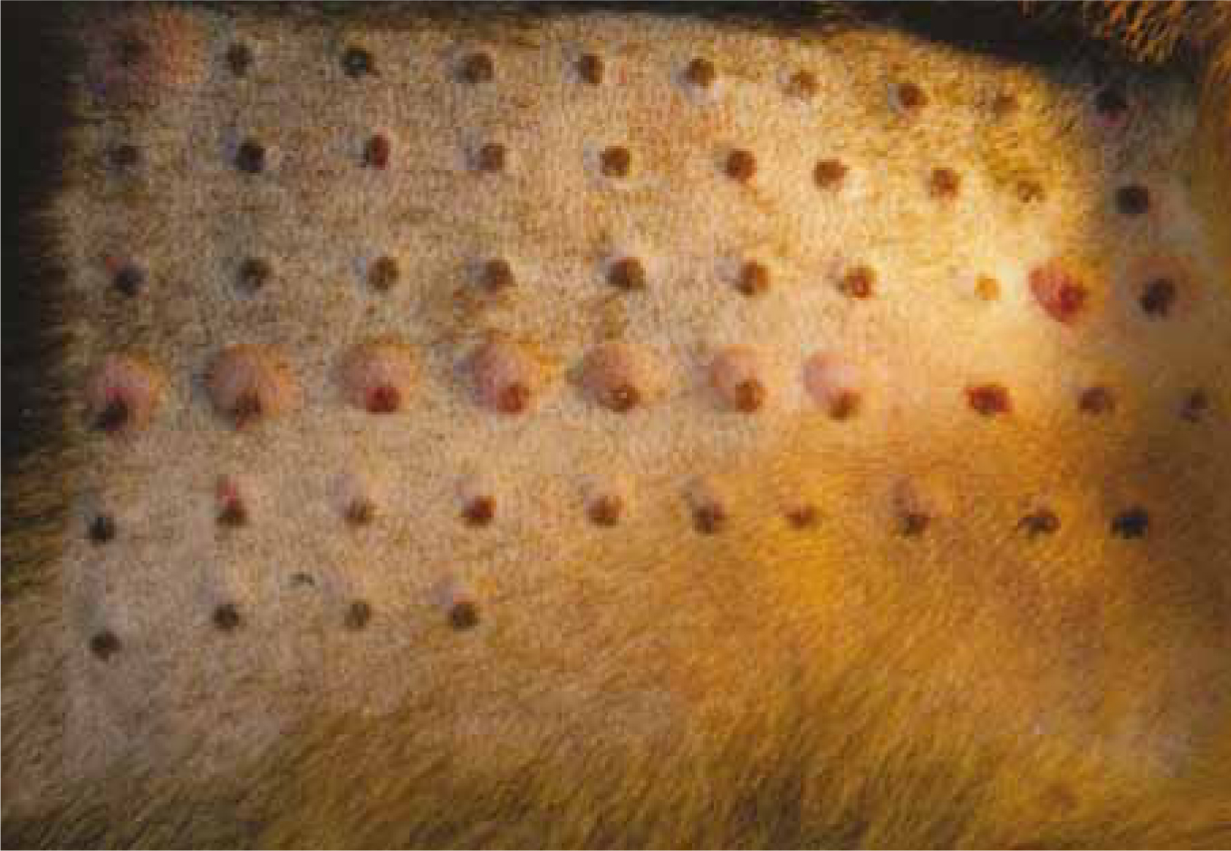
Testing involves the injection of histamine (positive control), saline (negative control) and various allergens — either individually or in combination; 0.05 ml of each is drawn up into an insulin syringe, it is important that no air bubbles are present in the syringe as this can affect the appearance of the skin. Within a few minutes of injection a wheal will be seen to develop in the area around the histamine injection. If this does not happen then the test is invalid, possibly due to the presence of corticosteroids in the circulation. Once all of the allergens have been injected each test will be evaluated for:
All of these parameters are recorded and compared with the positive and negative responses to decide on positive results. The whole process usually takes around 30 minutes, after which the sedation can be reversed.
Therapeutic options
A variety of different treatment options are available depending on the clinical presentation, underlying causes, specific allergens, patient temperament and owner considerations. The best approach is the one that the owner is able to follow and so time needs to be devoted to both explaining the options to the owner and finding out more about which methods will work best for them. A combination of different therapies might be used initially when the patient may have bacterial and yeast infections, pruritus and skin changes associated with scratching; as the condition improves the therapies will change to respond to the needs at the time.
Oral medication
Corticosteroids cause side effects such as polyuria, polydipsia and polyphagia. However they are useful to calm down severe pruritus, reduce self trauma and give the skin a chance to respond to other medication.
Cyclosporine has now been available for over 10 years and has been shown to be successful. Side effects such as gingivitis or skin lesions are possibilities which owners need to be aware of.
Antihistamines are not licensed for use in companion animals, but can prove effective. Different antihistamines have been used with varying success in individuals so an antihistamine trial might be needed. Owners will need to give informed consent for their use and again be warned about possible side effects such as medication making the animal drowsy.
Oclacitinib (ApoquelTM, Zoetis) is a relatively new medication which relieves itch and has been shown to be useful in cases of canine atopic dermatitis (Little et al, 2015).
Topical medication
If the owner and animal are willing, then topical therapy is very useful for atopic patients. Products need to be selected according to the condition (see Box 2). Bathing can be carried out at home, or in the practice if facilities allow. Owners may prefer to bring their pet to the practice for a bath so it is always worth considering offering this service.
Hyposensitisation therapy
Where serology or IDST has been carried out and specific allergens identified it is possible to develop a hyposensitisation vaccine. This works by exposing the immune system to low levels of allergen and through time altering the allergic response. It has been suggested by Griffin and Hillier (2001) that 50–100% of dogs receiving hyposensitisation will show at least a 50% improvement after 4 months or more of therapy. Owners need to be patient if considering this form of therapy. Where a good response is seen then monthly administration of the vaccine may be required for the rest of their life.
Hyposensitisation vaccines can either be given by injection (subcutaneously) or orally. Again anaphylaxis is rare, but is a possibility so initial doses should be given at the practice where treatment could be given if side effects developed.
Other therapies
While not a therapy, avoidance of allergens can help some patients. House dust mites are one of the most common allergens that dogs develop allergies to (Hill and DeBoer, 2001). While cleaning reduces their numbers it is impossible to totally eradicate these allergens from a house. Higher levels of allergens may be found in bedrooms, so it may be helpful if the dog or cat could sleep in another room. Being in a warm environment can also make pruritus worse — another reason to keep them out of the bedroom. However, owners may be resistant to this approach. Dogs with thick coats may also benefit from stripping the coat again to lower the temperature at the level of the skin.
Herbal medications and evening primrose oil have been used with varying success. If owners would like to try them, then they may be worth considering, although it would be unlikely that these therapies would be successful on their own.
As some atopic patients may also present with a food hypersensitivity condition then an exclusion diet may be of benefit. However, not all atopics are necessarily affected by gastrointestinal problems.
Role of the veterinary nurse
The veterinary nurse can play a central role in the successful management of cases of atopic dermatitis by undertaking laboratory work during a skin work up and providing owners with information and guidance.
Client support is vital when managing a case of atopic dermatitis. The condition is not static and owners need to understand that the clinical signs will get better or worse throughout the year depending on the underlying problem, allergen exposure and secondary problems such as ectoparasites. Taking time to explain the condition and the different treatment options means that owners are more aware of the problems they might face and what these will mean for their dog. Identifying problems early and altering the treatment to fit will also prevent the dog from developing secondary problems. The veterinary nurse needs to be able to respond to the needs of the animal and the owner.
Personal experiences of practice have shown that offering owners the services of a nursing clinic focused on atopic dermatitis will improve clinical outcomes. As the condition can change from day to day owners need to be able to contact someone in the practice who can provide advice and help decide if the veterinary surgeon needs to see the case again. Building links between the practice and the owners leads to client bonding, greater compliance and improved outcomes for patients.
Conclusion
In conclusion atopic dermatitis is a challenging condition both for the owner and the veterinary team. The veterinary nurse plays a central role in supporting the diagnosis of this condition and in helping owners to successfully manage these ever changing clinical cases.