References

Wounds UK. PHMB made easy. 2016. http://file:///C:/Users/a_j_c/Downloads/phmb-made-easy.pdf (Accessed 10.01.2020)

Wounds UK. Inhibition of biofilms through the use of manuka honey. 2011. http://file:///C:/Users/a_j_c/Downloads/inhibition-of-biofilms-through-the-use-of-manuka-honey%20(1).pdf (Accessed 13.01.2020)

Today's Veterinary Practice. Helpful tips for managing wounds in veterinary practice. 2013. https://todaysveterinarypractice.com/helpful-tips-for-managing-wounds-in-veterinary-patients/ (Accessed 13.01.2020)

Veterinary Practice. Tips for avoiding antibiotic use in wound care. 2018. https://veterinary-practice.com/article/tips-for-avoiding-antibiotic-use-in-wound-care-1 (Accessed 13.01.2020)

British Society for immunology. Biofilms and their role in pathogenesis. https://www.immunology.org/public-information/bitesized-immunology/pathogens-and-disease/biofilms-and-their-role-in (accessed 30th November, 2019)

Biofilms Made Easy. 2010. http://file:///C:/Users/a_j_c/Downloads/biofilms-made-easy-wint%20(1).pdf (accessed 30th November, 2019)

MSD Manual, veterinary manual. Initial wound management in animals. https://www.msdvetmanual.com/emergency-medicine-and-critical-care/wound-management/initial-wound-management-in-animals#v3300315 (Accessed 12.01.2020)

Clinimed. Phases of wound healing. https://www.clinimed.co.uk/wound-care/wound-essentials/phases-of-wound-healing (Accessed 12.12.2019)

Wound source. Wound source. Breaking the biofilm cycle: strategies for evaluating and managing wound bioburden. 2018a. https://www.woundsource.com/blog/breaking-biofilm-cycle-strategies-evaluating-and-managing-wound-bioburden (accessed 12th December, 2019)

Wound source. Wound source. Biofilm and wound healing. 2018b. https://www.woundsource.com/blog/biofilm-and-wound-healing (Accessed 05.12.2019)

Zhao R, Jackson C, Meilang X, Clarke E, Liang H. Inflammation in chronic wounds. Int J Mol Sci.. 2016; 17:(12) https://doi.org/10.3390/ijms17122085

Biofilms and their significance in veterinary wound management
02 March 2020
11 mins read
Volume 11 · Issue 2
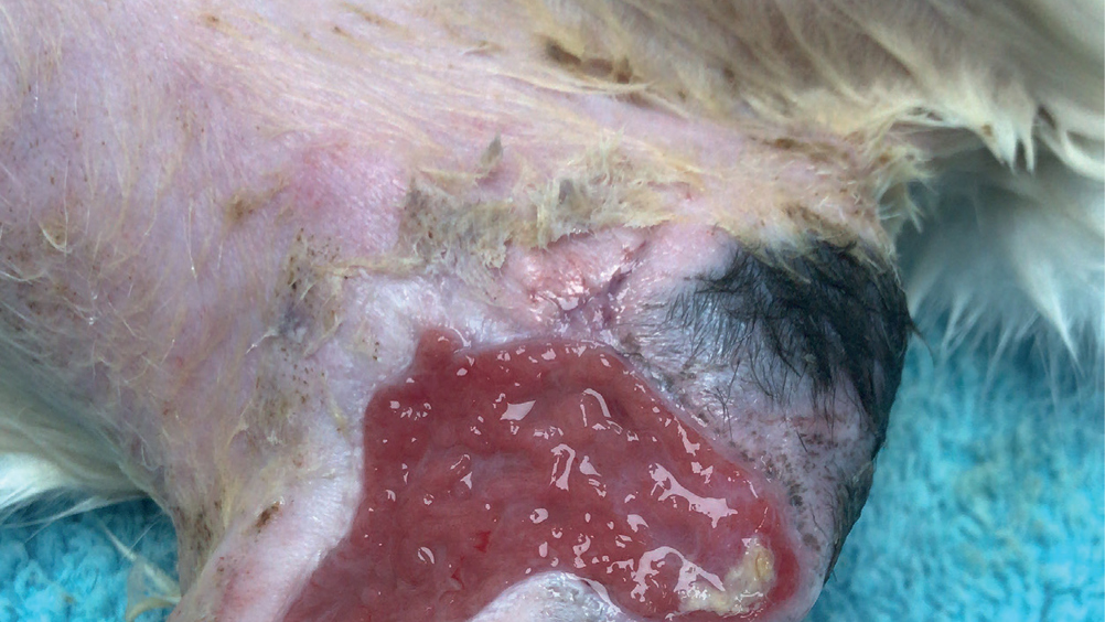
Figure 1. Biofilm shiny layer covering an unhealthy granulation bed.

Abstract

Wound management in veterinary practice can be challenging at times. With a variety of factors to consider when managing a wound, it can be further complicated if that wound starts to stagnate or regress. A process of elimination begins to identify the cause of the problem. Infection is a contributing factor to delays in wound healing, but biofilms may not always be considered, especially during the initial stages. These communities of bacteria need to be managed quickly and efficiently to prevent them becoming irreversibly embedded within a wound. As nurses it can be a useful wound management skill to be able to recognise biofilms and their signs and understand how early interventions and appropriate wound management can prevent a biofilm from becoming a problem.

Chronic wounds can present a variety of challenges in veterinary practice, and it can become time consuming and expensive when trying to eliminate what exactly may be causing a delay in wound healing. A chronic wound can be defined as a wound that has failed to progress through the natural stages of healing and remains in a continual and unmanageable inflammatory phase despite wound management interventions (Zhao et al, 2016). When presented with a wound that had been previously granulating, but seems to be regressing or showing signs of a prolonged inflammatory response, it is important to consider the reasons that this could be happening. Using the basic principles of wound management and considering the factors that contribute to delays in wound healing may help to highlight what might be contributing to the stagnation or prolonged inflammatory response within a wound bed. Presence of infection, movement, patient interference, ischaemia and tension are a few factors that can contribute to a delay in wound healing. Some of these can be eliminated more easily than others, such as patient interference, but when it comes to factors like infection, eliminating certain types of bacteria especially those found in biofilms can prove difficult.

Biofilms are complex communities that typically consist of multiple bacteria and/or fungi. These bacterial and fungal communities are suspended in a protective liquid matrix that allows them to firmly attach to a variety of surfaces in the environment and on living tissue (Phillips et al, 2010). Early recognition, intervention and management of biofilms found within the wound bed is becoming an increasingly important aspect of wound management (Wound Source, 2018a). These microbial communities can present during different phases of wound healing, most typically in the initial inflammatory phases causing this to become prolonged, but they can also contribute to delays in wound healing if they present during the proliferation stage (Wound Source, 2018a), this is why it is important that nurses are able to recognise biofilms when they present and understand how they can be effectively managed and prevented.

Formation of bacterial and fungal biofilms

Biofilms are found on a variety of living and non-living surfaces; recent studies have shown that biofilms can present on numerous medical consumables and devices, such as urinary catheters, endotracheal tubes, and orthopaedic implants, they are also considered to be a major contributing factor in hospital acquired infections in humans (Phillips et al, 2010). These complex communities of bacteria and/or fungi are suspended in an extracellular polymeric matrix, this ‘slimy’ liquid substance helps it to adhere to surfaces and creates a protective barrier for the embedded micro-organisms (Phillips et al, 2010). Most microbes are micro-scopic in size, and would typically require a microscope to view them, but as biofilms mature, they can be easier to spot due to this liquid protective matrix presenting as a shiny film covering the wound surface (Figure 1) (Phillips et al, 2010).

Figure 1. Biofilm shiny layer covering an unhealthy granulation bed.

It had been previously presumed that most microorganisms existed in isolation living in a planktonic state as free-floating single organisms, but under the right conditions the majority will establish colonies that will eventually adhere and form a biofilm on the wound surface. The removal of these microorganisms is easier during the initial stages of contamination as they have yet to begin producing the extracellular matrix required that allows them to form adhesive bonds to a surface (Hollmann et al, n.d.). If the microorganisms are not removed after the initial contamination, they will begin to form a bacterial monolayer across the wound surface that starts secreting an extracellular polymeric substance (EPS) that creates a firm bond; once this process begins it is irreversible and removing the newly formed colony becomes very challenging (Hollmann et al, n.d.). The EPS generally consists of structural proteins, polysaccharides (long chains of sugars), cell debris and nucleic acid, this can vary depending on the microorganisms involved. The primary formation of the extracellular matrix is initiated by the nucleic acid or extracellular DNA (eDNA found floating free inside the bacteria), and during the later stages structural proteins and polysaccharides take over. The matrix enables significant and rapid growth of the microorganisms involved and causes changes in their genetic patterns leading to a form of cell to cell communication known as quorum sensing (Phillips, et al, 2010). Quorum sensing allows the different bacteria within the colony to communicate; they can effectively share their abilities which helps aid in their survival (Phillips et al, 2010). As the biofilm becomes established the micro-colony will begin to form multiple layers of microorganisms that causes them to grown out from the wound bed in a three-dimensional manner — usually by this stage the biofilm may become visible and the attachment is now classed as irreversible (Hollmann et al, n.d.). During the final stages the mature biofilm will take on a ‘mushroom’ shape and some of the enveloped microorganisms and biofilm fragments can detach and be dispersed allowing for the potential growth of new biofilms in other areas of the wound bed (Figure 2) (Hollmann et al, n.d.).

Figure 2. Biofilm cycle.

Effects of biofilms on wound healing and the difficulties they pose in wound management

Depending on the type of microbes present a mature biofilm has been shown to form within as little as 2–4 days. Generally, single free-floating microbes will loosely attach to the wound bed within minutes of an injury occurring. If the bioburden is not removed then loosely attached micro-colonies begin to form, with these becoming firmly attached within 2–4 hours. The production of the initial EPS starts forming within 6–12 hours causing resistance within the micro-colony to biocides, and if lavage and debridement are inadequate, they have been shown to reform within 24 hours (Phillips et al, 2010).

Mature biofilms are very effective at protecting their embedded microorganisms, the formation of the EPS means these now colonised microorganisms can become highly resistant to the body's natural immune response and external environmental factors, such as traditional biocides (antibiotics, antiseptics and disinfectants) that would usually be effective at destroying the same microorganisms living in a planktonic state as free-floating single organisms (Phillips et al, 2010). Some of the methods employed by the EPS includes providing protection in the form of a physical barrier that blocks antibodies, inflammatory cells and sometimes smaller cells, such as antimicrobials, from entering the matrix (Phillips et al, 2010). Quorum sensing promotes the sharing of certain traits amongst the community of different microorganisms contained within the EPS. The ability to share certain qualities helps aid in the survival of the community, for example antibiotic resistant bacteria can secrete antibiotic binding proteins that protect non-antibiotic resistant bacteria within the matrix (Phillips et al, 2010). Microorganisms within the EPS can also enter a state of hibernation; the microorganisms can become metabolically inactive, causing topical or systemic antibiotics to be ineffective as the bacteria needs to be metabolically active for the treatment to be successful (Phillips et al, 2010). In human medicine some research has highlighted that the standard oral doses administered to patients with planktonic bacterial infections are completely ineffective against biofilm encased bacteria, and that the doses required to penetrate them would exceed maximum prescription levels (Phillips et al, 2010).

Biofilms have been shown to have a significant effect on wound healing times by causing a prolonged inflammatory response within the wound bed (Phillips, et al, 2010). During the initial inflammatory phase, the bloods vessels at the site of injury will dilate and begin to leak transudate fluid; this process allows for the flooding of leukocytes, antibodies, growth factors, enzymes and nutrients into the wound bed (White, n.d.). As all these essential elements flood the site and an immune response is initiated, macrophages and neutrophils will start autolysing devitalised and necrotic tissue, which when combined with the release of transudate fluid will cause exudate levels to rise (Figure 3) (White, n.d.). If a biofilm can form, due to the ineffective removal of microorganisms in the initial stages of presentation, the body will continue to respond by releasing increased numbers of neutrophils and macrophages, which will attempt to rid the wound of the biofilm by releasing high levels of reactive oxygen species (ROS) and proteases (Phillips et al, 2010). These aim to disrupt bonds between the biofilm and the wound bed, but the issue with this increased inflammatory response is that the ROS and proteases are not specific and will damage healthy tissue, proteins and immune cells essential to healing during this process. It is thought that this natural chronic inflammatory response is not generally effective at removing and detaching the biofilm, and that the increased exudate levels can instead act as a nutrient source for the biofilm (Phillips et al, 2010).

Figure 3. A wound in a prolonged inflammatory phase with suspected biofilm involvement.

However, it should be noted that not all biofilms are necessarily negative, Staphylococcus epidermis, a commensal bacterium commonly found on the skin, can prevent pathogenic bacteria from colonising through stimulation of a cell-mediated immune response and reducing available space and nutrients for pathogenic bacteria to thrive and grow (Hollmann et al, n.d.). Despite this most biofilms, particularly those found in the wound bed, will be pathogenic and problematic for wound healing.

Treatment and prevention

As previously discussed, biofilms can begin their attachment to the wound bed within a matter of hours, this highlights how important it is that nurses perform basic wound management techniques as quickly and effectively as possible after presentation so that potential complications can be prevented. Difficulties can arise with delayed presentation as the bacteria may have already become established and micro-colonies may have already started to form irreversible attachments.

When presented with a wound the animal should be triaged and if all body systems are deemed stable then effective wound management should begin immediately (Winkler, n.d.). The animal should be provided with adequate analgesia, and in most cases sedation will be required so that a thorough examination of the wound can take place; these requirements should always be discussed and implemented with the overseeing veterinary surgeon. Preparation of the wound bed is important at this stage; adequate clipping and lavage should be performed. If the animal cannot be dealt with immediately it is advisable to try and lavage the wound and then cover with a protective adhesive dressing to help reduce bacterial load and prevent further contamination while the patient is awaiting treatment (Winkler, n.d.).

Clippers should always be sharp and clean to prevent damage to the surrounding tissue; a wide area should be clipped to allow for full visualisation of the wound. It is also advisable to apply a viscous preparation to the wound before clipping, this can be a simple sterile lubricant which can help prevent further contamination of the wound bed while clipping is carried out (Figure 4).

Figure 4. Clippers with sharp and clean blades, size 40 in photo, size 50 can also be used. Also various types of sterile lubricants which can be used within the wound prior to clipping.

Once clipped the wound should be lavaged, ideally using an isotonic solution, such as compound sodium lactate or saline (Hollis, 2018). The use of antiseptic preparations is not generally advised, preferably lavage should be performed with a solution that can remove debris and has antiseptic properties but also causes minimal disruption and is non-toxic to healthy tissues and cells (Winkler, n.d.). Certain antiseptic solutions, such as chlorhexidine and iodine, have been known at certain dilution rates to be toxic to cells that are vital in the later stages of wound healing (Hollis, 2018). It is believed that the volume of solution used is important, most texts recommend either a volume of 500–1000 ml of solution is used or as a rule: 100 ml of chosen solution per centimetre of wound (Dycus and Wardlaw, 2013; Hollis, 2018). If antiseptic solutions are to be used for surrounding tissue or in the wound bed, manufacturer's guidelines should be followed regarding dilution rates, using incorrect dilutions could lead to potential issues with bacterial resistance, it should also be noted that some antiseptics are inactivated by organic material potentially rendering them ineffective (Hollis, 2018).

The use of surfactants containing the antiseptic polyhexamethylene biguanide (PHMB) for lavage has become a popular choice in the presence of biofilms over recent years. PHMB has broad spectrum antiseptic properties that is effective against a variety of Gram-negative and Gram-positive bacteria, it has been shown to have low toxicity within the wound bed and can help to prevent damage to the surrounding healthy tissue (Barrett and King, 2016). PHMB is available in a variety of formats (wound dressing/gels), but for biofilm removal during the lavage stage it can be purchased as a premade solution, with some also containing betaine, a gentle detergent that aids in the disruption of the biofilm and other debris (Bbraun, n.d.). PHMB could become a more mainstream treatment in veterinary medicine and should be considered when dealing with chronic or delayed healing wounds. In human medicine studies have shown these products to be effective at reducing and managing biofilms and when compared with other antimicrobials, including chlorhexidine, povidone-iodine, and silver, it was found to have superior antiseptic biocompatibility in comparative tests; this study measured antiseptic action versus the potential for the product to be cytotoxic to cells (Barret and King, 2016).

Another point to consider is the pressure at which the wound is lavaged — bacteria can be removed with pressure as low as 1.6 psi, but typically 7–8 psi is recommended (Dycus and Wardlaw, 2013). It is important to ensure that an appropriate method is used, poor delivery could potentially damage underlying tissue or not effectively remove the bacterial bioburden. This is a commonly debated subject as it is difficult to measure the exact pressure being delivered when lavaging a wound — one recommended method of delivery is using a 35 ml syringe with a 19 gauge needle attached to a giving set with a three-way tap, this can then be attached to a fluid bag so appropriate volume can be administered at approximately 7–8 psi (Figure 5) (Winkler, n.d.). Another study has suggested that using a pressure bag with a manometer set at 300 mmHg should consistently produce a psi of 7–8 irrespective of the needle size used (Dycus and Wardlaw, 2013).

Figure 5. Equipment prior to set up for wound lavage; 1 litre sterile Hartmann's, three way tap, 19 guage needle, 35 ml syringe. A rapid infuser bag can also be placed over the fluid bag.

After the wound bed has been assessed and prepared it may be necessary to carry out further debridement. If the animal has been presented after the initial injury and there are no delays then mechanical debridement may be adequate to ensure removal of any further bioburden, this can be carried out using debridement pads that help to lift organic material from the wound bed or a wet to dry dressing could be applied for 12–24 hours depending on the extent of contamination (Hollis, 2018). But, in cases of delayed presentation a more aggressive approach may be required. Sharp debridement involving the veterinary surgeon removing any devitalised and non-viable tissue can help to reduce biofilms, but because these colonies are difficult to visualise it is impossible to know if they have been completely removed or not, further research is required into which form of debridement is the most effective at reducing and removing biofilms (Phillips et al, 2010).

The frequency at which debridement or lavage should be performed to prevent reoccurrence of biofilms is unknown. During the inflammatory phases of healing the wound should ideally be redressed every 24 hours, however depending on the exudate levels more frequent changes may be required (Phillips et al, 2010). Other products have been suggested to aid in the removal or destruction of biofilms: manuka honey has been suggested to potentially reduce the inhibition of established biofilms but it needs to be maintained at appropriate concentrations in vivo, so dressings need to be changed regularly especially in highly exuding wounds as dilution of the honey may affect its efficacy (Cooper et al, 2011).


Table 1. Signs of biofilm formation and effective biofilm removal
Signs of biofilm formation
Poor-quality granulation tissue (dark red in colour and haemorrhages easily)
Excessive exudate
Reoccurring infection after antibiotics are stopped
Negative wound cultures despite signs and symptoms of localised infection
Slimy fluid present over wound surface
Prolonged inflammatory response
Signs of effective biofilm removal
Good-quality granulation tissue (pink in colour, often referred to as ‘strawberry jam’ consistency)
Progression from inflammatory phase into proliferation phase
Reduction in exudate levels
(Barret & King, 2016)

Prevention of future contamination of the wound bed should be of utmost importance; dressings and bandaging techniques should be used where appropriate and a thorough assessment of the wound carried out at each dressing change noting exudate levels, colour and odour, presence of necrotic and/or devitalised tissue, signs of inflammation and quality of wound bed. Good client communication and education is vital to ensure the smooth management of a wound, if the client is unaware of issues that can arise from poor management then there are more likely to be delays in healing and potential for contamination leading to biofilm formation.

Conclusion

Biofilms may become a bigger part of veterinary wound management over time, prevention is the best form of treatment for biofilms with a lack of laboratory testing to prove infiltration of biofilms within a wound bed, nurses must rely on clinical experience and the ability to spot signs and symptoms that could indicate the presence of a biofilm (Table 1) (Phillips et al, 2010). Care should be taken during the initial presentation of any wounds to ensure good basic wound management techniques are carried out. Appropriate lavage techniques and adequate volume of solution should be used, good hand hygiene and aseptic techniques should be followed whenever handling the animal or their wound. Also, the addition of surfactants containing PHMB and the responsible use of antimicrobials could all assist in the prevention and removal of biofilms.

KEY POINTS

  • Biofilms are complex communities that typically consist of multiple bacteria and/or fungi, these communities are suspended in a protective liquid matrix that creates a bond between the wound bed and the biofilm that makes their removal difficult.
  • Early intervention is the best treatment to prevent biofilm formation; minimising contamination, appropriate lavage and debridement techniques can all help to prevent biofilm formation.
  • Polyhexamethylene biguanide (PHMB) is a surfactant that contains an antiseptic and sometimes a detergent that can aid in the removal of mature biofilms by breaking down the bonds between the wound bed and the extracellular matrix.
  • Biofilms have been shown to have unique qualities, such as bacteria sharing their individual traits with other bacteria within the biofilm, this trait is called quorum sensing.
  • There are currently no commercial tests available to confirm the formation of biofilms and they can show resistance to traditional biocides, such as antimicrobials, antibiotics and antiseptics.