
Canine hip dysplasia is a developmental rather than a congenital disorder. Pups are born with normal hips, but in individuals with dysplasia, changes in development around the femoral head and acetabulum begin around 3 weeks of age (Kealy et al, 1992). Around this age, subluxation or complete luxation of the femoral head from the acetabular fossa occurs, as a result of joint and soft tissue laxity (Barrett, 2008). This leads to incomplete coverage of the femoral head by the acetabular fossa. Over time, this manifests as painful stretching of the joint capsule, and a reduced area for articulation leading to flattening of the femoral head. Resultant contact between the femoral head and the acetabular fossa causes wear of the protective layer of articular cartilage, which stimulates new bone deposition by the body and so the development of secondary osteoarthritis (King, 2017).
Genetics, environment and prevalence
Hip dysplasia is a complex inherited disorder, whereby both a number of different genes and environmental factors are influential in the disease process (Kennel Club, 2018). The condition is most prevalent amongst large or giant purebreds, but is also seen in smaller purebreds and crossbreed dogs. Certain breeds including German Shepherds, Labrador Retrievers and Boxers are over-represented. Many such breeds are subject to testing prior to breeding for registered litters under the hip scheme recommended by the Kennel Club and British Veterinary Association (BVA) (BVA, 2014). Radiographing potential parents to produce hip scores under the scheme, and only selecting those with lower than average scores for the breed, is likely to minimise the risk of producing clinically affected offspring. Monitoring the effectiveness of schemes such as this is often difficult, but is required to ensure the efficacy of selection (Wilson et al, 2006). Gender does not appear to have an influence on the likelihood of developing hip dysplasia (Barrett, 2008).
Much attention is currently being placed on environmental factors including excessive nutrient intake and exercise during development. Research unsurprisingly found a significant increase in prevalence amongst a group of adlib fed labrador retrievers compared with their limit-fed cohorts (Kealy et al, 1992). Excessive dietary consumption of calcium and vitamin D, and energy in the form of any food group in growing dogs is thought to lead to rapid skeletal growth and increase representation of the condition (Fries et al, 1995). Excessive bodyweight can be further demonstrated to be an associated factor as neutered animals have been shown to be 1.5 times more likely to develop hip dysplasia when compared with entire individuals, a statistic which is likely to be explained by the fact that neutering generally leads to weight gain within 6 months of the procedure (Rodrigues et al, 2011).
Controlled weight-bearing through restricted exercise programmes for growing dogs has also been suggested as a method of reducing prevalence as excessive loading of the hip joints through the growing phase is thought to be an additional environmental factor (Carver, 2016).
Discussion of both a suitable diet and exercise regimen with puppy owners in the veterinary clinic setting during routine appointments for vaccinations and health checks could be initiated by the veterinary nurse to help minimise the impact of environmental factors.
Clinical signs and diagnosis
Clinical signs in affected individuals generally develop at around 4–12 months of age, and include some or all of the following:
Veterinary examination findings are also likely to include pain or restriction on manual extension of one or both hip joints (Barrett, 2008).
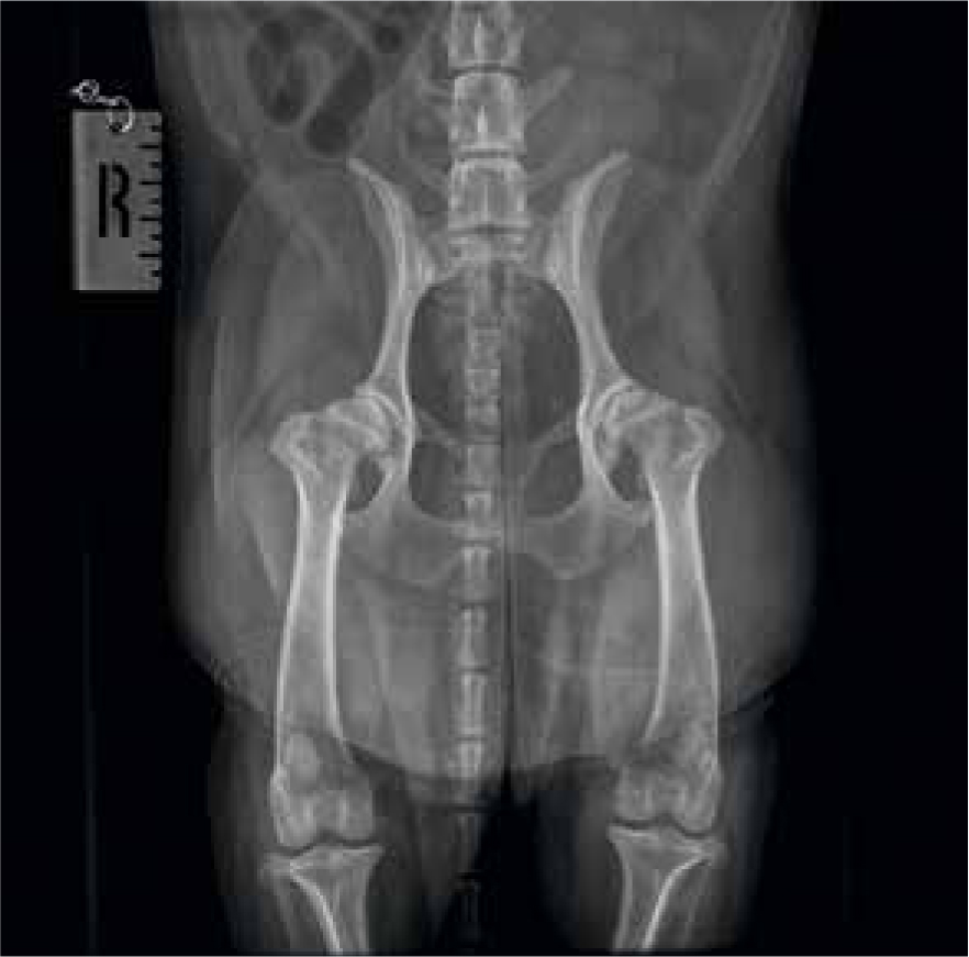
Diagnosis is confirmed radiographically, which should be performed under anaesthetic or heavy sedation to allow for accurate positioning. Standard practice is to lie dogs in dorsal recumbency with hind limbs extended, and stifles adducted. Care should be taken to ensure the pelvis is not rotated to allow for accurate comparison and interpretation. Radiographic changes do not always correlate with the severity of clinical signs displayed by the patient, and include (as shown in Figure 1) loss of femoral head coverage by the acetabular fossa and luxation or subluxation (Ginja et al, 2015). This is often paired with changes consistent with secondary osteoarthritis including:
In the 1980s, The University of Pennysylvania School of Veterinary Medicine developed the PennHIP screening method to assess dogs from as early as 16 weeks for the likelihood of developing hip dysplasia. The scheme, with proven accuracy and sensitivity, assesses the degree of laxity of the hip joints as dogs with ‘loser’ hips have been shown to be more likely to develop dysplasia than those with ‘tight’ hips. Specially certified veterinary surgeons take three different radiographic views of the hip joints, and use a specific tool to measure this degree of laxity under heavy sedation or general anaesthesia. The radiographs are submitted for assessment, which allows breeders to only select and breed from low risk individuals, with the hope of reducing prevalence of the condition (Antech Imaging Services, 2018).
The PennHIP screening method has not always been available in the UK due to regulations as two out of the three x-ray views required manual holding. A hands-free technique has now been developed meaning manual intervention is no longer necessary, and so UK veterinary surgeons can now train for PennHIP certification (Barrett, 2008).
Treatment
Treatment of hip dysplasia is focused on management as it is an incurable condition, options including surgical or non-surgical techniques (Kirkby and Lewis, 2011). Possible surgical interventions include the triple-pelvicosteotomy, a procedure where the pelvic bones are realigned in an attempt to improve acetabular coverage of the femoral head. Age is often the limiting factor in recommending this procedure, as it has a more successful outcome if performed below 7 months of age, at which point many affected dogs may be as yet undiagnosed. Other commonly available surgical procedures include total hip replacement, a procedure by which an artificial ball and socket joint replaces the existing joint; and femoral head excision (Norris, 2011). These techniques are often reserved for severe cases, or when non-surgical management alone is not successful in the control of associated pain. The success of any of the identified possible surgical procedures, described as a complete return to function, has been difficult to assess due to lack of follow-up (Bergh and Budsberg, 2014); but the likelihood is that there is not a ‘one size fits all’ approach, and a case by case selection, often dictated by age of the patient at time of presentation, is best.
Non-surgical options include the use of pharmaceuticals such as non-steroidal anti-inflammatory drugs (NSAIDs) and those affecting the progression of joint disease; weight management; exercise modification; nutraceuticals and rehabilitation therapies (Kirkby and Lewis, 2012). The veterinary nurse can play a vital role in each of these, as discussed below.
The role of the veterinary nurse in practice
Many owners are unaware of the changes in lifestyle which may positively assist in the management of dogs diagnosed with hip dysplasia, and the veterinary nurse can play a vital role in raising these issues and impacting management.
Pharmaceuticals
Several analgesics exist for the management of pain associated with hip dysplasia and secondary osteoarthritis, including NSAIDs, paracetamol/codeine based drugs, opioids, gabapentin and corticosteroids (Pettitt and German, 2015). Owners may not always be aware of the different presentations of pain and related behaviours in dogs. During weight clinics or routine appointments any behavioural changes, reluctance to exercise or alterations in gait the owners mention could first be picked up on by the veterinary nurse, for prompt referral to the veterinary surgeon for intervention with analgesics when necessary.
Nutraceuticals
Several nutraceuticals are now available which claim to assist in the management of secondary osteoarthritis. These include glucosamine sulphate and chondroitin sulphate, hyaluronic acid, avocado/soybean unsaponifiables and omega-3 fatty acids, many of which have anecdotally improved symptoms in human management (Henrotin et al, 2005).
Exercise modification
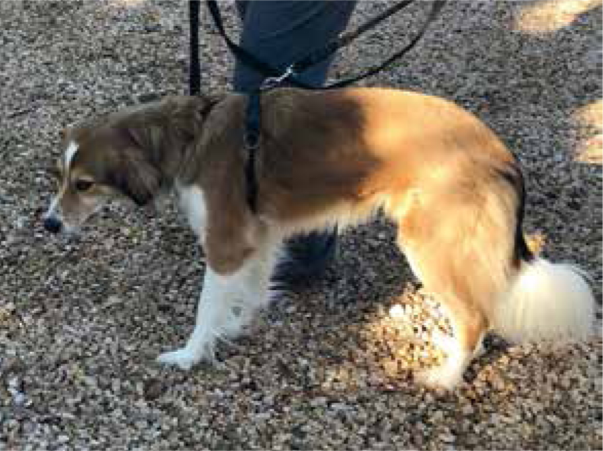
The goal of any exercise programme is to promote the maintenance of muscle mass, maintain joint range of motion and manage pain. Slow, controlled, leash walking (Figure 2) has been shown to promote hip extension when compared with trotting, which assists by helping to maintain as near normal range of motion within the hip joint as possible; and can be started immediately.
Initiating an exercise programme of short walks of 15–20 minutes' duration, two to three times daily, then progressing by adding 5 minutes to each walk per week to a duration of 35–40 minutes can be used to promote the development of muscle mass to support the hips and prevent stiffness by promoting range of motion. Once this level has been reached, this can be further progressed to include incline walking to promote hip flexion, and walking up and down stairs to promote extension.
Encouraging owners to allow their pets to warm up for 5–10 minutes before any form of exercise and cool down afterwards will help prevent painful flare-ups resulting from injuries (Dycus et al, 2017), and suggesting owners walk their dogs on more forgiving surfaces such as grass or sand rather than concrete will minimise forces applied through the joint.
Consideration should also be given to controlling high impact exercises such as ball throwing and walking on uneven terrain until it can be sure this will not result in painful flare-ups (Millis and Levine, 2014).
Weight management
Weight modification through dietary restriction has been demonstrated to decrease lameness score in dogs affected by hip dysplasia, and reduce the progression and clinical signs associated with secondary osteoarthritis in humans (Kirkby and Lewis, 2011). As such, the importance of the maintenance of normal body condition in management of canine hip dysplasia cannot be underestimated. Veterinary nurses are often responsible for running in-house weight management clinics and a few main considerations should be given to methods used to enhance compliance and increase success:
Rehabilitation
Techniques which can be performed in practice, at home by the owner, and by the rehabilitation therapist following referral will be discussed in detail in the next article.
Conclusion
This multimodal approach to conservative management of the hip dysplasia patient demonstrates the importance of communication with owner regarding a number of different options, many of which can be incorporated into the patient's daily routine to positively impact quality of life.