
A liver lobe torsion is when one or more of the liver lobes twist, with some reports documenting this including the gall bladder (Massari et al, 2012). Liver lobe torsions are documented in a variety of species including dogs (Schwartz et al, 2006), humans (Umehara et al, 2009), cats (Nazarali et al, 2014), horses (Bentz et al, 2009), guinea pig (Waugh et al, 2021), otters (Warns-Petit, 2001), camels (Ibrahim et al, 2021), ferrets (Vilalta et al, 2016) and rabbits among others.
The condition of liver lobe torsion is considered rare in rabbits, but the author believes that the condition is likely to be underdiagnosed, especially in this species because of the difficultly in recognising clinical signs. A study in 984 laboratory rabbits showed that 0.3% of asymptomatic rabbits had suffered from a liver lobe torsion (Weisbroth, 1975), highlighting that these can be subclinical. A rabbit's liver comprises four lobes, the caudate lobe and three cranial lobes called the right, medial left and lateral left lobes. The most commonly affected lobe is the caudate lobe, accounting for 62.5% of liver torsions documented at one clinic (Graham et al, 2014), however, torsion of other lobes is documented (Graham and Basseches, 2014). The reason for the torsion is not fully understood, but in dogs suggested causes include large breed predisposition, previous gastric dilatation and volvulus, neoplastic changes, trauma and vigorous jumping (Bhandal, 2008). Predisposing factors are not fully described in rabbits, however, theories include the absence of hepatic supporting ligaments, trauma, episodes of gastrointestinal stasis resulting in gastric dilatation and stretching of the left triangular ligament, and bacterial or parasitic hepatitis (Summa and Brandão, 2017).
Clinical signs of liver lobe torsion in rabbits are often vague. They may include acute anorexia, abdominal pain, pale mucous membranes and sudden death (Pignon et al, 2013). As a prey species rabbits often hide clinical signs, especially when brought into the veterinary clinic, so obtaining a detailed history from the owner regarding the animal's behaviours and clinical signs at home, as well as information on husbandry, is vital.
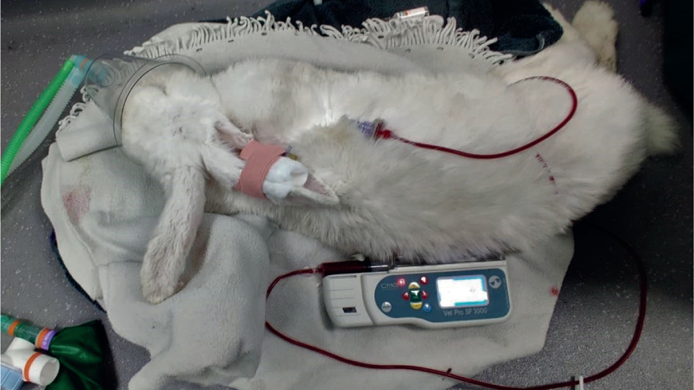
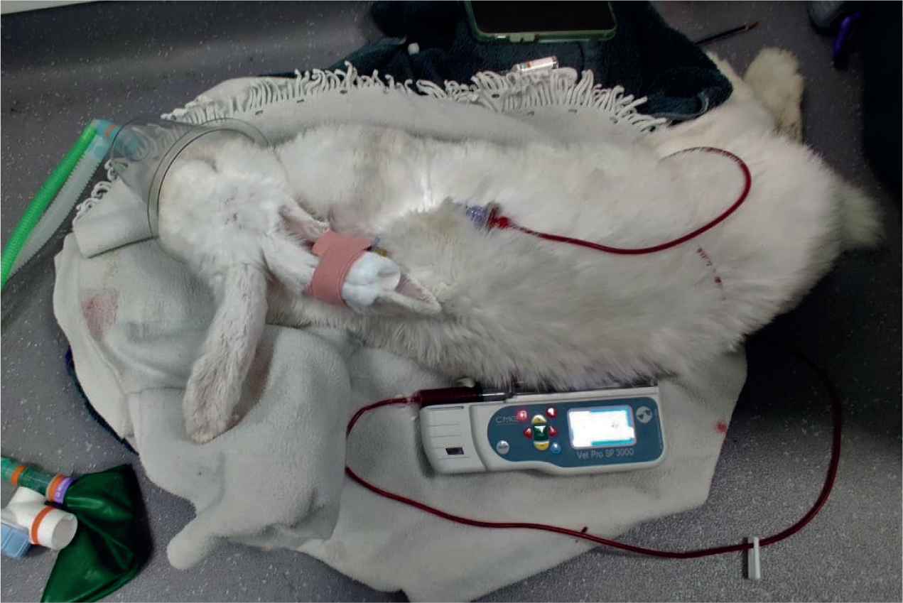
Diagnosis of a liver lobe torsion is based on a combination of clinical signs, physical examination and diagnostic imaging. There are reports of the twisted lobe being detected on abdominal palpation, or eliciting a pain response in the patient (Taylor and Staff, 2007), but this may not always be the case. Biochemistry and haematology will often prove informative in these cases. In many animals a reduced packed cell volume (PCV) and an elevation in alanine aminotransferase (ALT), aspartate aminotransferase (AST), and gamma-glutamyl transferase (GGT) will be seen (Wenger et al, 2009). Ultrasonography of the abdominal cavity may be used to confirm the diagnosis of a liver torsion, with the enlarged lobe visible with absent blood flow on colour flow Doppler, and in some cases free fluid around the affected lobe. One report used contrast enhancing ultrasonography to diagnose a liver torsion (Stock et al, 2020). However, visualising the affected lobe using colour flow Doppler technique to recognise lack of blood flow, is often all that is required to confirm diagnosis (DeCubellis and Graham, 2013). Haemoabdomen is a common finding in liver torsion with free fluid visualised on ultrasonography. Abdominocentesis can confirm the presence of blood if required (Oglesbee and Lord, 2020). In severely anaemic cases, where significant blood loss is seen, a blood transfusion can be considered (Figure 1), this is often done so by obtaining a donation of blood from a companion rabbit. Radiography can be used to aid diagnosis and may show hepatomegaly, free fluid or an obscured liver boarder (Figure 2). Many cases also have gastrointestinal gas, suggestive of gut stasis as a result of anorexia (Graham and Basseches, 2014). Computed tomography (CT) has also been described as a useful tool for the diagnosis of this condition (Daggett et al, 2021).
Treatment of liver lobe torsion involves surgical removal of the affected lobe and is described as a successful treatment for these species (Stanke et al, 2011). When treated medically these patients have a much lower survival rate, in one report only 43% survived compared with 100% survival in those treated surgically (Graham and Basseches, 2014). However, the author has seen several severely compromised individuals fail to survive surgical intervention as a result of being haemodynamically and systemically unstable prior to anaesthesia. Aggressive and intensive nursing care of sick rabbits is essential to a successful outcome in these species whether treated surgically or medically.
Rupert's case
Rupert was a 6-year-old, male neutered, crossbreed rabbit belonging to one of the clinic's veterinary surgeons. He was fully up to date with vaccinations against myxomatosis and rabbit haemorrhagic disease (RHD) 1 and 2 and lived in a large outdoor enclosure with his neutered female companion. Rupert had had a previous episode of gut stasis 2 years before this presentation, but had recovered well with supportive nursing care given at home. Rupert had an acute onset of anorexia around 18 hours prior to presentation, and had been given meloxicam 0.6 mg/kg per os (PO) (Loxicom 1.5 mg/ml solution for dogs, Norbook, UK) and ranitidine 4 mg/kg PO (Ranitidine 25 mg tablets, Summit UK), cisapride 0.5 mg/kg PO (Cisapride 5 mg/ml solution, Summit UK) and syringe feeding 10 ml/kg at home (Critical care, Oxbow USA), however had failed to make any improvement overnight. He was therefore brought into the practice for further care.
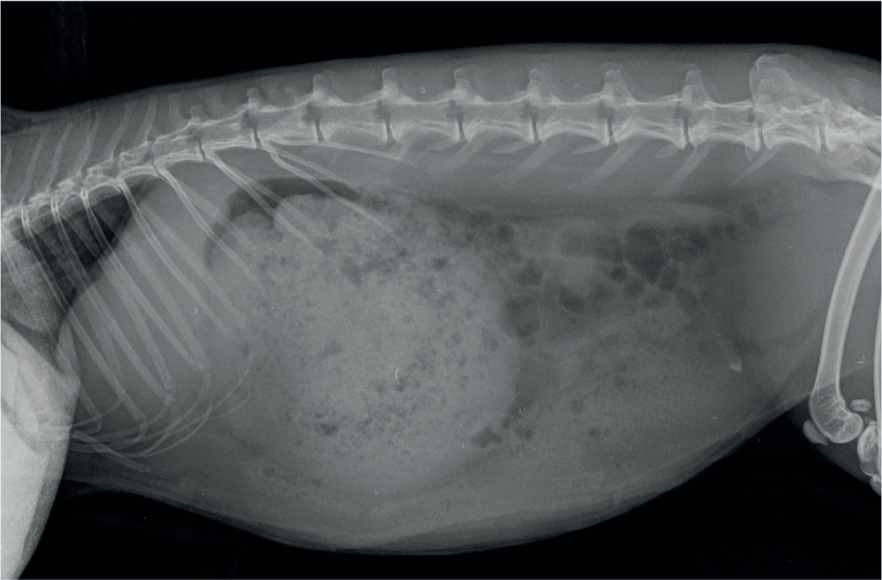
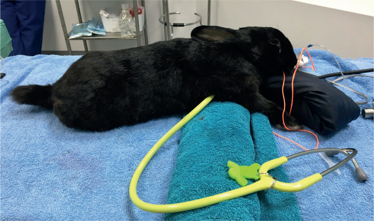
On presentation Rupert weighed 3.13 kg and had a body condition score of 4/5. He was quiet but alert and responsive, his respiratory rate was 120 breaths per minute (normal range 32–60 (Varga, 2014)), heart rate was 250 beats per minute (normal range 130–325 (Varga, 2014)), he had pale mucous membranes (Figure 3), and his temperature was slightly low at 37.8°C (normal range 38.5–40.0 (Varga 2014)). Lower temperatures in rabbits on admission are associated with poorer outcomes (Di Girolamo et al, 2016), however because he was not substantially hypothermic, Rupert was not housed in an incubator. On being placed into a walk in kennel with his companion, he was seen nibbling on strands of timothy hay and was not exhibiting signs of severe illness. As a result of the mild hypothermia and subdued temperament of the patient, treatment was administered as described in Table 1. The rabbit was initially given buprenorphine 0.05 mg/kg subcutaneously (SC) (Buprecare 0.3 mg/ml, Animal care Ltd, UK), but after diagnosis and surgery this was replaced with methadone 0.5 mg/kg (Comfortan 10 mg/ml, Dechra, UK).
Table 1. Medications used to treat the rabbit
| Drug | Dosage | Frequency | Route of administration |
|---|---|---|---|
| Meloxicam | 0.6 mg/kg | 12 hourly | Per os (PO) |
| Ranitidine | 4 mg/kg | 8 hourly | PO |
| Cisapride | 0.5 mg/kg | 8 hourly | PO |
| Methadone | 0.5 mg/kg | 4 hourly | Subcutaneous (SC) |
| Hartmann's | 50 ml/kg/24 hour | 4 hourly | Intravenous (IV) bolus |
| Lidocaine | 0.05 mg/kg/min | Constant rate infusion | IV |
| Oxbow critical care syringe feed | 10 ml/kg | 4 hourly | PO |
After application of lidocaine topical cream (EMLA cream 5%, Aspen Pharmacare, South Africa) a 24G intravenous catheter (Jelco IV catheter, Smiths Medical, UK) was placed into the marginal ear vein to administer intravenous fluids and medications. A blood sample was obtained from the lateral saphenous vein and a full biochemistry and manual PCV and total protein (TP), which were run in house. This revealed an elevated ALT of 407 U/litre (normal range 31–53 (Varga, 2014)), raised urea of 12.9 mmol/litre (normal range 3.6–8.6 (Varga, 2014)), elevated blood glucose of 14.81 mmol/litre (normal range 4.17–8.06 (Varga, 2014)) and a low PCV of 18% (normal range 30–50 (Varga, 2014)). As a result of the raised ALT and severe anaemia, liver lobe torsion was considered a differential diagnosis in this case, and the veterinarian requested an abdominal ultrasound. The ultrasound revealed a congested liver lobe with lack of circulation demonstrated by colour flow Doppler and free fluid within the abdomen. These findings were consistent with a torsion of the liver lobe, and the patient was prepared for emergency surgery. All emergency drugs were calculated prior to anaesthesia. This is good practice for all anaesthetics regardless of ASA classification (American Society of Anesthesiologists, 2014), and can save vital seconds during an emergency. Rupert would be considered an ASA classification of 4 — status 4 animals are considered a high risk, significantly compromised by disease. Rupert could be classed as a 4E — the E denoting that the anaesthesia is an emergency.
The rabbit was premedicated with midazolam 0.5 mg/kg intravenously (IV) (Midazolam 5 mg/ml, Hameln Pharmaceuticals, Germany) before induction with alfaxalone 2 mg/kg IV (Alfaxan 10 mg/ml, Jurox, UK) to effect. Intubation was achieved using an otoscope for a visualised technique. A 4F urinary catheter was used as an introducer for a 3.0 mm uncuffed Murphy's endotracheal tube. Placement was confirmed using a side stream capnograph (Solaris handheld capnograph, Burtons veterinary, UK). The ventral abdomen was clipped and prepared using chlorhexidine surgical scrub solution. A bupivacaine 2 mg/kg local anaesthesia line block was placed SC at the site of the surgical incision on preparation. This helps to provide multimodal analgesia to the patient. Pain and stress in these animals can lead to further complications (Varga, 2016), and will significantly affect the patient's welfare.
The patient was monitored under anaesthesia using heart rate and respiratory rate as well as end tidal CO2 (EtCO2), continuous rectal temperature, electrocardiogram (ECG) and pulse oximetry (SPO2), this was achieved using a multiparameter monitor (Mindray iMEC 8 Vet Multi Parameter Monitor, Burtons Veterinary, UK). Indirect blood pressure was monitored using a Doppler over the radial pulse (Model 811-B Doppler Ultrasonic Flow Detector, Parks Medical Electronics, Inc. USA), and a size 2 cuff on the forelimb. Systolic blood pressure was maintained between 78–102 mmHg throughout the anaesthesia. Normal systolic blood pressure in small mammals varies between 80–120 mmHg, however caution should be taken when taking a reading in small species, as large cuffs can falsely lower blood pressure, this is especially evident in ferrets (Lichtenberger, 2007). Reflexes are not reliable in prey species, so these were not monitored. The rabbit was connected to a mechanical ventilator for the procedure as apnoea is common in rabbits for various reasons including drug induced, breath holding, too deep plain of anaesthesia or impending cardiac arrest (Sibbald, 2018). In this case the patient maintained spontaneous ventilation throughout the surgery, and intermittent positive pressure ventilation (IPPV) was not required. The patient's thorax was elevated during anaesthesia and in recovery to aid ventilation. Rabbits are diaphragmatic breathers and elevating the thorax decreases pressure from the large abdominal organs on the diaphragm (Figure 4).
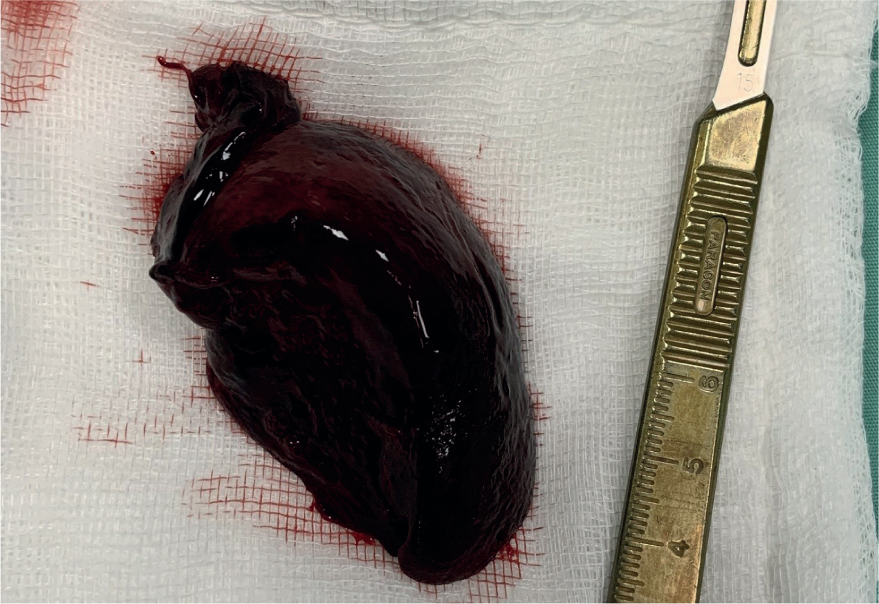
Exploratory surgery revealed heamoabdomen and confirmed the diagnosis of a torsion of the caudate liver lobe (Figure 5). The affected lobe was successfully removed using haemoclips and a biopsy was taken from the remaining liver because of a pale appearance. This sample was sent for histopathology and later results showed hepatic lipidosis. The abdomen was lavaged with warm saline and sutured using a routine three layer closure with monofilament suture.
The rabbit's temperature at the end of anaesthesia was 37.2°C. It is common for rabbits to suffer hypothermia during anaesthesia, this is likely a result of the high surface area to body size ratio of these animals, as well as other factors such as exposed viscera, cold preparation solutions and intravenous fluids. Various methods have been described for active warming and patients should be recovered in a warm ambient temperature of ideally 32°C (Druce, 2015). Rupert was placed into an incubator set at 30°C (Pet brooder ICU, Rcom, USA) and monitored closely into recovery.
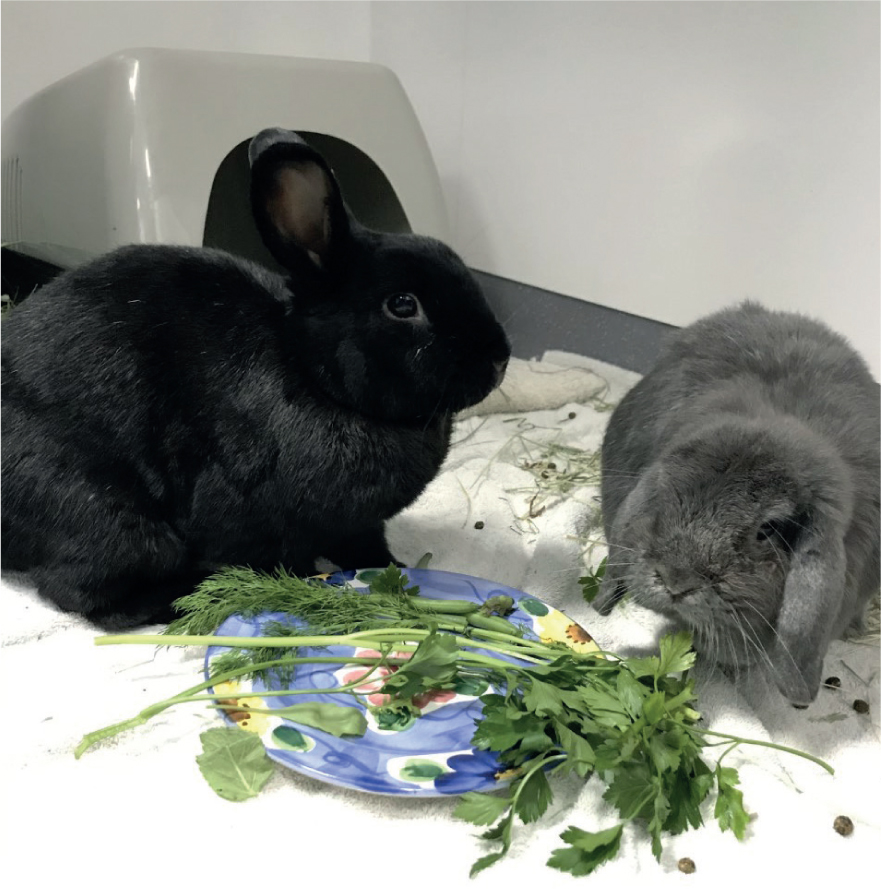
Within 24 hours of surgery the patient began to eat hay and small amounts of green vegetables and was considered to be recovering well (Figure 6). Methadone was replaced with buprenorphine 0.05 mg/kg 6–8 hourly SC and the lidocaine constant rate infusion (CRI) was ceased. He was started on vitamin K 2.5 mg/kg 24 hourly intramuscularly (IM), this vitamin plays a vital role in coagulation, and patients with hepatic dysfunction are likely to suffer from coagulopathies (Amitrano et al, 2002). This was continued at 1.5 mg/kg 12 hourly PO, and a hepatic support nutraceutical containing milk thistle was also given.
During postoperative recovery and times of illness rabbits often require more intensive nursing care than many species such as cats and dogs. Because of the need for constant grazing, rabbits are at risk of developing gastrointestinal hypomotility (gut stasis) during times of illness and recovery. It is therefore essential that rabbits that are hyporexic or anorexic are supplemented with syringe feeding to ensure gut motility is maintained. In this case Rupert was syringe fed Critical Care Herbivore (Oxbow Animal Health, USA) at a dose of 10 ml/kg five times during the day (every 4 hours with a break given overnight); this was gradually reduced in frequency as Rupert began to eat more. High fibre food is essential to maintain the motility and gastrointestinal flora health of a rabbit's digestive system (Hamlin, 2011). As a result of being prey animals, rabbits are also often harder to monitor for signs of pain, this can be achieved by the use of validated scales such as the rabbit grimace scale developed by Newcastle University. The author uses their own modified pain scoring scale (Table 2) and assesses both a facial grimace as well as other symptoms of pain and stress in these species, giving the patient two scores. This was monitored at regular intervals throughout Rupert's recovery to ensure adequate analgesia was being administered.
Table 2. Rabbit and rodent pain scoring
| Pain score | 0 | 1 MONITOR | 2 REASSESS PAIN RELIEF | 3 REASSESS PAIN RELIEF | 4 IMMIDIATE VET ATTENTION |
|---|---|---|---|---|---|
| Grimace scale |
|
|
|
|
|
| Behaviour |
|
|
|
|
|
You must be familiar with the appearance and behaviour of your species and your individual patient's temperament before scoring. Stress can be associated with many of these behaviours such as tachypnoea and anorexia
In the author's opinion rabbits are not known to interfere with surgical wounds provided that they are given adequate analgesia. However, Rupert was closely monitored for wound interference. His surgical wound was also checked twice daily for signs of inflammation or breakdown and no complications were noted.
Rupert was also housed with his bonded companion ‘Phoebe’ in a large walk in run. It has been shown that the presence of a companion is rated as highly as food, and will reduce the stress of hospitalised rabbits (Stapleton, 2016). Wherever possible, companions should be housed during a rabbit's stay. Rupert was separated for 6–8 hours every night, this allowed time to monitor his urine and faecal output and allowed time for eating without competition. Rabbits with particularly delicate bonds will require a grid divide during this separation to avoid any time without visual or physical contact risking the breakdown of a bond.
By day 7 of hospitalisation, Rupert's injectable and intravenous medications and fluids had ceased and syringe feeding was stopped because of a return of normal appetite. He was sent home on oral cisapride at 0.5 mg/kg and meloxicam 0.6 mg/kg twice daily for 5–7 days. Because he was owned by one of the practice veterinary surgeons he was not presented back to the clinic for postoperative checks. In normal circumstances these are usually at 3 and 10 days after returning home. In some cases these checks can be done via a telephone call if the owner and veterinary surgeon/veterinary nurse are competent and experienced in the species. This can also be advantageous if the rabbit has a particularly easily stressed personality, avoiding unnecessary travel. Stress can reduce healing and recovery times. However, if there is any difficulty at home during recovery, or an unsure owner or veterinary team, it is better to see rabbits back sooner because of this species' ability to hide signs of illness and pain. Veterinary nurses should take a full history, question the owner on how well the rabbit is eating at home and how well they are managing medications and treatments. A full physical examination, including examining surgical wounds, is essential during a postoperative check to ensure the patient is recovering well and no complications, such as ongoing gut stasis or surgical wound infection, are occurring.
Rupert has since made a full recovery from his liver lobe torsion.
Conclusion
Liver lobe torsions may be difficult to recognise in rabbits because of the non-specific clinical signs or in some cases subclinical presentation. This differential should be considered in any acutely unwell or collapsed rabbit. Rupert's case highlights the importance of performing routine biochemistry and haematology in rabbits presenting with vague signs of gut stasis, especially if anaemia is suspected on clinical examination. His case also demonstrates that severely compromised patients may continue to eat, showing no obvious signs of an acute abdominal emergency or haemorrhage. Failure to rule out severe systemic disease, could result in increased morbidity or mortality of the patient. To provide a high standard of veterinary care, acutely unwell rabbits should always be seen promptly and assessed quickly to ascertain a working diagnosis. This will ensure that conditions such as liver lobe torsions can be recognised quickly and more rabbits' lives saved as a result.
KEY POINTS
- All rabbits presented for an acute illness should be fully and thoroughly examined including rectal temperature, as because they are a prey species they hide signs of illness well.
- Although considered rare, liver lobe torsion should remain a differential in an acutely unwell rabbit until a further diagnosis is made.
- Liver lobe torsions may have vague or no clinical signs, the best way to diagnose this condition is the presence of raised liver enzymes and anaemia with confirmation via ultrasonography examination of the liver.
- The cause of liver torsions in rabbits is not fully understood, however, underlying causes have been suggested such as congenital lack of hepatic ligaments, continual gastric dilatation stretching ligaments, hepatic parasites, trauma and bacterial hepatitis.
- Surgical intervention holds a higher survival rate than medical management in liver lobe torsions in rabbits.