

While the colour, appearance and odour of an otic discharge can give strong clues as to the presence of and type of infection, visual inspection of the ear cannot replace cytological examination. It is impossible to decide on the need for further diagnostic tests or select appropriate therapy without undertaking cytology of the ear discharge. A cytology sample can be taken, processed and interpreted by any competent veterinary nurse. The results of cytology can then help in the selection of appropriate therapy by the attending veterinary surgeon.
Cytology is also important to assess the response to therapy and decide on the end point for medication. Therapy should not be discontinued until there is no evidence of infection or an inflammatory infiltrate on cytology. It is therefore important that cytology should be used to help in the initial management of disease and also to ensure that therapy has been successful and the infection has resolved.
Assessing the ear prior to sample collection
An assessment of the dog's ear condition prior to attempting to take a sample is important to try to gain an understanding of the severity of the otitis. While some infections such as with Malassezia spp. are highly pruritic and many dogs enjoy sample collection from their ears as it satisfies their need to scratch, in other cases for example where Gram-negative infection is present, the ear can be acutely painful. In these latter cases it is important that the dog is given pain relief, which may be combined with sedation or a general anaesthetic, in order that the ear may be examined without frightening the dog.
Visual inspection of the discharge
Although the colour or odour of an otic discharge should never be used as the sole means to diagnose a particular infection, it can act as a useful starting point as part of a wider investigation. Table 1 demonstrates the different colour changes that can be seen with different infective processes. This table should be used as a guide and should never replace cytological examination.
Table 1. Colour of otic discharge as a rough guide to the type of infection that may be present
| Colour of discharge | Interpretation |
|---|---|
| No discharge there is unlikely to be infection. If the ear pinna and canal are erythematous and pruritic this may be early signs of allergy | |
| Dark brown ceruminous discharge. Discharge is likely to be mostly wax and there may be no bacterial infection | |
| Pale brown ceruminous discharge. Discharge is likely to be mostly wax often with secondary infection with Malassezia spp. yeast. Where there is no inflammatory infiltrate it is likely that bacteria are present | |
| Pale brown–yellow ceruminopurulent discharge. Discharge likely to be wax mixed with an inflammatory infiltrate consistent with secondary infection with Malassezia spp. yeast and Gram-positive cocci | |
| Yellow–pale green purulent discharge. Discharge likely to be mostly an inflammatory infiltrate with little wax consistent with mixed bacterial infection (Gram-positive and negative). Yeast an inconsistent finding | |
| Pale green muco-purulent discharge. Discharge is likely to consist mostly of mucus mixed with an inflammatory infiltrate and containing green (fluorescein/pyoverdine) and blue (pyocyanin) pigments from Pseudomonas aeroginosa |
If there is no discharge there is unlikely to be infection. If the ear pinna and canal are erythematous and pruritic this may be an early sign of allergy.
Investigation of otitis
Collection of the samples from the ears
Sample collection from the ear canal is low cost, quick and easy to undertake, consistently yielding diagnostic information that can help with the management of the case (Miller et al, 2013). Both plain unstained samples and stained samples should be examined from the external ear canal. This procedure can be used to make initial decisions for treatment, possibly pending cultures and can also be used to monitor the progress of the disease. Cytology samples can be taken from the ear using a cotton tipped swab or if the animal is sensitive and the ear is painful, a gloved finger may be gently inserted into the ear to collect discharge. The sample should be taken if possible from the junction of the horizontal and vertical canals. Otodectes cynotis and Demodex spp. mites may be found in discharge, however when sampling for Demodex spp. it is useful to rub the cotton swab firmly along the wall of the canal to collect discharge; there is no need to scrape the wall of the canal with a scalpel blade. The swab or gloved finger can be rolled along the slide. Care should be taken to ensure the sample is not wiped too vigorously across the slide as this can lead to cellular damage and difficulty in interpretation of the sample. Similarly the sample should not be too thick or visualisation of ectoparasites or infective organisms can be difficult. The sample may be air dried or carefully warmed to fix it (e.g. a hair dryer or hand drier). Generally it is useful to examine the unstained sample under a lower power (x4 or x10 objective) for ectoparasites before staining the fixed sample. The slide can be stained using a modified Wright's stain (Diff Quik) before being gently rinsed across the reverse of the slide and dried again. Examination of the stained sample under low power (x10 objective) helps to select areas for close inspection subsequently on high power (x40) or under oil immersion (x100). The application of a cover slip allows the sample to be checked under oil immersion (x100) to look for the presence of an inflammatory infiltrate and pathogens (Chickering, 1988).
Interpretation of results
While veterinary nurses do not undertake the diagnosis of dermatological problems they are capable of taking diagnostic samples and reporting the findings to the veterinary surgeon. These findings can then be verified, and used to help in the selection of further tests or the institution of therapy. It is essential for the veterinary nurse to be able to identify the two most important ectoparasites that are found in ears, notably Demodex canis and Otodectes cynotis, as well as the different types of infectious organisms in the form of bacteria and yeast.
Ectoparasites found on ear samples
Otodectes cynotis
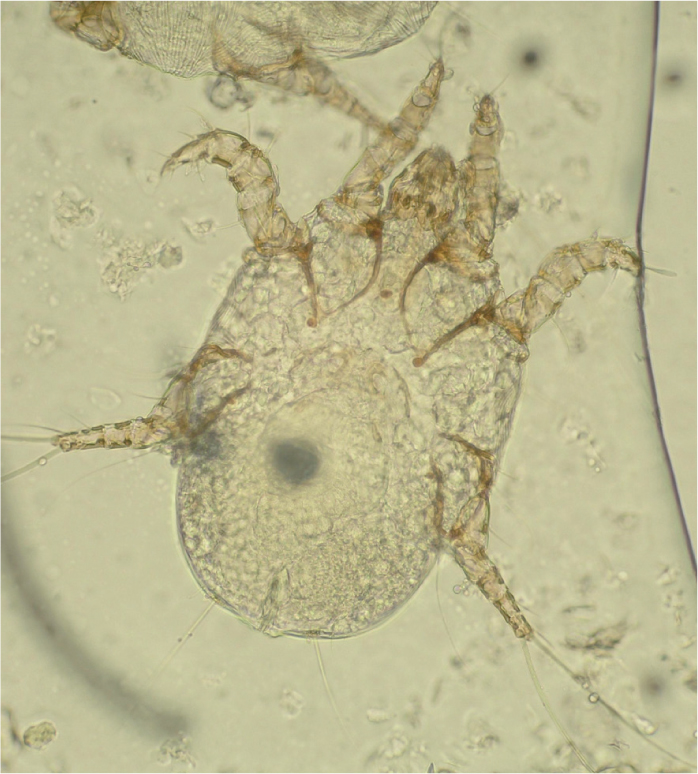
Otodectes cynotis is a psoroptiform mite that is found in the ears of cats and dogs. It is a large oval mite (adult females are 350–450 μm in length, adult males are 275–360 μm in length) (Figure 1). The adult male has short unjointed pedicles and wine glass shaped caruncles on the distal ends of each leg. The female has unjointed pedicles with caruncles on the first two pairs of legs. The eggs are typically oval. Six legged larvae hatch from the eggs and develops through two nymph stages before becoming an adult. Eggs, larvae, nymphs and adults can all be found within ear wax. Mites are photophobic and can be observed as small white dots moving away from the light source when the canal is examined with a hand held otoscope.
Demodex canis
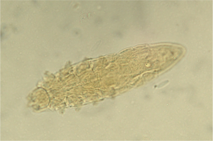
Demodex spp. mites are considered to be normal skin commensals in the dog, however the presence of mites, intermediate stages or eggs should be regarded as suspicious when they are identified on cytology samples taken from ears. Demodex canis is the most common of the Demodex spp. mites to be found in the ear and ranges in size from 180–201 μm in length (Figure 2). All stages of Demodex spp. mites can be identified in ear wax. The adults are eight legged slender ‘cigar shaped’ mites. Nymphs and larvae have six legs, eggs are lemon shaped. Demodex spp. mites cannot be seen with the naked eye.
Infection found on ear samples
Bacteria
A coccus (plural cocci) is any bacterium that has a spherical, ovoid or generally round shape. It is one of the two distinct bacterial shapes found in ears, the other being bacillus (plural bacilli) which are rod-shaped.
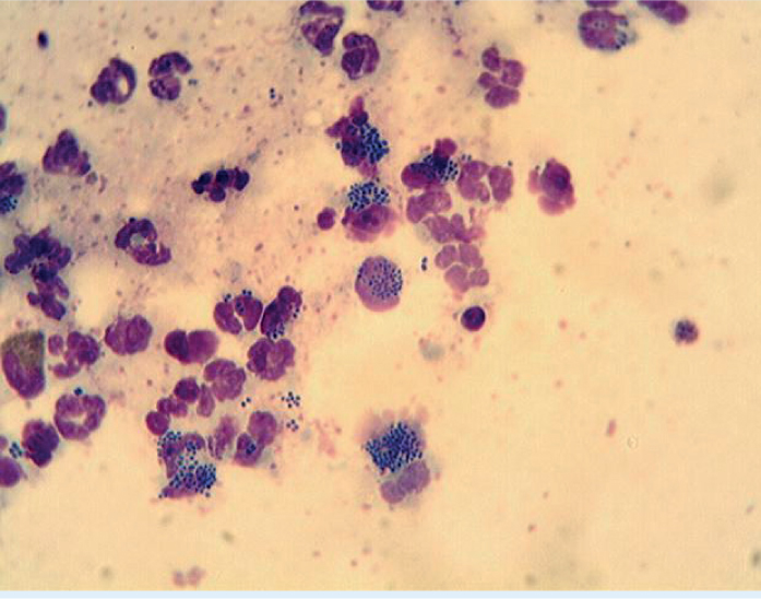
Cocci can be arranged in pairs, e.g. Neisseria spp. (which is not normally a skin pathogen) or long chains which is the normal arrangement of Streptococcus spp. or in clusters, e.g. Staphylococcus spp. (Figure 3). Cocci are most commonly associated with acute otitis in the dog and where clusters of cocci are found on ear cytology they are frequently identified as Staphylococcus pseudintermedius. In acute cases where the dog is presenting for the first time or has only had the occasional previous episodes of otitis, culture is not usually indicated as the bacteria is unlikely to be a multiply resistant isolate. In these cases empirical therapy is, in the author's opinion, appropriate with an antibiotic with good Gram-positive activity. Where cocci are identified with a different morphology, or where the disease is longer standing, or the dog has had multiple courses of antibiotics, then culture may be necessary. This may identify a more unusual organism, e.g. Enterococcus spp., or a resistant staphylococcal isolate. In these cases although empirical therapy may still be prescribed, culture is useful and recheck is essential to ensure the dog has responded to therapy.
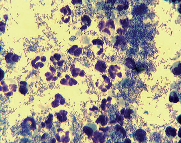
Bacilli appear as elongated rods when viewed under the microscope, they usually appear as single cells. All bacteria and yeast stain dark blue with a Diff Quik stain. Diff Quik stains are quick, easy and low cost to perform and demonstrate the morphology of an organism, but by themselves do not allow speciation. While this is not problematic when cocci are identified, as all of the principal pathogens found in the ears are Gram-positive, it is more of an issue if rods are identified on cytology. Gram-negative bacteria stain red and Gram-positive bacteria blue with a Gram stain which can help to differentiate some types of bacilli, but these types of stains are more difficult and time consuming to perform. Corynebacterium spp. which are Gram-positive rods look almost identical morphologically to Gram-negative organisms such as Pseudomonas spp. when stained with Diff Quik (Figure 4). Therefore the author would always culture a sample where bacilli are identified on cytology or where a mixed population of cocci and bacilli are identified. This is particularly pertinent when it comes to the selection of therapy, as the spectrum of drugs used to treat Gram-negative infections such as Pseudomonas spp. is very different to those used to treat Gram-positive infection with organisms such as Corynebacterium spp. While empirical therapy may be started if the dog is very uncomfortable, culture is useful to ensure the organism can be identified and can give the clinician the option to change topical therapy if the culture dictates this may be necessary.
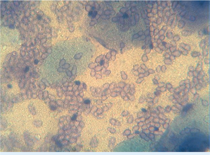
Yeast
Yeast are much larger than bacteria on cytology. Malassezia pachydermatis is the principal yeast identified from the ears of dogs (Figure 5). It appears as a dark blue/purple staining peanut shaped organism. As the discharge associated with yeast infections is commonly very waxy and may not contain an accompanying inflammatory infiltrate, cytology samples often do not stain well. Therefore it may be necessary to adjust the collimation, when examining samples microscopically, to create some shadow to be able to visualise yeast on samples. Occasionally Candida spp. can be found in ears; these has a more rounded shape with a more defined waist almost like a ‘baker's bun’.
Assessing the end point of therapy in otitis
Follow-up appointments are as important as the initial consultation in cases of otitis. The therapeutic use of potent topical glucocorticoids will always improve the appearance of the ear canal due to their ability to reduce swelling, erythema and the amount of discharge that is present. However it is important at recheck appointments that not only is the ear canal assessed visually to ensure the inflammatory changes have resolved but that cytology is repeated to ensure that no bacteria or yeast are present and the inflammatory infiltrate has gone. Topical therapy should only be discontinued when the ear canal is completely clear of disease. At this stage treatment may be switched to maintenance therapy which may be treatment to resolve the underlying primary cause together with topical therapy which may be an antiseptic ear cleaner.
Conclusion
The collection of samples for cytology can easily be undertaken by the veterinary nurse. Cytology is a low cost diagnostic test that provides useful information to identify both ectoparasites, bacteria and yeast. Cytology can help in decision making to select empirical therapy or the need for further more diagnostic tests and is essential to decide on the end point of any therapy.
KEY POINTS
- Ear cytology is quick and easy to do and can be used to identify potential pathogens.
- Bacteria can be identified on cytology, a knowledge of their morphology can allow decisions to made on the need for further investigation such as culture and susceptibility.
- Yeast can be identified on cytology without the need for culture.
- Cytology can help in the selection of appropriate ear cleaners and ear drops.
- Cytology should be used to assess an end point in therapy to ensure that the infection has resolved.