
Chronic diarrhoea, vomiting, and weight loss are common conditions seen in general practice. There are a number of underlying causes, including poorly understood conditions such as inflammatory bowel disease (IBD). IBD is a collective term that describes a number of different inflammatory conditions of the mucosa in the large and/or small intestine. It is a condition diagnosed histologically when no other etiology can be found. There are a number of different forms of IBD that can be diagnosed but the most common is lymphocytic-plasmacytic enteritis, and is often idiopathic in origin. The condition is common in dogs and cats of any age and is associated with chronic intermittent episodes of vomiting and/or diarrhoea as well as anorexia, decreased intestinal functioning, intestinal mal absorption, and weight loss. The exact cause of the inflammation is poorly understood and treatment can prove challenging. Controlling clinical signs is the primary concern throughout treatment and it is important that clients are supported and educated to ensure the best success in managing the disease (Hall and German, 2010).
Aetiology and clinical signs
IBD can present with an array of clinical signs including chronic intermittent vomiting and/or diarrhoea, anorexia and weight loss. In severe cases, haematemesis, haematochezia and protein losing enteropathy can also be present along with subsequent hypoproteinemia and ascites (Table 1). Clinical signs fluctuate intermittently and in addition, faecal characteristics may be altered by stress or changes in the environment (Matz and Leib, 2006). Diarrhoea can originate from the small bowel, large bowel or be mixed in origin. Vomiting and weight loss suggest upper intestinal disease, while haematochezia and mucoid stools are often a sign of lower intestinal disease (Table 2). The presence of blood in the vomit or diarrhoea is seen in more severe disease and is also commonly associated with weight loss. Anorexia can occur with severe inflammation (Ferguson and Gaschen, 2009; Hall and German, 2010).
Table 1. Clinical signs seen in animals with inflammatory bowel disease
| Polyphagia |
| Anorexia |
| Weight loss/poor body condition |
| Vomiting |
| Bile |
| Food |
| With or without hair (cats) |
| With or without grass (dogs) |
| Haematemesis |
| Diarrhoea |
| Increased frequency |
| Mucochezia |
| Haematochezia |
| Melaena |
| Tenesmus |
| Flatus (dogs) |
| Increased borborygmi |
| Abdominal discomfort/pain |
| Thickened bowel loops or enlarged mesenteric lymph nodes on abdominal palpation (cats) |
Table 2. Differences in clinical signs between small and large bowel disease (adapted from Hall and German, 2010)
| Signs | Small intestinal disease | Large intestinal disease |
|---|---|---|
| Stool volume | Large | Small |
| Mucoid stool | Rare | Common |
| Presence of blood | Melaena | Haematochezia |
| Steatorrhoea | Sometimes | Absent |
| Stool colour | Variable | Normal |
| Undigested food in stool | Occasionally | Absent |
| Tenesmus | Rare | Common |
| Frequency of defecation | 2–3 times per day | 3+ times per day |
| Urgency of defecation | Uncommon | Common |
A definitive cause of IBD is unknown. Onset may or may not follow an obvious event such as a change in diet or behaviour. Once triggered, disruption of the mucosal barrier leads to dysregulation of gut associated lymphoid tissue and a disturbance in the intestinal microflora leading to subsequent immunologic intolerance to dietary or endogenous antigens (Hall and German, 2010).
IBD can present in dogs and cats of any age but there is a predisposition for middle to older aged animals to acquire the disease. There are several different types of IBD classified according to their anatomic location and the type of inflammatory cells involved and lymphocytic-plasmacytic enterocolitis (LPE) is the most common. While any breed of cat or dog can acquire the disease, a breed predisposition is suspected for certain forms with purebred dogs appearing more likely to acquire the condition than mixed breed dogs (Table 3). There is some evidence to suggest that IBD occurs in animals that are prone to nervous behavioural conditions, and similarly that environmental stress can exacerbate the condition, but the role of behaviour in the pathogenesis and therapy of canine and feline gastrointestinal disorders remains largely unexplored (Washabau, 2009).
Table 3. Forms of IBD, characteristics and breed predispositions (Fogle and Bissett, 2007; German, 2008)
| Characteristics/signs | Breed predisposition | |
|---|---|---|
| Lymphocytic-plasmacytic gastroenteritis and enterocolitis | Inflammatory agents such as endoparasites or bacterial infections. Less common causes include trauma, llergies, internal organ dysfunction. May be idiopathic in origin. Vomitingand odorous yellow, greenish, black, or tarry diarrhoea that is watery in consistency and are passed frequently. |
|
| Eosinophilic gastroenteritis and enterocolitis | Clinical signs as above. Allergic reaction to diet or intestinal parasites. | Any but Rottweilers and German Shepherds appear more likely to present with the disease. |
| Granulomatous (regional) and transmural granulomatous gastroenteritis and enterocolitis | Thickened obstructive lesions often associated with Trichuris vulpis. Chronic diarrhoea with mucus and blood. | Any but German Shepherds appear more likely to present with the disease. |
| Neutrophilic enterocolitis | Infiltration of mature white cells into tissue and blood vessels causing acute and chronic diarrhoea. | Any (rare). |
| Immunoproliferative enteropathy | Often triggered by stress. Alopecia and hyper-pigmentation of the skin, especially the pinnae. Episodic or constant diarrhoea. | Occurs almost exclusively in Basenji aged around 3 years old. |
| Histiocytic ulcerative enterocolitis | Severe diarrhoea containing mucus and blood, also can result in anaemia nd weight loss. | Occurs almost exclusively in Boxers. |
Success of treatment for IBD in dogs is variable. A poor outcome is more likely when dogs with IBD present with concurrent pancreatitis, hypocobalaminaemia and hypoalbuminaemia. Treatment of the cat is much more successful and remission is often for a longer period of time (German, 2008; Hall and German, 2010). The prognosis of LPE is variable and depends on the disease severity. Some cases respond poorly to therapy and require prolonged treatment to manage clinical signs (German, 2005).
Diagnosing the condition
Diagnosis of IBD is ultimately a diagnosis by exclusion although even with a diagnosis many cases are still idiopathic in origin. Typically non-invasive tests are carried out first to eliminate the possibility of bacterial, parasitic or allergenic causes in order to reach a definitive diagnosis once intestinal biopsy is performed (Hall and German, 2010). Dietary trials are useful for identifying food allergies and there are a number of other tests and diagnostic imaging and surgery that can be useful for identifying contributing causes and the type of intestinal cellular inflammation.
Laboratory tests
Serum biochemistry and haematology are commonly performed to rule out other disorders that may cause gastroenteritis such as renal or hepatic disease, hypoadrenocorticism and exocrine pancreatic insufficiency; however, some cases of IBD show no haematological or biochemical abnormalities at all (Ferguson and Gaschen, 2009). A low cobalamin level can indicate intestinal malabsorption resulting from severe intestinal disease.
Faecal samples should be assessed for endoparasites and faecal culture can be performed to rule out bacterial conditions although these conditions are rarely the cause of chronic diarrhoea in animals (Hall and German, 2010).
Diagnostic imaging
Diagnostic imaging is a minimally invasive technique that can help isolate the cause of many clinical signs of IBD. Abdominal ultrasound is useful to identify intestinal thickening, mesenteric lymphadenopathy and gastric or intestinal obstructions. It can also help rule out possible causes related to neoplasia, intussusception or extraintestinal disease such as pancreatitis. Ultrasound can also help assist with needle aspirates of lymph nodes (Ferguson and Gaschen, 2009). Most patients will lie still for abdominal ultrasound yet sedation or anaesthesia may be required if the procedure is prolonged or the patient is in discomfort. The hair has to be clipped from the abdominal area, and owners should be made aware of this, prior to the procedure taking place.
Endoscopy is useful to investigate diffuse changes in the mucosal layer of the oesophagus, stomach, small intestinal tract and colon. Endoscopic abnormalities identified in patients with IBD can include focal or obstructive lesions, ulceration, de-pigmentation of the mucosa, rigidity of the mucosa and loss of normal mucosal architecture. During endoscopy, multiple small biopsies are commonly taken for histological examination (Ferguson and Gaschen, 2009).
Surgical
Exploratory surgery can be performed in order to collect full thickness gastrointestinal biopsies; however, if there is significant intestinal inflammatory disease there is a greater risk of wound breakdown subsequently leading to increased likelihood of complications such as septic peritonitis. Endoscopy is the method of choice in cases where wound dehiscence is a concern (German, 2005).
Intestinal biopsies are necessary for a diagnosis of intestinal inflammation. Biopsies can histologically reveal IBD with evidence of inflammatory infiltrate (neutrophillic, eosinophillic, lymphocytic, plasmacytic or granulomastous), associated mucosal pathology (villus atrophy, fusion, crypt collapse), distribution of the lesion (focal or generalized, superficial or deep), severity and mucosal thickness (mild, moderate, severe) and the site of primary inflammation (gastric, duodenum, jejunum, ileum, caecum, colon) (Washabau, 2009).
Disease management
Treatment of IBD involves reducing diarrhoea and vomiting, promoting a healthy body condition and decreasing inflammation of the intestinal tract. Any identified causative agents such as parasites, food allergies or bacterial infections should be eliminated. Dietary management can be effective in some cases both to reduce dietary allergens and to minimize the severity of clinical signs. Antibiotic and immunosuppressive medications may also be used.
Drug therapy
There are numerous types of drugs used in the treatment of IBD to target clinical signs and complications of the disease (Table 4). Metronidazole is a common drug used in the treatment of IBD. It has both an antibacterial and anti-inflammatory effect as well as an antiprotozoal effect. A major advantage of using combination therapy of an antimicrobial with an immunosuppresive drug like prednisolone is that the corticosteroid dose can usually be decreased from the initially high dose decreasing the likelihood of significant corticosteroid-related side effects. Side effects of prednisone can be significant and include severe polyuria/polydipsia, panting and lethargy. In severe cases of IBD, prednisolone can be administered parenterally, since oral absorption may be poor (German, 2008). Mild cases of IBD frequently respond to diet change and metronidazole, especially in cats. However, early intervention with immunosuppressive therapy is frequently unavoidable in severe cases (Hall and German, 2010). In dogs azathioprine is commonly used when initial treatment has failed or steroid side effects are marked. However the onset of action may be delayed for up to 3 weeks, and its myelosuppressive potential means that haematological monitoring is necessary. Azathiaprine is not recommended for cats but chlorambucil is a suitable alternative (German, 2005).
Table 4. Some common drug therapies used in managing irritable bowel disease (adapted from Hall, 2005; German, 2011)
Antimicrobials
|
Immunosuppressive therapy
|
Other
|
Duration of drug treatment varies. In some dogs with severe lymphocytic-plasmacytic enteropathy and marked hypoproteinemia, therapy can be successfully discontinued as early as 6 months to 1 year. In others, lifelong treatment is required (Tam, 2001).
Dietary therapy
Dietary therapy, is extremely important in the long-term management of chronic intestinal inflammation and in some patients, signs can be well controlled with dietary therapy alone (Jergens and Zoran, 2005). If dietary sensitivity is suspected, an exclusion diet trial should be undertaken using a diet with a single novel (i.e. the animal has not been exposed to before) protein source and a single novel carbohydrate source (Table 5). Dietary trials should be reserved for cases where there is a strong association between clinical signs and a particular diet, or when all other diagnostic tests have proved inconclusive. It can be extremely difficult to identify a specific allergen especially in cases where owner compliance is poor or when animals have had access to unregulated feeds.
Table 5. Dietary therapy (adapted from Boyle and Bissett, 2007)
| Diet characteristics | Protocol |
|---|---|
|
|
There are a number of hypoallergenic commercial diets available (Table 6). Some of these diets contain hydrolyzed proteins (proteins that have been broken down into smaller units to reduce antigenicity and enhance digestibility). Gluten is perceived to be a common food allergen, so most commercial diets are gluten free, however there are a number of studies investigating gluten intolerance in dogs and it is still unclear whether there is an abnormal immune response or a direct toxic effect caused by gluten (German, 2005).
Table 6. List of a small selection of Hypoallergenic and Hydrolysed diets commercially available
| Diet | Protein source |
|---|---|
| Purina HA Hypoallergenic® Canine Formula | Soy |
| Purina DRM® diet | Blue whiting |
| Royal Canin® hydrolyzed diet dry only | Soy |
| Hills z/d® hydrolyzed diet wet or dry | Chicken |
| Eukanuba® Dermatosis F/P wet or dry (hypoallergenic) | Fish |
| Hills d/d® Hypoallergenic wet or dry | Various (venison, salmon) |
| Royal Canin® Sensitivity control hypoallergenic wet or dry | Chicken, duck |
Some owners may choose to make their own homemade diet and there are advantages and disadvantages to both methods (Table 7). Regardless of the method chosen, if the feeding of a single protein, single carbohydrate source diet has a good outcome (decrease or absence of clinical signs) this could be considered diagnostic of food responsive enteropathy.
Table 7. Table of advantages and disadvantages of homemade or commercially prepared diets (adapted from Harvey and Hall, 2009)
| Homemade diet | Commercial diets | |
|---|---|---|
| Advantages |
|
Consistency of content of high quality brands |
| Disadvantages |
|
|
Encouraging the patient to eat
When faced with a hyporexic or anorexic patient, the temptation is to encourage the patient to eat anything. With dietary sensitivity, even the smallest amount of the wrong food can cause ongoing clinical signs thus it is important to develop a feeding strategy that is centred around the hypoallergenic diet but not forgetting the individual needs of the patient.
Getting the hospitalized patient to eat a hypoallergenic diet is an even bigger challenge. Some patients will prefer to eat from small plates or saucers rather than deep bowls and others have a preference for porcelain or glass over plastic or metal bowls (Chandler, 2002). If a patient ignores the food, it may be because it is protesting at the situation or distrustful of the person feeding it or the environment it is in. This can often be overcome by establishing a rapport with the patient (Killner, 2008). A comfortable content patient is far more likely to eat. Grooming them and spending time with them can make a significant impact on the animal's stress levels and willingness to eat. Encouraging the owners to visit and bring along home comforts such as the animal's bed can also help as can simulating a home environment like playing the radio and having access to their favourite toy. To prevent food aversion, it is beneficial to encourage voluntary eating and avoid offering food if the animal is overly nauseated. Avoid offering multiple bowls of different acceptable diets all at once as this can further increase the chances of nausea-related food aversion.
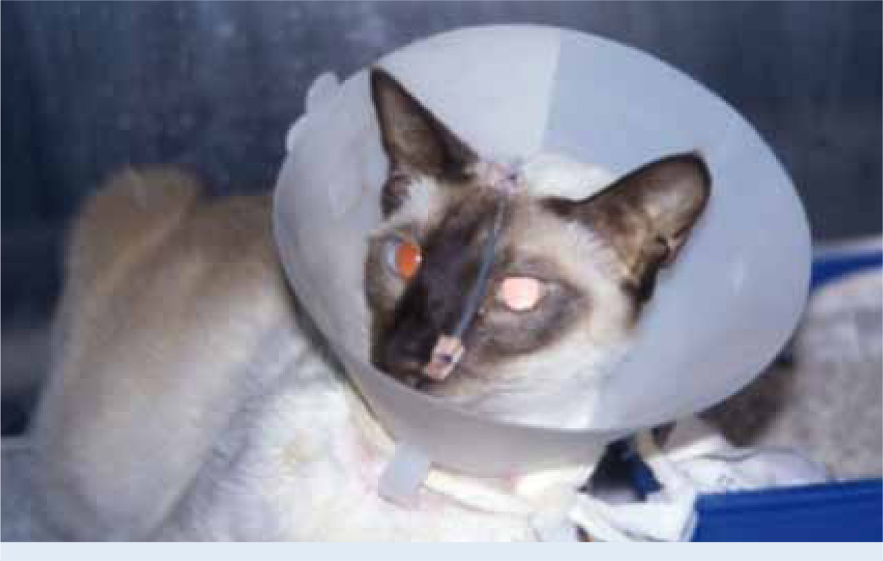
Case study.Signalment: Jack is a 10-year-old male neutered domestic short hair cat.Presenting condition: Jack was presented with a history of chronic intermittent vomiting and more recent watery diarrhoea.Diagnostics: Physical examination revealed dilated thickened loops of small intestine, there was evidence of abdominal discomfort on palpation. Repeat examination under sedation revealed a possible intussusception that was manually reduced. Routine biochemistry revealed a mild increase in total protein which may suggest dehydration. Haematology revealed a left shifted neutrophilia. Serum folate and cobalamin were within the reference range. Total T4 was normal. A faecal sample was submitted for parasitology and was negative for endoparasites. A right lateral abdominal radiograph revealed dilation of the small intestine and proximal colon with areas of gas build up. Abdominal ultrasound showed areas of dilated small intestine, with normal layering, the ileocolic junction was prominent with a dilated fluid filled distal colon. Mesenteric lymph nodes were prominent. Gastroduodenoscopy revealed a grossly normal stomach and oesophagus. The duodenum had patchy abnormalities with pallor and nodular changes. Histopathology of endoscopic biopsies revealed a moderate lymphoplasmacytic inflammation and mild eosinophilic and neutrophilic inflammation. A diagnosis of moderate, chronic, inflammatory bowel disease was made.Treatment: Jack was treated with a hypoallergenic diet, metronidazole, omeprazole, prednisolone and chlorambucil. While hospitalized, Jack was weighed daily to monitor any losses. He had very watery diarrhoea so he was supplemented with intravenous fluid therapy to replace fluid loss. Throughout the period of his treatment his weight ranged from 4.4 kg to 6.6 kg when he was well. Food that was offered was weighed so his calorie intake could be calculated.
Photo 1. Jack.
Outcome: Jack responded very well to the regimen and the medications were progressively tapered and stopped. Jack will remain on the hypoallergenic diet as it appears to be helping.
Energy requirements
When calculating the energy requirement for patients that are ill or underweight it is very tempting to increase the calorie intake to encourage the patient to put on weight. However, in terms of providing dietary support, there are many complicating factors for patients with IBD so initially, the aim of nutritional support is to maintain body condition and avoid exacerbating any digestive upsets (Zoran, 2005).
The resting energy requirement (RER) should be calculated for each patient and a feeding plan should be developed to meet its nutritional needs. It can be difficult to determine the actual energy requirements for patients with IBD because they not only have a number of digestive upsets, they may also have an increase in protein catabolism and an overall increase in the resting metabolism rate associated with stress and disease (Remillard and Thatcher, 1989; Bartges, 2001). In general, current recommendations for nutritional support of ill animals are to use more conservative energy estimates to avoid overfeeding (Michel, 2006; Chandler, 2008). As a result, it is sufficient to use the normal RER for the patient as a starting point to meeting its nutritional requirements and adjust if necessary if the animal fails to maintain body condition (Table 8).
Table 8. calorie requirement calculation (Zoran, 2005)
| For animals weighing between 2–30 kg | For animals weighing less than 2 kg or more than 30 kg |
| RER (Kcal per day) = (30 × body weight in kg) + 70 | RER (in Kcal per day) = 70 × (body weight in kg)0.75 |
In patients with IBD, especially those with a history of vomiting and diarrhoea, calorie intake must be gradually increased to full calorie intake over approximately 2 or 3 days to avoid vomiting and abdominal distension (Abood, McLoughlin, & Buffington, 2006). A good strategy is to provide one third of the calories on the first day. If this is well tolerated, the amount can be increased to two thirds of the calories on the second day and a full feed on the third day (Chandler 2002). It is important to record the total calories that the patient has actually eaten by measuring the amount offered to the patient and then subtracting the amount that is left over after feeding. It is also essential to weigh the patient daily during this time as well as objectively evaluate the body condition and muscle mass using a standardized body condition scale.
Assisted feeding methods
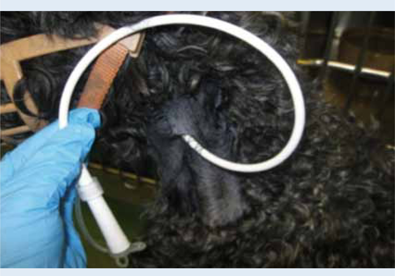
In polyphagic patients, keep meals small and frequent, and avoid increasing their food intake as this can result in vomiting and diarrhoea. In patients that are unwilling to eat regularly, assisted feeding may be necessary. Some patients tolerate hand feeding very well, but in many cases it can cause undue stress to the animal. Syringe feeding has many limitations including food aversion and the risk of aspiration pneumonia if the animal struggles so other methods should be considered in some cases (Figures 1–4).
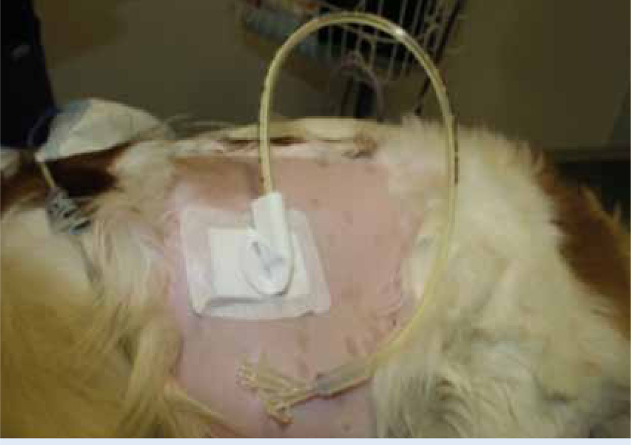
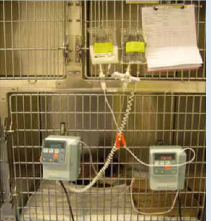
A patient should be considered for a feeding tube if they have not eaten for 72 hours (Abood and McLoughlin, 2006). The feeding tube chosen must be appropriate to meet the individual needs of the patient and if the patient will be discharged with the tube in place, the tube must be manageable for the client (Zoran, 2005).
Many feeding tubes can be managed at home. Owners must be shown how to look after and maintain the tubes. Encouraging the owners to visit the practice while the animal is still an inpatient is a good way to teach them how to use the feeding tubes. Clients should be advised to keep their pet indoors at all times and dogs must be on a lead when exercising to avoid the feeding tube becoming dislodged or contaminated. This is often a very difficult decision for the client as restricting their pet's outdoor activity may affect their quality of life.
Success and monitoring
Success in treating IBD is variable. Most cases will partly respond to treatment but only about 25 % will achieve complete remission of the disease leaving as many as 50 % of all cases with some intermittent signs for the rest of their life. As a result, euthanasia is common in unresponsive cases (German, 2008). In terms of ongoing care and management, the patient should be monitored regularly to ensure that he/she is responding to therapy; patients should be monitored for a number of things including body weight, changes in appetite, presence of vomiting, stool consistency and frequency and presence of any other signs that could indicate abdominal discomfort. Blood counts and serum biochemistry are also frequently indicated for patients with anaemia, inflammation, or hypoproteinemia as well as those receiving drug therapies that may be toxic to the liver or other organs. In addition, patients receiving some immunosuppressive therapies may be at risk for keratoconjunctivitis sicca so a Schirmer tear test can also be performed regularly.
Successful home management requires a commitment on the part of the client and the clinic to ensure good communication is maintained and any problems can be identified and addressed promptly. There must be strict adherence to dietary recommendations as well as careful observation of the patient's clinical signs. Client compliance with prescribed medications is essential to ensure success.
Conclusion
IBD is a challenging condition to diagnose and manage. Long-term management of IBD is often required and many patients may need regular adjustments of treatment. Some animals' clinical signs may not completely resolve and the animal may not achieve full remission. Patients with IBD can require a great deal of care and effort to treat the clinical signs. Dietary management is a critical aspect to managing IBD and this can pose many challenges in the nursing care and ongoing management of these patients at home.
A dedicated and compliant owner is essential to the success of the therapy (Ferguson and Gaschen, 2009). The client should be made aware from the beginning of treatment that there is no cure for IBD. Management and treatment alterations will likely be required for the remainder of the patient's life. The client's life style should be considered when discussing the management of their pet and careful instructions and advice given to ensure ongoing success. Client support is very important in long-term management of IBD and all the information must be given to the owners at the beginning of treatment so they are fully prepared.
Key Points
- Inflammatory bowel disease (IBD) is defined as a spectrum of gastrointestinal disorders characterized by inflammation to the lining of the intestinal tract.
- IBD can affect dogs and cats; the cause of IBD is largely unknown.
- Clinical signs of IBD include chronic reoccurring episodes of anorexia, diarrhoea and/or vomiting and weight loss.
- Dietary trials can help identify food allergies but conclusive diagnosis of IBD usually requires intestinal biopsies.
- Common treatments involve dietary management and may include a range of medication to target specific clinical signs.
- A complete cure for IBD is unlikely but the condition can usually be controlled with careful patient management.