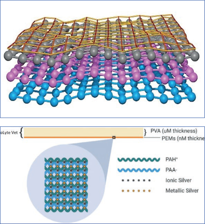
MicroLyte Vet contains a microscopic ‘active’ layer and a visible ‘handling’ layer. These layers consist of an advanced polyelectrolyte matrix (PEM) combined with the antimicrobial properties of ionic silver that are laid on an ultra thin hygroscopic polyvinyl film (PVA) membrane.
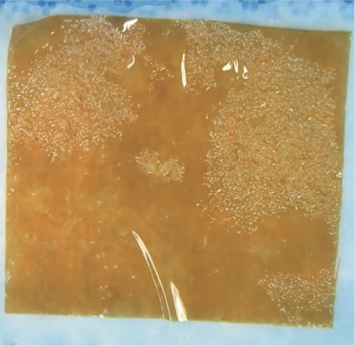
The dressing (Figure 1) presents as a distinctive transparent orange film that when placed into the wound visibly begins to cling to the wound bed covering the full surface and its contours.
Wound compatible anti-microbial technology
The structure of Microlyte Vet combines anionic and cationic charged polymeric components that complement the complex chemistry of the wound to support the formation and reorganisation of granulation tissue. Additionally, a low but precise level of bioactive silver is incorporated into the layers to create a very safe but effective antimicrobial environment at the wound bed (Figure 2).
Why intimacy is important
Many primary dressings with antimicrobial properties are made in textile form perhaps as a woven polymer, synthetic polyurethane or spun fibres. These dressings are often designed for human use for exuding and chronic wounds where capacity to absorb exudate is prioritised; while antimicrobials impregnated into these products act on microbes absorbed into the dressing via wound exudate.
A limitation of dressings of this type is that they may vary in their capacity to conform to the wound bed meaning that pockets of dead space may be left without contact to the dressing and any microbes in those areas may still be able to colonise and develop into more challenging biofilms (Figure 3).
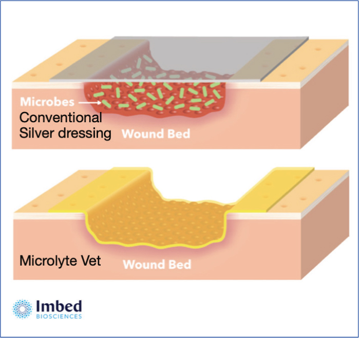
Microlyte's unique film matrix behaves very differently to the more traditional primary dressings. Its hygroscopic nature means that once placed on the wound it literally ‘shrink fits’ to meet the surface of the wound. This intimate contact achieved on a microscopic level ensures that the silver matrix follows the contour of the wound bed rendering pockets of colonising microbes sterile (Figure 3).
When to use Microlyte Vet
Microlyte Vet can be used for a range of presentations where contamination could lead to colonisation, biofilm formation and wound infection.
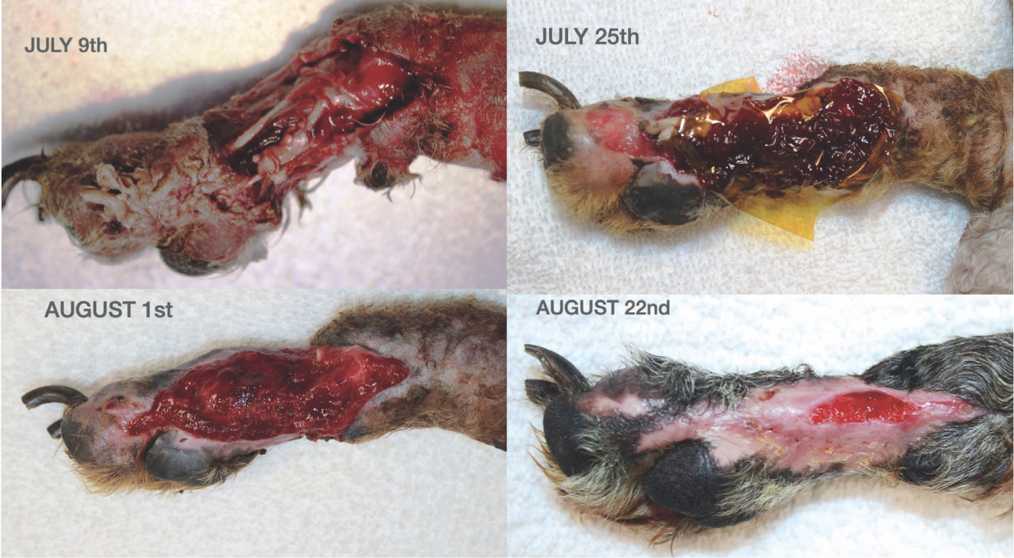
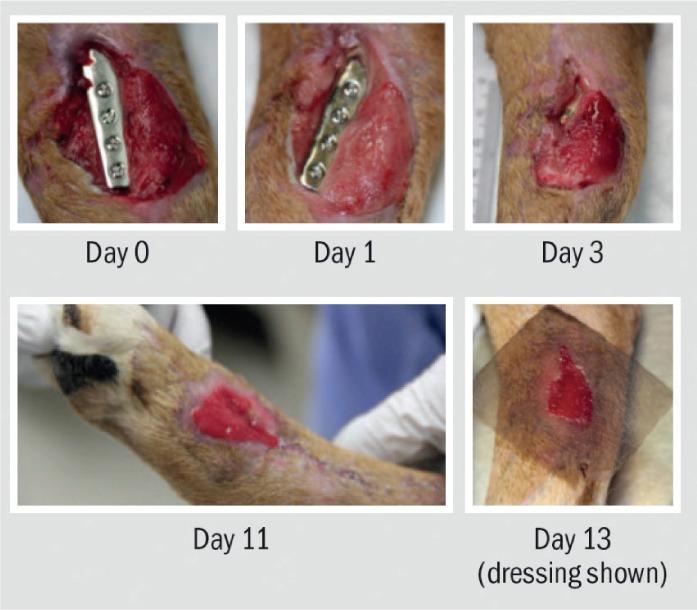
As presented in the workshop, and outlined by the 3P's of Prepare, Promote, Protect, Georgie Hollis explained that fresh, traumatic, contaminated wounds should be clipped, lavaged, and debrided. Microlyte Vet could be used post debridement to prevent bacterial colonisation and potential biofilm formation. This is particularly important where orthopaedic implants are in place (Figure 4).
For wounds with established complications as a result of bacterial colonisation or infection, patient care should include proper medical management, where necessary complemented by Microlyte Vet used topically to reduce bacterial proliferation and support granulation tissue formation after the wound is thoroughly cleansed.
Microlyte Vet can been used without a secondary dressing where bandaging and dressing of wounds is causing more trauma than benefit.
When to change the dressing
Mircolyte Vet can stay in place for 4 days and dissolves in situ, therefore avoiding any need to interfere with the wound bed at dressing change. Simply apply another layer and your choice of secondary dressing to avoid interference (Figure 5).
Conclusion
Even with best efforts to lavage, debride and adhere to aseptic standards it is likely that some contamination will occur. Where wounds are already challenging because of the extent of tissue loss or implanted devices, the use of Microlyte Vet offers a novel, truly intimate contact layer with the power of ionic silver. It has the potential to be used in a sophisticated way alongside established secondary dressings to further prevent the development of colonies of biofilm forming bacteria, while surrounding and neutralising those that have already begun to take hold.