
Tumours of the nasal planum are relatively uncommon on dogs, representing approximately 1% of all canine cancers (Lana et al, 2004). Nasal planum tumours are much more common in cats, representing between 14–18% of all feline cancers (Williams, 2006): the incidence is much higher in white cats and those living outdoors and it is much more prevalent in sunny countries, reflecting the role of UV light in causing the malignant changes, through a process known as actinic metaplasia (Fuchs and Marmur, 2007). By and large, the most commonly occurring neoplasm of the nasal planum is the squamous cell carcinoma (SCC) (Dobson et al, 2006).
Typically, patients are presented with lesions on the nasal planum which vary from scabby, dry plaques, through to erosive ulcers (Murphy, 2013). Occasionally, patients may present with acute epistaxis, when erosion through a nasal planar blood vessel has occurred.
Various treatment options are available for SCC, including surgery (Lascelles et al, 2004; Ladlow, 2014), radiation therapy (Lana et al, 2004; Melzer et al, 2006), photodynamic therapy (Magne et al, 1997), intralesional chemotherapy (Kitchell et al, 1995), immunomodulation (Gill et al, 2008) and cryotherapy (Fernandes de Queiroz et al, 2008). For superficial, or early SCC lesions, a combination of curettage and diathermy has been shown to be very effective in providing remission (Jarrett et al, 2013). However, surgery is considered to be the treatment of choice with larger SCC lesions, greater than 5 mm deep (Ladlow, 2014).
With any oncological resection, the aim is to remove the entire tumour, with ‘clean’ margins, at the time of the first surgical procedure. This remains the most effective means of controlling cancer and gives surgery the greatest single cure rate of any treatment modality for many tumours (Liptak, 2013). However, surgical excision of a nasal SCC is often disregarded as an option due to perceived difficulties in surgical technique and concerns re cosmesis, i.e. the post-operative appearance of the patient. Certainly, in order to obtain a clinical margin, surgery necessitates the removal of the nose (nasal planectomy, or, more colloquially ‘nosectomy’) and occasionally the premaxilla (Ladlow, 2014; Haar and Hampel, 2015). The surgery itself is not particularly onerous, although requiring moderately intensive post-operative nursing management; the appearance of the pet following surgery is acceptable to most owners and the prognosis, providing meticulous attention to pre-operative planning is adhered to, is very good.
With any cosmetically-altering procedure, it is a very good idea to warn the owner in advance how the pet's appearance will change after the surgery. These days, there are many sources, such as the internet, for obtaining photos of dogs and cats that have undergone similar surgery. Some owners of pets who have had similar surgeries may be happy to talk to clients who are considering their pet having nosectomy.
Pre-operative planning for nosectomy should include routine biochemical and haematological investigations (ideally with urine examination also). A full oncological workup should be performed, in order to define the margins required for excision and to rule out metastatic disease (Fowler and Williams, 1999). Examination of the nostrils, using an otoscope, will give an idea of extent, but skull radiographs should also be taken, along with three lung views. If there is any suspicion that deeper invasion has occurred, referral for magnetic resonance imaging (MRI) or computed tomography (CT) should be considered. Aspiration of the submandibular lymph nodes should also be performed, although SCC usually has a very low metastatic rate. With white cats, meticulous examination of the ear tips should also be performed: any dry, or flaky skin may well be an indication of early actinic keratosis; in these cases, it may be prudent to consider removing the pinna, in part, or whole, at the same time as nosectomy.
Generally, the nasal tumour should be staged according to World Health Organization Comparative Oncology TMN system (Table 1).
| T (tumour) |
| T0 (no sign of tumour) |
| N (lymph node) |
| N0 no evidence of lymph node metastasis |
| M (metastasis) |
| M0 no distant metastasis |
Patient preparation
Patient preparation is fairly self-explanatory — the care team should discuss analgesia prior to surgery and be aware that hospitalisation is generally required for 48–72 hours following surgery. An intravenous cannula must be placed prior to surgery and it is a good idea to pack the pharynx with saline-soaked swabs to prevent any blood from entering the trachea. There is no requirement for specialised instrumentation, although it is recommended that electrocautery be available, as blood flow from the surgical site may be brisk.
The surgical clip should be fairly generous, allowing for a surgical margin of at least 1 cm from the visible edge of the tumour. However, wherever possible, the whiskers should be left in place: they take a long time to regrow and contribute to a poor quality of life post surgery if clipped. The skin is prepped as normal, ideally using povidone-iodine to prep the oral cavity. A gag should be placed in the mouth to allow for a slight opening and drapes are placed in the mouth to cover the endotracheal (ET) tube.
Tumour excision
The nose is excised using a number 10 or 15 blade: depending on the extent of the SCC, a premaxillectomy (or maxillectomy (Lascelles et al, 2004)) is performed. Haemorrhage, as mentioned, may be brisk and should be controlled by a combination of electrocautery and direct compression. If performing a maxillectomy, it may be necessary to ligate the palatine arteries (Gallegos et al, 2007).
Reconstruction
Reconstruction of the surgical site will depend on the extent of resection: in cases where the planum has been removed, without a premaxillectomy, the skin may be closed around the nasal opening with a simple purse-string suture, although the author prefers to place simple interrupted sutures through the nasal cartilage and mucosa. Where a more extensive resection has been carried out, it is necessary to elevate labial flaps to close the oral defect (Worley, 2016): palatine mucosa must also be closed. The nose should be sent to a histopathology laboratory to confirm margins.
Post-operative care
Post-operative care involves monitoring for adequate analgesia (the use of a pain-score chart is strongly recommended) and gentle flushing of the wound site to ensure the patient has a patent airway. In most cases, this can be achieved consciously, but it may be necessary to consider sedation if the patient becomes anxious or painful during the flushing process. Once immediate post-operative inflammation has receded and the airway remains clear, the patient may be discharged. The author would recommend reminding the owner again prior to discharge that their pet's physical appearance will have changed (Hoad, 2014).
Examples:
Case 1: Baloo, 11-year-old male neutered Retriever
Baloo presented with low grade intermittent epistaxis. Examination of the nasal planum revealed a small erosion adjacent to the philtrum which had exposed a blood vessel. Examination of the nostril consciously was not possible, so Baloo was sedated with medetomidine and buprenorphine and the nostrils examined with an otoscope. An irregular, ulcerated mass could be seen extending approximately 1 cm medially into the left nostril. The patient was anaesthetised (using propofol for induction and maintained on isoflurane). A small incisional biopsy was obtained of the mass and a small ligature was placed around the bleeding vessel. Skull and intranasal radiographs were taken, as were inflated lung views. No abnormalities were seen. Aspiration of the submandibular lymph nodes bilaterally were taken. Routine haematology and biochemistry samples were taken: results were unremarkable. The results of the biopsy confirmed SCC.
Options were discussed with the client: radiotherapy and chemotherapy were declined, on the basis of the client's previous experience with these treatment modalities. The option of a nasal planectomy was discussed and the owner was shown several pictures of similar surgical procedures to give an idea of expected outcome. The owner agreed to go ahead with nasal planectomy. It was felt that premaxillectomy would not be necessary, due to the limited visual extent of the tumour.
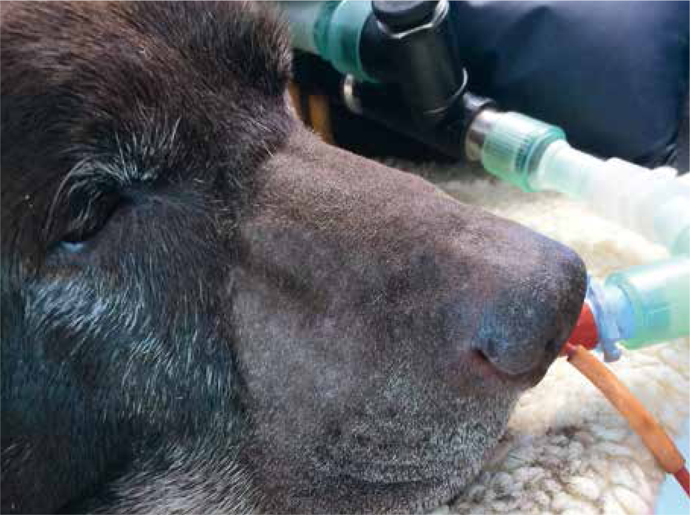
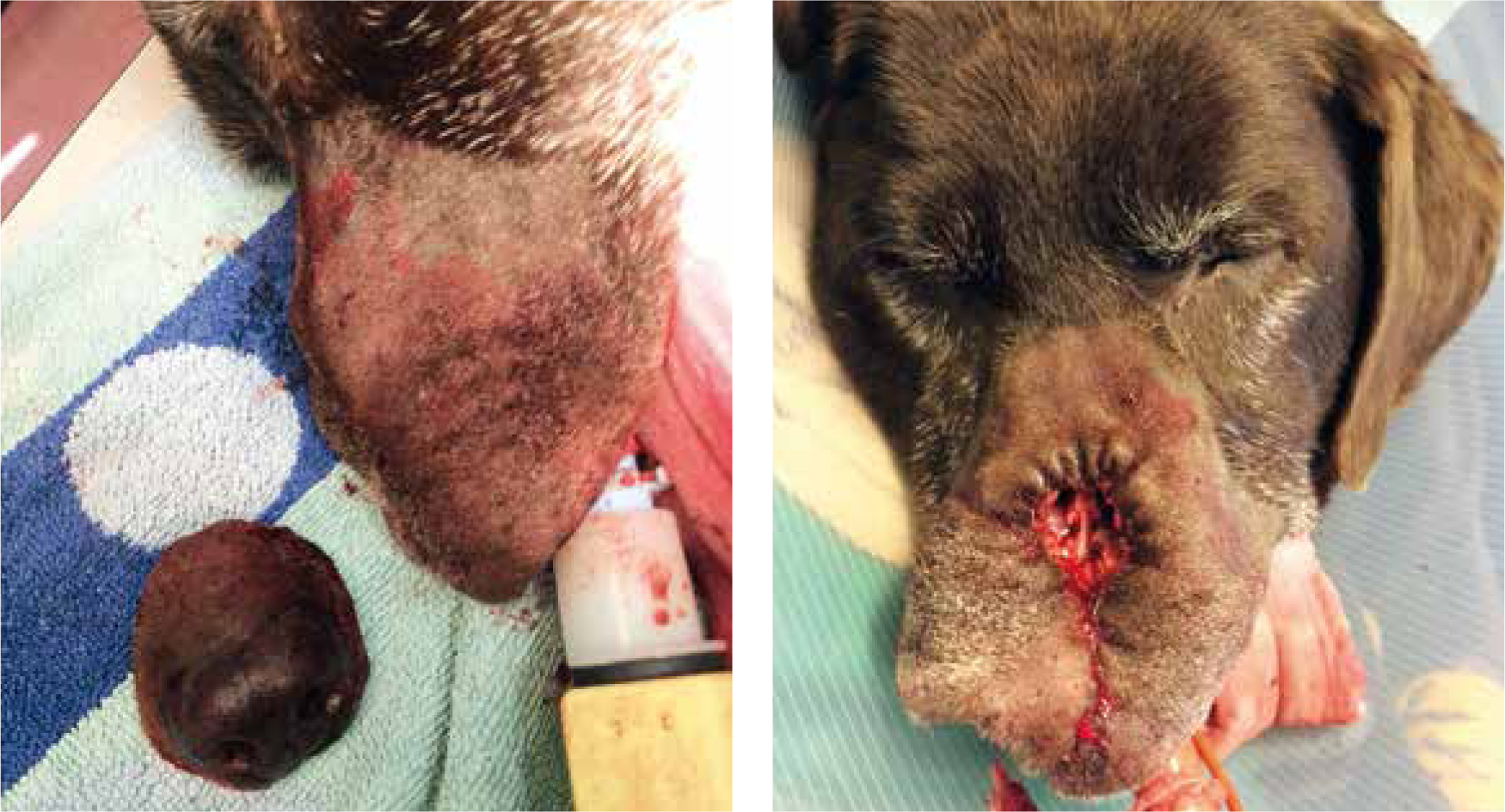
Transdermal Fentanyl (Recuvyra, Elancovet, UK) was applied prior to surgery and the patient was placed in ventral recumbency. The surgical site was prepared as described above (see Figure 1) and a nasal planectomy was performed. Haemorrhage was light and easily controlled with bipolar electrocautery and the skin was closed around the nasal opening with simple interrupted sutures of 4/0 poligecaprone 25 (Monocryl, Ethicon, UK) (Figures 2a and 2b).
Post-operatively, Baloo was kept in for 48 hours. A standard pain scoring chart (Colorado State acute canine pain scoring chart; http://www.biomedcentral.com/content/supplementary/s12917-015-0338-4-s2.pdf) was used to monitor analgesia. Generally, pain scores remained low post operatively: the transdermal fentanyl appeared to be very effective in controlling postoperative pain. A moderate amount of crusting, serosanguineous discharge accrued over the wound site over the next 48 hours: this was removed by gently flushing with a dilute solution of lidocaine in sterile saline (1 ml lidocaine in 5 ml sterile saline) and then cleaning very gently with saline-soaked sterile gauze swabs. There was a little fresh haemorrhage from the wound edges, but this was self limiting.
Baloo ate within an hour or so of surgery. His appetite was a little reduced the following day, which may well have been due to the transdermal fentanyl. There was a moderate amount of swelling around the muzzle the following morning, but this cleared within 3–4 days of surgery.
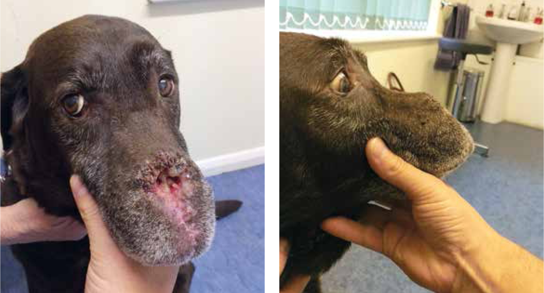
Baloo was discharged after 48 hours. The owner was instructed to continue bathing the wound gently with saline-moistened gauze swabs, to ensure the airways were kept clear. An Elizabethan collar was given to the owner, with instructions to place it if the patient rubbed at the wound, but it was not necessary. The wound healed well and although a slight mucous discharge continued until the 3 week post-operative check (Figure 3a and 3b), this cleared up within the following 3 weeks. At this time, Baloo seemed very comfortable and the owner thought he was ‘his old self.’ The histopathology confirmed complete excision, with moderate clear excisional margins. Baloo went on to survive a further 18 months, eventually being euthanased due to nonrelated illness.
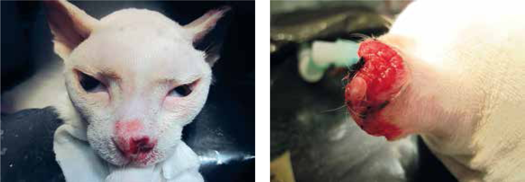
Case 2: Moon, 10-year-old female neutered Domestic Short Hair
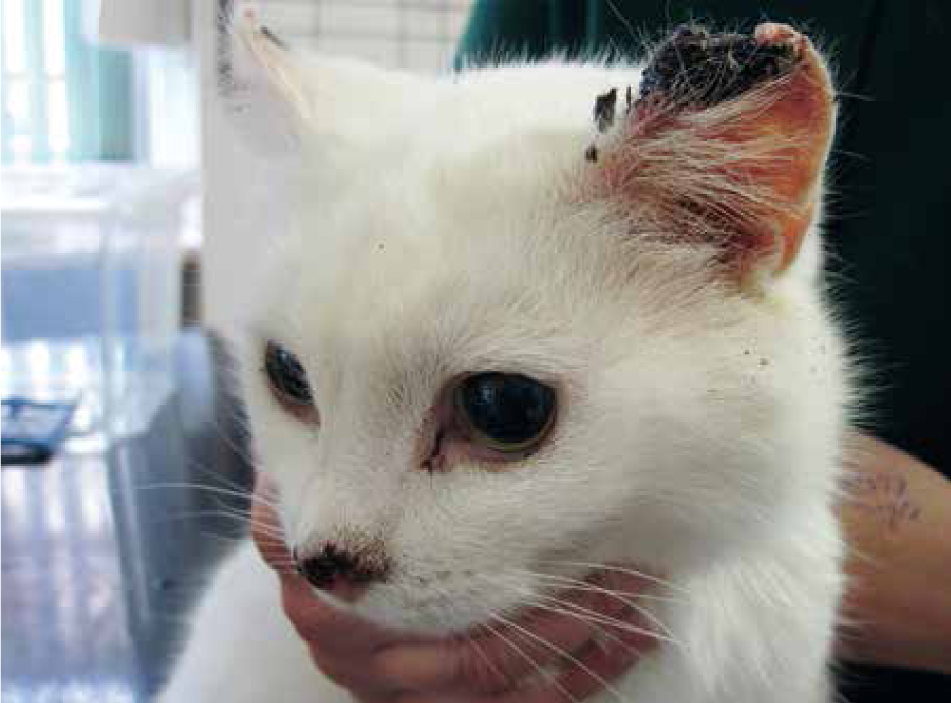
Moon was presented as a recently rehomed cat with obvious ulcerated growths to both ears and the nasal planum (Figure 4). Routine bloods and urine analyses showed no significant abnormalities. The owner was keen for surgery to go ahead, having seen a friend's cat that had undergone similar surgery previously. Due to the characteristic appearance of the lesions, a presumptive diagnosis of SCC affecting the pinnae and nasal planum was made. Biopsies were not taken, as it was felt this would not affect the course of action taken.
Moon was premedicated with methadone (Comfortan, Dechra), medetomidine (Medetor, Virbac, UK) and meloxicam (Metacam, Boehringer, UK) at standard dose rates. Skull and thoracic radiographs were unremarkable. Examination of the nares did not reveal any likely extension of the tumour within the nasal chambers. Aspiration of the submandibular lymph nodes was performed.
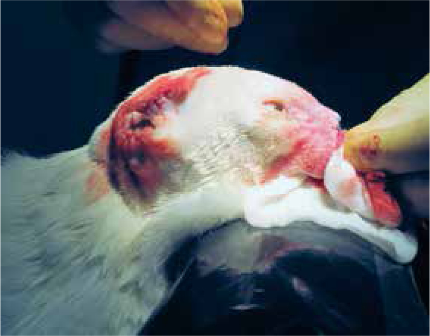
The head was surgically prepped (Figure 5a and b) and a bilateral pinnectomy was performed, in addition to a nasal planectomy. The nosectomy was reconstructed as above with 4/0 poligecaprone 25 using simple interrupted sutures: the pinnectomy wounds were closed using 4/0 poligecaprone 25 in a simple continuous pattern (Figure 6). Any haemorrhage was minor and controlled with application of pressure via sterile swabs. Lidocaine was applied to the wounds in the form of a splash block (1 ml of lidocaine in 5 ml of sterile saline: 1 ml sprayed or dripped over wound site every 3–4 hours). An Elizabethan collar was placed prior to full recovery, to reduce the risk of self trauma. However, this was removed the following morning, as it was felt that the collar was rubbing on the pinnectomy sites, increasing pain and irritation in these areas.
Moon made an uneventful recovery. Pain scoring was carried out for the following 72 hours (Colorado State feline acute pain scoring chart; http://www.vasg.org/pdfs/CSU_Acute_Pain_Scale_Kitten.pdf) and initially methadone (for the first 24 hours) and then buprenorphine was used every 4–6 hours depending on pain score. The pinnectomy wounds appeared to be significantly more painful than the nosectomy site. The lidocaine splash block on the nose was repeated every 3–4 hours for the first 48 hours, and then twice daily to facilitate cleaning of the nasal wound. Quite marked dried serosanguineous discharge (‘scabbing’) tended to build up over the 48 hours following surgery and it was important to clear this in order to allow Moon to breathe easily. Also, importantly, any nasal discharge reduced the ability of the patient to smell food.
Moon refused food on the day of surgery: a small amount of soft food was syringed the first day. Although initially reluctant to eat the day after surgery, gentle clearing of the discharge and stroking stimulated Moon to commence eating. No further problems were noted with appetite, although the owner was instructed to warm food gently in order to increase the aroma, thus increasing appetite. Moon was discharged 3 days post surgery.
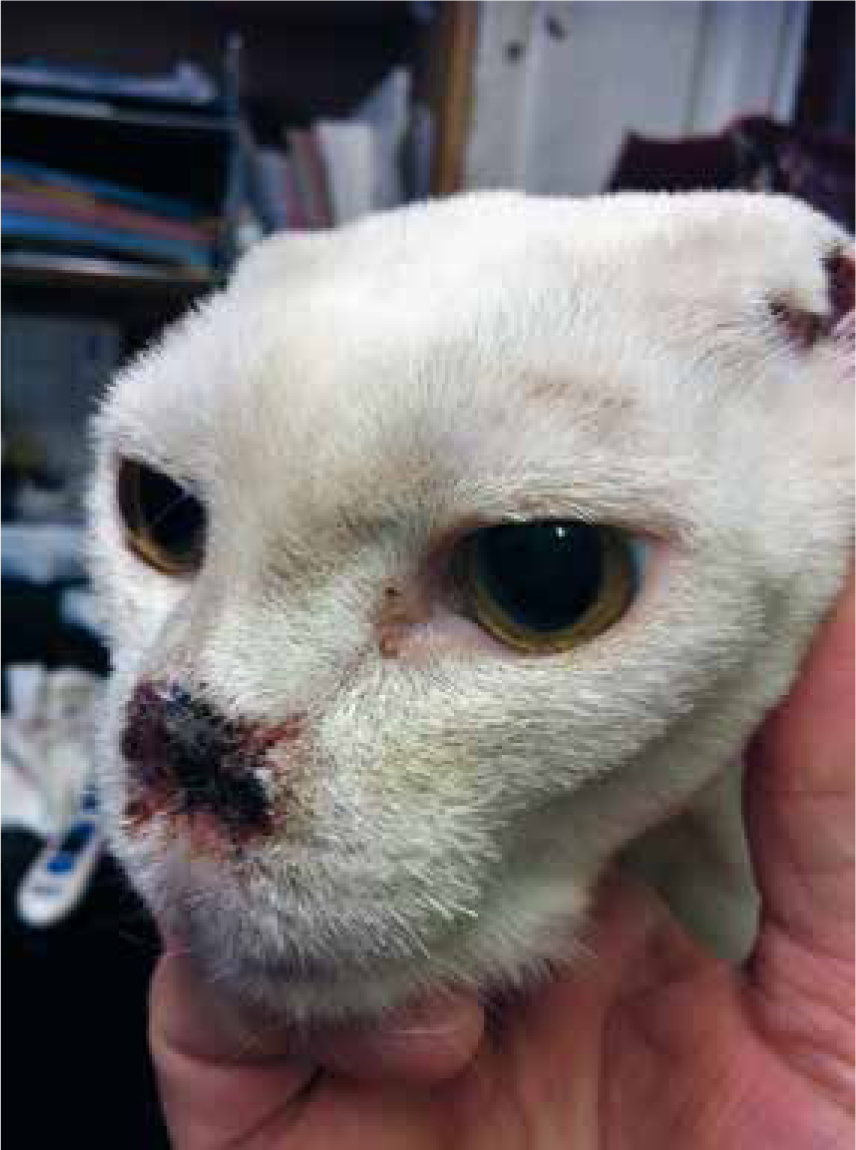
A 3 week postoperative follow up showed acceptable healing and the owner was pleased with the appearance (Figure 7). Histology results confirmed SCC of the nasal planum and pinnae, with no sign of lymph node involvement. Moon continues to do well 30 months post surgery.
Discussion
Baloo and Moon recovered well from nasal planectomy. Immediate postoperative care was not onerous and pain management was fairly straight forward. The most important aspect of postoperative management is to ensure that the wound site is kept clear of discharge. This is for several reasons:
In both cases above, overall recovery time (time to progress ‘back to normal’) was reasonably short — certainly within the space of around 3 weeks. Owner satisfaction was excellent and the surgical appearance (‘cosmesis’) was felt to be very acceptable. Despite the lack of advanced imaging, in both cases a clear surgical margin was obtained and the prognosis was thus favourable. It must be pointed out, however, that the ‘gold-standard’ of surgical cancer therapy must really be to consider the use of CT and MRI to more accurately determine the extent of tumour invasion prior to excision.
Analgesia is a crucial aspect of planning for nasal planectomy: in the canine example above, a preanaesthetic application of transdermal fentanyl seemed to provide excellent analgesia throughout the postoperative period. In the feline case, methadone worked very well. However, these examples should not be taken as a ‘one-size-fits-all’ approach to analgesia: different drugs, or classes of drugs should be chosen on the basis of surgical team familiarity. For example, if attempting this, or another, procedure for the first time, do not be tempted to try a brand new analgesic protocol — better to stick to one that the surgical and nursing team is used to, as it will be easier to monitor ‘standard’ protocols (provided these work, of course!). The use of a standard ‘pain chart’, to monitor, record and respond to different pain states, cannot be too highly stressed. As mentioned already, the author uses the Colorado State pain scoring scheme. However, other excellent schemes exist and the reader is encouraged to become familiar with a scheme that works within their own practice setting.
Both of the cases presented here had no nutritional problems post operatively: although the cat was syringe fed on the day of surgery, it ate normally the following day. In some cases there may be more reluctance to eat following surgery, especially surgery of this nature. Generally, good and conscientious nursing will encourage most patients to eat: ensuring that pain is controlled; that the nasal passages are kept clear; and that the patient is in a calm, secure and peaceful environment will all help. In some cases, particularly where there may have been pre-operative anorexia (either due to pain, nasal discharge, or other factor), it may be necessary to consider placing a feeding tube, such as an oesophagostomy tube, at the time of surgery (or even a few days before if the patient has been inappetent for a while, or is in a poor plane of nutrition.
Finally, it should be reiterated that, although veterinary professionals may be used to seeing cosmetically-altering surgical procedures, many clients are not; it is not unusual for clients to feel very nervous, even unwell, immediately before being reunited with their pets. It may be worth considering getting the client to remain seated while the pet is brought back to them, in case they feel faint. It would certainly be advisable to reunite them with the patient in a private room, or consulting room, rather than a busy waiting room.
Conclusions
Radical surgery for the treatment of cancer in pets is often contemplated for limb-based tumours, less so for tumours of the face and nose: in part because of actual or perceived owner concerns regarding a change in appearance of their pet. However, surgery remains the best chance of curing tumours such as SCC and it is to be hoped that this article may encourage veterinary care teams to consider nasal planectomy as an option for cats and dogs presenting with these. If doing so, or if referring for radical surgery, it is a good idea to allow the client access to photographs of pets having undergone similar procedures, or even to allow them to discuss a procedure with a client whose own pet has had it done.
Finally, to answer a question that the clients may have: ‘my dog has no nose: how does he smell?’ the answer is, of course, olfactory function is unaffected.