Sepsis is the systemic inflammatory response to infection. There are a myriad of diseases or injuries that can cause a patient to become septic. In 1991 the American College of Chest Physicians/Society of Critical Care Medicine (ACCP/SCCM) Consensus Conference Committee met and established standards that defined sepsis in human medicine (Balk et al, 1992). Up until that time the use of words such as infection, bacteraemia, septic shock and sepsis were being used interchangeably. While there are no of-ficial set of standards used to diagnose sepsis in veterinary medicine, there have been numerous studies in recent years working on creating such standards.
The 1991 the ACCP/SCCM Consensus Conference Committee identified common abnormalities in septic human patients with regards to temperature, heart rate, respiratory parameters, and the leukogram (Balk et al, 1992). In 2001 these criteria were expanded to include parameters for systemic inflammatory response syndrome (SIRS) (Otto, 2011). During the ACCP/SCCM Conference several definitions were de-fined (Box 1). While these terms have not been of-ficially defined in veterinary medicine for the purposes of this paper the ACCP/SCCM terminology will apply.
In 1997 veterinarians Hauptman et al looked at temperature, heart rate, respiratory rate, white blood cell count, percent of immature neutrophils, platelet count, and serum glucose concentration as markers of clinical canine sepsis, in an effort to establish veterinary parameters. Based on the 1997 Hauptman recommendations, veterinary medicine currently has a set of parameters that defines SIRS. SIRS can be diagnosed in a dog or cat when two or more of the parameters in Box 2 are met. Sepsis, defined by human medicine, is the presence of an infection combined with SIRS.
Pathophysiology
In order for sepsis to occur there first must be a source of infection. The first step in the initiation of the in-flammatory response is for the body to recognize there is an infection. The body will attempt to contain and destroy the pathogen through controlled inflam-mation (Raffe and Wingfield, 2002a).
The inflammatory response results in the activation of endothelial cells, neutrophils and monocytes. In response, the inflammatory cascade becomes activated. The inflammatory cascade is mediated by cytokines, which are macrophage derived (Maki and Tambyah, 2001). There are several types of cytokines with the most important being tumor necrosis factor (TNF) and interleukin-1 (IL-1) (Murtaugh, 2002). Both of these will have direct and indirect effects on all organ systems (Murtaugh, 2002). When TNF and IL-1 are released the body will see a change in temperature (hyper or hypothermia), depressed cardiac output and increased vascular permeability (Murtaugh, 2002).
During sepsis, the body will elicit an inappropriate inflammatory response resulting in hypercytokine-mia (Campbell, 2005). Hypercytokinemia is a potentially fatal reaction which occurs between cytokines and immune cells. This results in too many cytokines being released which results in too many immune cells damaging the body. It is the hypercytokinemia that will cause the SIRS (Campbell, 2005). Ultimately the inflammatory mediators can overwhelm the organs leading to multiple organ dysfunction.
It is important to note that the coagulation system and inflammatory cascade work together (Maki and Tambyah, 2001). Thrombin, a key player in the coagulation cascade, also has important inflammatory and cellular proliferative properties (Maki and Tambyah, 2001). Cytokines themselves also help to activate the coagulation cascade (Maki and Tambyah, 2001). It is for this reason that septic patients are at risk of clotting disorders such as disseminated intravascular coagulation (DIC).
Another common problem in septic patients is hypoglycaemia. Hypoglycaemia likely occurs due to the increase in glucose consumption that is triggered by the inflammatory mediators (Koenig, 2010). Other causes of hypoglycaemia during sepsis may be secondary to decreased food intake, decreased liver function, hypotension or hypoxic-induced anaerobic glycolysis (Koenig, 2010).
Signs/symptoms
Certain diseases may cause an animal to be predisposed to developing sepsis. These generally include diseases that have caused a weakened immune system or are infection based (Raffe and Wingfield, 2002a). Patients receiving chemotherapy or those that have immune dysfunction are at a greater risk (Norkus, 2012). Patients diagnosed with peritonitis, pneumonia or those with traumatic infected wounds are predisposed to developing sepsis (Raffe and Wingfield, 2002a).
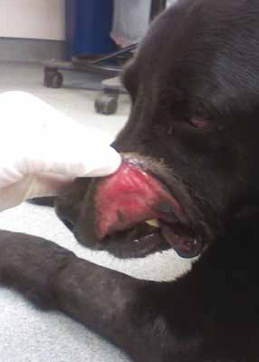
During the early stages of sepsis the body will experience high cardiac output, low systemic vascular resistance, hypoglycaemia, metabolic acidosis and normal to increased systemic blood pressure (Murtaugh, 2002). Appropriate inflammatory signs such as localized pain, fever, redness, swelling and brick red/ muddy mucous membranes will also occur (Raffe and Wingfield, 2002a) (Figure 1).
Ultimately if the infection cannot be controlled the patient may experience decreasing cardiac function, capillary leaking, organ dysfunction, organ failure and death (Raffe and Wingfield, 2002a). As the sepsis worsens patients will develop tachypnea, tachycardia or bradycardia, severe hypotension, marked mental depression, gastrointestinal issues (hematachaezia/ vomiting), multiple organ dysfunction and DIC (Murtaugh, 2002).
Treatment
The veterinary septic patient can prove to be one of the toughest nursing challenges in veterinary medicine. A veterinary nurse typically spends more time with the patient than the veterinarian, which allows for nurses to notice subtle changes in the patient. Ultimately the treatment of sepsis depends on the inciting cause. Treatment may be relatively simple with just intravenous antibiotics and fluid therapy, such as in the case of a bacterial infection. In more complex infections, such as septic peritonitis, surgery may be required to remove the infected source. Due to the numerous complications arising from sepsis, patients may experience everything from respiratory complications requiring ventilatory support to DIC which requires plasma transfusions (Raffe and Wingfield, eds 2002b).
One of the key factors in treating sepsis is ensuring that the patient is on an appropriate antibiotic that will target the specific infection. In order to ensure an appropriate antibiotic is chosen, an infected sample must be submitted to a laboratory where it will be cultured. It is important to ensure an infected sample is collected before any antibiotics are started (Raffe and Wingfield, 2002a). This will yield the best results from the laboratory. Studies in humans have shown that administering antibiotics within 6 hours of being admitted into the hospital improves the patient's survival (Dellinger et al, 2008). So while obtaining a sample is important, antibiotic therapy should not be delayed because of sample collection, and antibiotics should be started before the laboratory sample is back.
Treatment in all septic patients should be aggressive. The goal is to rid the body of the infection and to support and treat multiple organ failures resulting from the sepsis (Daniels and Nutbeam, 2010). Early goal-directed therapy is imperative when treating septic patients (Daniels and Nutbeam, 2010).
In human medicine it is recommended that every septic patient should have serum lactates measured, and blood cultures obtained prior to antibiotic administration; broad spectrum antibiotics are used until culture results are back and early goal directed therapies are initiated (Daniels and Nutbeam, 2010). These early goal directed therapies include fluid therapy and blood pressure maintenance.
Specifc nursing care
Depending on how critical the patient is, nursing care may be directed towards fluid therapy only or include more involved care like ventilatory assistance. Septic patients should be immediately admitted into an intensive care unit if one is available (Daniels and Nutbeam, 2010).
Basic monitoring
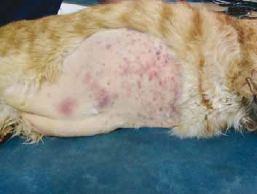
Septic patients should minimally have a full physical examination performed every 4–6 hours. This should include heart rate, respiratory rate and effort, mucous membrane colour, capillary refill time, rectal temperature and neurological status. If there is any change from normal parameters, the veterinarian should be notified. Because patients are at risk from DIC it is important to look for early signs, which includes excessive bleeding after venipuncture sticks and/or pe-techiae on the gums, pinna or abdomen of the patient (Figure 2).
Monitoring the septic patient's fluid therapy is important. Patients should be weighed at the beginning of treatment and then at least twice a day to determine fluid losses and gains. Rapid changes in body weight are usually a result of fluid gains or losses. Septic patients can often experience large fluctua-tions in weight due to fluid shifting, retention or loss of fluids through vomiting and/or diarrhoea. A 0.5 kg weight gain is equivalent to a 0.5 litre fluid gain (Davis, 2008).
Urine output should be monitored and recorded. Quantifying urine output is key in monitoring fluid therapy as well as in patients with renal disease. Since septic patients may experience multiple organ dysfunctions (MODs), kidney failure may occur in these patients. In both dogs and cats, 1 to 2 ml/kg/hr of urine should be produced if the patient is not on flu-ids (Raffe and Wingfield, 2002b). If the patient is on fluids then the total volume being given to the patient should ideally be urinated out (Raffe and Wingfield, 2002b). If possible place non-absorbent litter in cages with cats and catch the urinations of canine patients in a bowl for quantification.
Advanced monitoring
Since septic patients often require frequent blood draws a central line should be placed. This will allow for faster and pain-free blood collection from the patient. Without a central line veins often become overused and obtaining blood can be a real, if not impossible, challenge. An arterial line should be considered in patients experiencing respiratory problems. This will allow for measurement of partial pressure of oxygen in arterial blood (PaO2), which is the gold standard when measuring overall oxygenation ability (Crowe, 2008).
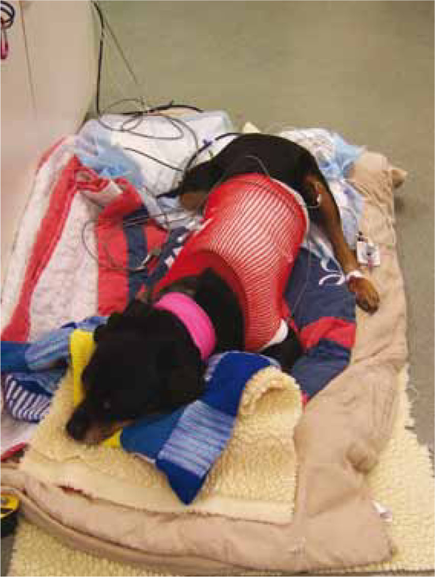
Besides a stethoscope and thermometer to monitor vitals there are generally a number of other tools necessary for appropriately monitoring the septic patient; blood pressure, blood glucose, lactate and central venous pressure (CVP) should all be measured (Figure 3).
It is important that septic patients have their blood pressure monitored minimally every 6–8 hours. Septic patients are at risk of developing hypotension. If the mean arterial pressure (MAP) falls below 60 mmHg, the kidneys and other organs are not appropriately perfused putting them at risk of organ failure. Arterial hypotension is defined by a MAP (diastolic + 1/3(systolic–diastolic)) less than 60 mmHg or by a Doppler ultrasonic blood pressure with a systolic reading less than 80 mmHg (Bryant, 2009). Normalization of blood pressure, defined by a MAP of 80–120 mmHg or systolic between 110–160 mmHg, is the goal in any septic patient (Bryant, 2009).
Septic patients are at risk for developing hypoglycaemia (Raffe and Wingfield, 2002a). Blood glucose should be monitored at least once a day or when hy-poglycaemia is suspected; symptoms of hypoglycae-mia include lethargy, bradycardia, disorientation and hypothermia. When hypoglycaemia is present intravenous dextrose should be added to the intravenous fluids to help ensure the patient does not seizure from a low blood sugar.
Blood pressure can be monitored either directly or indirectly. Direct arterial pressure monitoring is gold standard (Battaglia, 2007a). It requires the placement of an arterial catheter, which can also be used to obtain arterial blood gas samples to monitor PaO2. An electronic transducer is placed at the end of the arterial catheter and measured continuously. If an electronic transducer is not available then the blood pressure can be measured using a CVP manometer (Battaglia, 2007a). Indirect methods include oscillo-metric devices or Doppler ultrasound flow detectors. Indirect readings are less accurate, but require less skill and are non invasive (Battaglia, 2007a).
CVP is generally used when a patient is prone to changes in blood pressure or when aggressive fluid therapy is being utilized, which is the case with septic patients. CVP is considered a measurement of cardiac pumping ability, circulating blood flow, vascular tone and intrathoracic pressures (Battaglia, 2007a). A central line is placed into the jugular vein and is fed down until it rests on the right atrium. The pressure in the right atrium indicates how the heart handles the volume of fluid presented to it. The measurement helps to determine how much fluid can be administered to a patient without causing fluid overload or dehydration. Depending on the literature normal CVP measurements vary, but most will agree it is somewhere between 1–10 cmH2O (Battaglia, 2007a).
Lactate accumulates in the tissues and blood as a result of inadequate oxygen availability, which is generally caused by tissue hypoperfusion. In some cases increases in lactate may be the only indication that hypoperfusion still exists. Increases in lac-tate are commonly seen in septic patients. A value of under 2 mmol/l is normal. In human medicine monitoring lactate trends is common. Studies in human medicine have shown that lactate values that remained above 4 mmol/l are a predictor of poor outcome with mortalities of greater than 40% (Daniels and Nutbeam, 2010). Lactate can be measured using a simple hand-held device similar to a blood glucose machine. It is important to normalize lac-tate concentrations through fluid therapy and blood pressure normalization.
Nutritional support
Nutritional support must be considered in septic patients that are hospitalized for more than 48 hours. Sepsis causes an increase in resting energy expenditure and an increased protein consumption (Battaglia, 2007b). Providing nutritional support to these patients early is essential in order to minimize weight loss and to provide adequate energy for metabolic support (Battaglia, 2007b). In both human and veterinary patients a better outcome is seen in those that receive nutritional support earlier (Khalid et al, 2010). Enteral feeding is the best. Often these patients will not eat on their own, so placement of a feeding tube should be considered (Battaglia, 2007b). Certainly if the patient continues to vomit or enteral feeding is not appropriate, parenteral feedings should occur.
Pain management
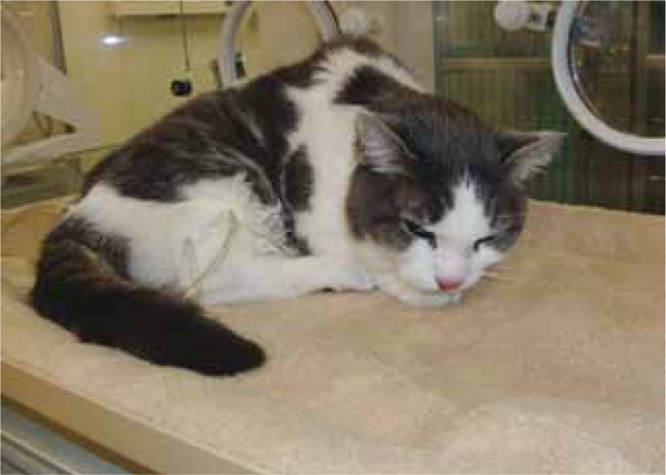
Most septic patients are in some level of pain. This is particularly true for post-operative septic peritonitis patients. The patient's nurse should watch for signs of pain. In dogs this may include vocalizing, shaking, aggression and panting (Battaglia, 2007c). In cats, more commonly, this will include aggression or hiding (Battaglia, 2007c) (Figure 4). If a patient is in pain it should receive pain medication. Opioids are the choice of drugs for septic patients (Battaglia, 2007). They offer excellent analgesia with limited effects on the haemodynamic system. Multimodal and continuous rate infusion analgesia should be considered in these patients (Battaglia, 2007).
The post-operative surgical patient
There are many reasons a septic patient may require surgery. The sepsis could be from a necrotic tumor, perforated intestine from a foreign body or other similar causes. Perforating trauma (such as a stick or bullet) into the abdomen may also introduce bacteria causing peritonitis. The infection in the abdomen may lead to septic peritonitis.
Septic peritonitis is the inflammation of the peritoneum (the membrane which lines the abdominal cavity) due to the introduction of bacteria into the abdominal cavity from a perforation or rupture of a hollow viscous (Raffe and Wingfield, 2002c). Depending on the cause, many patients may have limited symptoms such as anorexia or restlessness. As the disease progresses vomiting, diarrhoea, abdominal pain and/or abdominal distention may be noted (Raffe and Wingfield, 2002a). Generally, in advanced stages of peritonitis the abdomen becomes very painful. These patients often require surgery to remove the source of infection.
Post operatively the abdomen may be left open or closed, depending on the degree of infection and the cause of the peritonitis. Open abdominal drainage allows for septic material to passively drain out of the abdomen. It also allows for easy evaluations of the area and decreases the survival of anaerobic bacteria (Battaglia, 2007d). The incision is covered with highly absorbent sterile towels or pads and is held in place by an abdominal bandage or umbilical tape that has been woven through stay-sutures in the abdomen (Battaglia A, 2007d). Because the incision is open, a urinary catheter must be placed to ensure the patient does not soil the bandages. The dressing should be evaluated every 6–8 hours for discharge and changed aseptically at least three times a day or whenever the bandage is soiled (Battaglia, 2007d). The amount of fluid lost can be estimated by weighing the bandages after they are changed with the dry bandages. Fluid should be examined microscopically to look for any cellular changes (such as reduction of bacteria). Open abdomens are associated with a high number of complications including hypoproteinemia, hypothermia, ascending infection and evisceration (Battaglia, 2007d).
Closed abdomens create less intensive care challenges for technicians. Closed suction drains are placed in the abdomen and the incision is closed around the drain. This technique does not allow for complete drainage of the abdomen, but the fluid that is removed is confined to the drain itself. The drain must be kept in place until the amount of flu-ids is within physiologic limits and the fluid cytology shows no evidence of infection (Battaglia, 2007d). Studies have shown survival rates of patients following closed and open abdomens to be about the same (Robledo et al, 2007).
It is interesting to note that despite advances in medicine, the survival rate for dogs that undergo surgery for septic peritonitis has not improved in the last 10 years. A study performed at the University of Pennsylvania concluded that there was no signifi-cant difference in survival among dogs treated surgically for septic peritonitis between 1988 and 1993 (64%) and 1999–2003 (57%) (Bentley et al, 2007). It is important for owners to understand the level of critical nursing care their pet will need post op-eratively and the high mortality rate associated with septic peritonitis.
Ventilation patient
In some cases, septic patients may be put on a ventilator if they are experiencing severe respiratory distress due to poor overall oxygenation ability. The veterinary nurse will be responsible for the maintenance of the ventilator machine. Besides the machine, the patient itself will require considerable nursing care. Ventilation patients often require a round-the-clock nurse assigned to only them.
It is important that the air entering the patient is humidified. Oral mucous membranes and eyes should be moistened and lubricated to avoid drying out. The oral cavity itself should be cleaned and rinsed every 4 hours (Hopper, 2009).
The endotracheal or tracheotomy tube should be suctioned sterilely every 4 hours and the endotrache-al tubes should be replaced every 24 hours (Hopper, 2009). In addition the cuff on the tubes should be de-fated, repositioned and reinflated every 4 to 6 hours (Hopper, 2009).
Since the patients are recumbent, passive range of motion must be performed every 4 to 6 hours and patients should be repositioned at that time as well (Hopper, 2009). Since patients are not moving, it is best to place a urinary catheter in them to keep them as clean and dry as possible. Adequate soft bedding should be used and bedding should be cleaned and replaced once a day.
Conclusion
Most veterinary nurses will encounter a patient that is septic at some point in their career. It is important to understand not only how sepsis affects the body, but also the unique nursing care the patient will require. Providing excellent nursing care will mean the differ-ence between life and death to the septic patient.