
Immediate pet travel considerations most frequently concern tapeworm, ticks, rabies and pet passports. It is essential that pet owners abide with legislation when travelling abroad with their pets, but prevention against parasites, disease and spread into the UK goes far beyond legal requirements. The fluid nature of parasite distributions means that an increasing range of parasites need to be considered and general principals in parasite control and biosecurity implemented. This article considers some of these control measures as well as some of the more novel parasites that have entered the UK in travelled and imported pets in the past 12 months.
The Pet Travel Scheme
The Pet Travel Scheme (PETS) is designed to protect countries against the introduction of zoonotic disease. This legislation also serves two-fold to protect pets from disease, but there is significantly more that can be done to protect pets. For example, tick protection was removed from PETS in 2012 after the risk of Rhipicephalus sanguineus ticks entering and establishing in the UK was deemed negligible. The ability of mandatory tick treatment to prevent zoonotic tick-borne diseases entering the UK was also considered questionable. However, removing the tick prevention from PETS has contributed to the UK border being unprotected against the entry of tick-borne parasites such as Babesia spp. and Ehrlichia canis, both of which pose significant risk to UK pets. Pet travel advice should consider both mandatory and advisory parasite control measures in order to fully protect both the pet and the UK from exotic disease. The current PETS legislation is summarised in Table 1. It is important to always check the pet travel requirements of the country being visited and remember that the Republic of Ireland, the Channel Islands and the Isle of Man have their own regulations for pets travelling from outside of the UK.
| Pet Requirement | Additional details |
|---|---|
| Before travel | |
| Microchip | Before rabies vaccination |
| Rabies vaccination | At least 21 full days before travel (day of vaccination counts as day 0). |
| EU Pet Passport (or Official Veterinary Certificate) | |
| During travel | |
| Pet must travel within 5 days of owner/accompanying person | If the pet is not travelling with the owner, the owner must authorise the person responsible for the pet in writing prior to travel |
| Owner must sign a declaration stating there is no intention to sell or transfer ownership of the pet | |
| Echinococcus multilocularis (tapeworm) treatment with praziquantel or equivalent | 24–120 hours (1–5 days) before re-entry time into the UK (or Finland, Ireland, Malta or Norway) |
| Travel with an approved transport company via an authorised route | |
The usual suspects
Although they are not the focus of this article, it is important not to forget those commonly considered parasites and the diseases they cause while travelling abroad. Namely, Echinococcus multilocularis, Dirofilaria immitis, Leishmania infantum and various tick-borne diseases (these, and other important parasites and diseases are summarised in Table 2). Perhaps that of greatest threat is E. multilocularis. The ecological theory of biological invasions states that the risk of a disease establishing in the UK is determined by the propagule pressure and the habitat suitability (both climate and vector availability). Habitat suitability for E. multilocularis is very high in the UK due to an abundance of foxes and the optimal habitat for the microtine vole intermediate hosts (Craig, 2014). There is a 98% risk of one dog out of 10 000 returning to the UK with E. multilocularis if no tapeworm treatment is administered (Torgerson and Craig, 2009). The 5-day window following the mandatory tapeworm treatment allows increased opportunities for reinfection and for pet passports to be signed in a greater number of countries before (re)entry to the UK, thus increasing the risk of entry into the UK.
| Parasite | Disease | Definitive hosts | Intermediate hosts | Transmission | Possible clinical signs | Control |
|---|---|---|---|---|---|---|
| THE USUAL SUSPECTS | ||||||
| Echinococcus multilocularis | Alveolar echinococcosis in humans | Foxes, dogs, (cats) | Voles, rodents, humans | Intermediate hosts — ingestion of eggs passed in the faeces of canids. Definitive hosts predation of rodents (microtine voles) | No clinical signs in the canine or feline definitive host. Metastatic and infiltrative spread primarily in the liver in intermediate host | In endemic areas, pets should be treated at 30 day intervals with an effective anthelmintic containing praziquantel. Dogs should be treated again with praziquantel within 30 days of return to the UK. This is in addition to the compulsory treatment |
| Dirofilaria immitis | Dirofilariosis | Dogs, cats, foxes, ferrets humans | Mosquitoes — vector-borne disease (VBD) | L3 infective larvae in the body of the mosquito are transmitted to definitive hosts during feeding. The larvae undertake an extensive migration through body tissues to reach the pulmonary arteries and the right side of the heart where they develop to the adult stage and reproduce | Primarily cardiac signs in dogs, respiratory in cats. Infections may be subclinical | Monthly treatment with a macrocyclic lactone to prevent adult heartworm infection developing (many efficacious products licensed). Treatment should begin at least 1 week prior to exposure to infected mosquitoes and end at least 1 month after last exposure to infected mosquitoes |
| Babesia spp. | Babesiosis | Dogs, wolves, foxes, cats (Babesia felis cases mostly in South Africa to date) | Dermacentor reticulatus and Rhipicephalus sanguineus ticks depending on Babesia species — vector unknown for Babesia felis | After a tick feeds on an infected host, Babesia spp. stages penetrate the gut of the tick, multiply and migrate. Sporozoites are then transmitted to new hosts through the tick's saliva when they take a blood meal | May be subclinical. Signs include fever, lethargy, anorexia, depression, anaemia, sickness, red coloured urine, renal failure | Risk of infection can be significantly reduced by effective tick control. Chemoprophylaxis with a product that repels, expels or rapidly kills ticks. Monitor pets for ticks every 24 hours and remove any ticks found with a tick hook using a ‘twist and pull’ action to avoid leaving in mouthparts |
| Ehrlichia canis | Canine monocytic ehrlichiosis | Dogs, wolves, foxes, (cats) | Rhipicephalus sanguineus ticks — VBD | After feeding on an infected host, the bacteria are then transmitted to new hosts through the tick's saliva when they take a blood meal | Various. Including apathy, depression, anorexia, dyspnoea, anaemia, fever epistaxis and vomiting. Dogs may appear clinically normal | Risk of infection can be significantly reduced by effective tick control. Chemoprophylaxis with a product that repels, expels or rapidly kills ticks. Transmission has been demonstrated to be within hours so a repellent should be used where possible and where E. canis is endemic. Monitor pets for ticks every 24 hours and remove any ticks found with a tick hook using a ‘twist and pull’ action to avoid leaving in mouthparts |
| Leishmania spp. | Leishmaniosis | Dogs, cats, foxes, humans | Sandflies — VBD | Parasites present in the superficial dermis of infected dogs fall into the well of blood created by the sandfly's mouthparts when feeding. After migration to the salivary glands of the sandfly, promastigotes are released into the superficial dermis of the dog during a subsequent blood meal. These then form amastigotes in macrophages that infect a wide variety of tissues | Many infected hosts will not show clinical signs. Cutaneous lesions, enlargement of lymph nodes, weight loss, anorexia and muscle weakness, potential for kidney and heart failure | Prevention of sandfly bites via application of a licensed pyrethroid fly repellent in dogs, deltamethrin collar or licensed permethrin spot on preparation every 2–3 weeks. No product is licensed for sandfly protection in cats but a flumethrin collar has been shown to be efficacious and safe. Avoid hosts being outside during dawn and dusk which are optimal sandfly feeding times. Vaccine is available in dogs which can help prevent disease |
| Dirofilaria repens | Dirofilariosis | Dogs, foxes, humans (cats) | Mosquitoes — VBD | Mosquitoes ingest L1 microfilariae while feeding and L3 larvae develop within the vector, which are then transmitted into the subcutaneous tissues of new hosts via their saliva while feeding. Here they reach maturity | Dermatitis, conjunctivitis, skin nodules, aberrant worm migration to a variety of organs | Monthly use of a licensed moxidectin/imidacloprid spot on product while in endemic countries. Avoidance of peak mosquito feeding times |
| Thelazia callipaeda | Thelaziosis | Dogs, foxes, cats, humans | Fruit flies — VBD | The intermediate host fly ingests L1 larvae from a definitive host while feeding on lacrimal secretions from the eyes. L1 larvae then develop over a period of 14+ days to L3 larvae which are deposited to a new host when feeding and mature in the conjunctival sac | Conjunctivitis, keratitis, epiphora and tear overflow onto the face, eyelid odema, corneal ulceration, blindness | No licensed treatment for prevention although milbemycin oxime may be beneficial as it has some efficacy in treatment. Fly repellents will reduce exposure |
| Spirocerca lupi | Spirocercosis | Dogs, foxes, (cats) | Coprophagous beetles | Definitive hosts become infected through the ingestion of coprophagous beetles infected with L3 S. lupi larvae. This may occur through direct predation or through the consumption of contaminated foodstuffs. The larvae penetrate the stomach wall and migrate to the thoracic aorta via the gastric arteries. The larvae then moult to L4 larvae and immature adults. There is then a further migration after 3–4 months to the oesophagus where they mature in granulomatous nodules that form around them as part of the host inflammatory response | Retching, regurgitation, signs associated with oesophageal sarcoma, sudden death from aortic aneurism, spinal pain | Monthly use of a licensed moxidectin/imidacloprid spot on product while in endemic countries or in dogs with a predilection for eating invertebrates in countries where cases have been reported |
| Linguatula serrata | Linguatulosis | Dogs, foxes, humans | Ruminants, horses, rabbits, humans | Definitive hosts — infected through consumption of raw offal. Intermediate hosts — infected through ingestion of eggs in environment or on contaminated pets or foodstuffs | Nasal discharge, coughing, retching, aberrant larvae in anterior chamber of eye, visceral pain | Avoiding eating raw or undercooked offal in endemic countries, good hand hygiene, avoid facial licking from infected dogs |
| Tick-borne encephaltitis | Encephalitis | Dogs, foxes, cats, ruminants, horses, wildlife, humans | Ixodes ricinus ticks | Viral infection transmitted when ticks feed | Central nervous systemn (CNS) signs, fever, death | Risk of infection can be significantly reduced by effective tick control. Chemoprophylaxis with a product that repels, expels or rapidly kills ticks. Monitor pets for ticks every 24 hours and remove any ticks found with a tick hook using a ‘twist and pull’ action to avoid leaving in mouthparts. Avoidance of prolonged time in woodland in endemic countries |
Another parasite advantaged by the 2012 amendment to PETS is the tick. The removal of the mandatory tick treatment in PETS caused much controversy before, and after, the legislative amendment, with associations such as European Scientific Counsel Companion Animal Parasites (ESCCAP UK & Ireland), British Small Animal Veterinary Association (BSAVA) and British Veterinary Association (BVA) all opposing the change. It was predicted that with the removal of the mandatory tick treatment it would only be a matter of time before exotic tick-borne diseases established in the UK. The removal of the compulsory tick treatment was one of a number of factors that has contributed to the increased risk of novel ticks and tick-borne diseases entering the UK. Recent surveys have shown that the incidence of ticks is increasing throughout Europe, with Dermacentor reticulatus extending its range south and into Scandinavia, while R. sanguineus is extending into Eastern and Central Europe. This is bringing both of these ticks into popular UK holiday destinations such as France and countries from which pets are frequently imported such as Romania (Wright, 2016). In spring 2016, Babesia canis was diagnosed in a number of untraveled dogs in Essex and DNA testing showed that there were endemic D. reticulatus ticks carrying the parasite (Swainsbury et al, 2016).
The lesser known threats
Climate has previously dictated that serious fly-borne parasites are not of concern in the UK. However, with increased pet travel and the migration of fly-borne diseases migrating further north in Europe, these parasites are becoming very real risks to the UK's biosecurity. Two such parasites are Dirofilaria repens and Thelazia callipaeda.
Dirofilaria repens
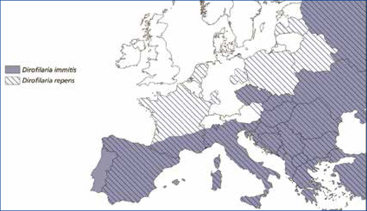
The vectors for D. repens (Aedes, Culex and Anopheles mosquito species) are already endemic in the UK, however, the UK has typically not had a suitable climate for the establishment of D. repens; the development of Dirofilaria species L1 microfilariae to L3 larvae requires 29 days at a constant 18°C (Lloyd, 2011). D. repens is widespread across Europe, as far north as France and Belarus (Figure 1). D. immitis and D. repens both have similar development requirements, although D. repens does seem to have less stringent temperature requirements meaning that it is more likely to establish in the UK (Morgan, 2016).
Definitive hosts include dogs, cats and other domestic and wild carnivores. Humans can also be infected and D. repens is considered the most significant Dirofilaria species responsible for human infections in Europe (ESCCAP UK & Ireland, 2016). Transmission occurs when L1 microfilariae are excreted into the blood stream of the host by adult worms already present. Mosquitoes ingest L1 microfilariae while feeding and L3 larvae develop within the vector, which are then transmitted into the subcutaneous tissues of new hosts via their saliva while feeding. Here they reach maturity, sometimes causing pruritus, dermal swelling or subcutaneous nodules. In rare cases, adult worms migrate to the eyes of the host where they may be visible and may cause conjunctivitis. Prevalence of dirofilariosis in cats tends to be only one tenth of that in dogs and typically occurs in areas of high canine infection rates (ESCCAP, 2012).
Thelazia callipaeda
T. callipaeda, also known as the ‘oriental eye worm’ due to its high incidence in Asia, is a zoonotic vector-borne nematode which resides in the conjunctival sac of definitive hosts (domestic and wild carnivores, rabbits and humans) (Mihalca et al, 2015). The parasite is found widely in Asia and has been spreading through Europe in recent years. The vector in Europe is the drosophilid fruit fly, Phortica variagata. The intermediate host fly ingests L1 larvae from a definitive host while feeding on lacrimal secretions from the eyes. L1 larvae then develop over a period of 14+ days to L3 larvae which are deposited to a new host when feeding and mature in the conjunctival sac (Graham-Brown et al, 2016). Although often apathogenic, ocular thelaziosis can commonly cause conjunctivitis, keratitis, epiphora and tear overflow onto the face, eyelid oedema, corneal ulceration and, in serious cases, blindness (Graham-Brown et al, 2016).
T. callipaeda was first reported in Europe in Italy and since 2007 autochthonous cases have spread east and northwards, being reported in France, Germany, Switzerland, Spain and Portugal, and more recently into Eastern Europe, including Romania, in 2014 (Mihalca et al, 2015). In endemic areas 60% of dogs have T. callipaeda adults in their eyes (Lloyd, 2011), but clinical cases of ocular thelaziosis have also been reported in cats in many endemic countries (Mihalca et al, 2015).
In 2016, the UK saw its first case of ocular thelaziosis in a dog imported from Romania 6 weeks previously (Graham-Brown et al, 2016). Although this case was considered to be imported, it illustrates how easily T. callipaeda can enter the UK. PETS has never included a requirement for nematode treatment and the P. variagata intermediate host is widespread in the UK (Morgan, 2016). Increasing pet movement, especially from Eastern Europe, means that without rapid detection and treatment of cases, it is only a matter of time before T. callipaeda establishes in the UK.
Linguatula serrata
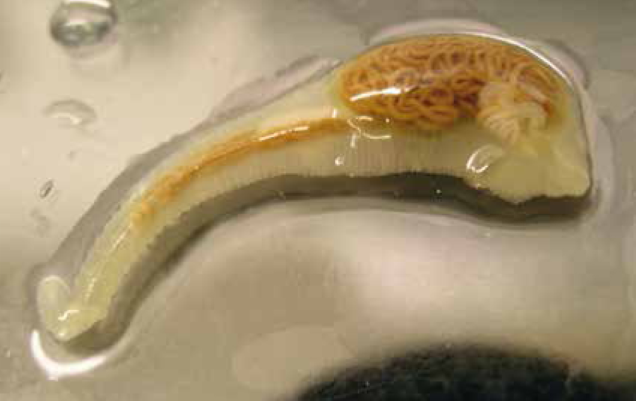
This parasite is known as a ‘tongue worm’, but is actually a pentastomid and is now thought to be more closely related to arthropods than true worms. The adult parasite is an elongated tongue-shape (Figure 2) and is found in the nasal cavities or sinuses of dogs and foxes. Infection occurs through the ingestion of nymphs in raw offal of infected intermediate hosts such as ruminants, rabbits and horses. Eggs from the adult parasite are passed in the faeces or nasal secretions of infected dogs and are immediately infective. Adult parasites are large with females typically 30–130 mm in length. Although the parasite has been reported in UK foxes (Lapage, 1968), it is thought to be rare, however, there has been a sharp increase in clinical cases reported in UK dogs in 2016. These have been in imported stray dogs from Romania, where raw meat is routinely fed. The concern with this sharp increase in cases is the zoonotic potential of the parasite. Although in endemic countries zoonotic infection occurs primarily through the ingestion of raw or undercooked viscera, it can also occur through ingestion of eggs in the environment or in mucoid discharge from infected dogs’ noses. This can lead to a variety of clinical presentations including naso-pharyngitis, blocked nasal passages, visceral pain and aberrant larval migration to the anterior chamber of the eye (Koehsler et al, 2011).
Spirocerca lupi
Spirocerca lupi is a spiruroid nematode inhabiting granulomatous nodules in the oesophagus of dogs and rarely cats. Transmission occurs through the predation or accidental ingestion of coprophagus beetles. It is found widely through the Southern USA, India, Israel, Japan and South Africa (Van der Merwe et al, 2008). Infections may be subclinical, but can lead to significant and potentially fatal disease syndromes including aortic aneurisms, oesophageal obstruction, progression of nodules to oesophageal sarcoma and thoracic spondylosis. S. lupi has not been considered a significant pathogen for pets travelling from the UK to Europe due to its low prevalence in most European countries. Although it is known to be prevalent in Greece, this appears to be predominantly among hunting dogs rather than pets, and wildlife reservoirs are known in more temperate parts of Europe such as Poland. However, cases have now been reported in untraveled domestic dogs in Spain, France and Italy (Giannelli et al, 2014). The threat that this represents to travelling dogs from the UK is unquantified and it is unclear whether this represents genuine spread or increased reporting. If cases continue to occur in countries where UK dogs frequently travel then the risk of UK dogs developing clinical infections, or introducing the parasite into the UK, increases. A case in an untraveled dog and cat in the UK has been reported (Wright et al, 2016), and while not thought to be endemic in the UK, increased vigilance for this parasite is required.
Tick-borne encephalitis (TBE)
TBE is a virus transmitted by feeding Ixodes ricinus ticks, although it has also been transmitted through unpasteurised milk and through exposure to infected tissues in abattoirs. It may infect a variety of mammalian hosts including dogs, foxes and ruminants. It is a potentially severe zoonosis with infections most commonly resulting in a transient fever, but sometimes progressing to menigioencephaltitis and central nervous system (CNS) signs. Although human infection is uncommon with 1 case per 10 000 hours spent in woodland activity, it can be fatal and so concern about its spread through Europe has been high (Amicizia et al, 2013). In Europe, it is a parasite predominantly of Eastern Europe and the Mediterranean but it has been moving north and west in its distribution with cases beginning to emerge in Scandinavia, Austria and Holland (Pettersson et al, 2014). I. ricinus is endemic in the UK so infected ticks or pets entering the country could lead to establishment of infection in native ticks.
What precautions can be taken?
Specific control measures for the parasites described are summarised in Table 2. To limit the risk of the introduction of novel parasitic infections into the UK and keep travelling pets safe it is also vital that a number of general preventative control measures are in place:
The role of the veterinary nurse
The veterinary nurse plays a vital role in delivering the aforementioned services in practice and maintaining UK biosecurity and travelling pet health as a result. Veterinary nurses are usually the first point of call for pet owners vising the practice, whether for routine check-ups, pet travel advice or veterinary diagnosis. They are therefore likely to be at the forefront of recognising clinical signs in newly imported pets, carrying out diagnostic tests, communicating risks and compliance with pet owners.
Conclusion
PETS is only designed to keep the UK free from specific zoonotic parasitic diseases and veterinary professionals and pet owners have an important role to play in ensuring that the UK and its pets are protected. It is important to consider that not all pets infected with exotic diseases will present clinical signs and so screening of all travelled and imported pets is essential to fully understand the health of the pet. Just as biosecurity requires cooperation and consistency of message at a national and international level, it also requires teamwork and vigilance within every veterinary practice in the UK. Within these teams, veterinary nurses play a vital role.