
The following article looks at the basic principles of rabbit anaesthesia and how they can differ significantly from those of other species. Recognising and providing species-specific care is essential to ensuring patient safety and reducing the risk of a negative outcome. Before any animal is put under general anaesthetic it should be physically examined, and if necessary pre-anaesthetic tests carried out. This is becoming more common in rabbit medicine and as many rabbits may have underlying pathologies, such as subclinical Pasteurella multocida, it is imperative the rabbit has been examined and risk assessed before anaesthesia. The American Society of Anaesthesiologists (ASA) has a useful physical status scale which can be applied to any animal, categorising them into anaesthetic risk groups (Table 1).
| Risk level 1 — minimal risk | Normal healthy animal with no underlying disease | 6-month-old rabbit for routine neutering |
| Risk level 2 — slight risk | Mild systemic disturbance but able to compensate | Obese rabbit for routine neutering |
| Risk level 3 — moderate risk | Moderate systemic disease with clinical signs | Rabbit suffering chronic dental disease |
| Risk level 4 — high risk | Significantly compromised by disease | Rabbit with complete gut stasis |
| Risk level 5 — severe risk | Animal is moribund, anaesthetic as last resort to preserve life | Collapsed rabbit with liver lobe torsion |
Adapted from the American Society of Anesthesiologists, 2014
A study into the unexpected death of 160 rabbits undergoing general anaesthesia for ovariohysterectomy or castration revealed the incidence of anaesthetic-related deaths was 1.9% (Ishida et al, 2014). The study retrospectively looked at 160 rabbits all categorised according to the ASA scale and undergoing general anaesthesia. Two of the rabbits died within 1 hour of being administered a pre-medication and the other died 18 hours after surgery after suffering a cardiac arrest (Ishida et al, 2014). These rabbits were all categorised as risk level 3–4 as imaging and diagnostic testing revealed one was suffering from uterine disease, one had urinary calculus and the other had a gastrointestinal blockage; there was no information about the age of the animals. Although the sample number is small, this highlights the importance of assessing and evaluating the health status of the animal prior to anaesthesia and why anaesthetic protocols should be flexible to accommodate the patient.
Accurate weight measurement is also extremely important in rabbits as even small inaccuracies can result in an under/over dosage of large percentages. Rabbits should not be starved prior to anaesthesia as the risk of vomiting is nil due to the rabbit's inability to vomit (Varga, 2014). Regurgitation has (rarely) been reported (Parkinson et al, 2017). Fasting can actually be disastrous as there is the potential for slowing of gut motility and subsequent stasis.
Pre-medications
Pre-medications are routinely given to canine and feline patients but may be overlooked in small mammals. The aim of pre-medication is to calm the animal, promote smooth induction of anaesthesia, provide pre-emptive analgesia, promote smooth recovery, reduce the dose of maintenance drugs needed, and in small mammals reduce airway secretions and provide gut motility stimulation; therefore, the benefits far outweigh the risks (Girling, 2013). Pre-medication in any animal can cause marked hypothermia and due to the high surface area to bodyweight ratio of the rabbit, this may be exacerbated further. Supplemental heat can be provided in the form of heat pads, foil blankets, forced air warming devices and incubators, and these are extremely useful especially in the pre, peri and post-operative periods.
After pre-medication, but before induction is an ideal time to place an intravenous catheter, in the author's opinion. The patient is usually sedated (depending on the pre-medicant) and this should allow for easy restraint and cause less stress to the patient. In most rabbits, a small gauge catheter can be placed into the marginal ear vein with relative ease. Other sites include the cephalic and saphenous veins, however restraint for placement may be more difficult and patient interference can be a problem. The area should be clipped and prepared as with dogs and cats and topical local anaesthesia can be applied to the skin to further reduce the chance of patient non-compliance. The benefits of securing a vein at this stage are invaluable. They allow the anaesthetist or surgeon to provide intravenous fluid therapy throughout the anaesthetic, provide more pain relief if required, and allow the administration of emergency or anaesthetic drugs.
Fluid therapy
Fluid therapy is extremely important as rabbits have a high metabolic rate and their daily fluid requirement is 100 ml/kg/day maintenance. During anaesthesia it is recommended that normovolaemic patients receive 6 ml/kg/hour when undergoing diagnostic procedures, and rabbits undergoing abdominal surgery for example, should receive 10 ml/kg/hour to compensate for evaporative losses and the higher risk of haemorrhage (Eatwell, 2014). Fluid therapy can be administered via intravenous, subcutaneous, intraperitoneal and intraosseous routes. There are advantages and disadvantages to each, however, these are outside the scope of this article.
Pre-oxygenation and induction
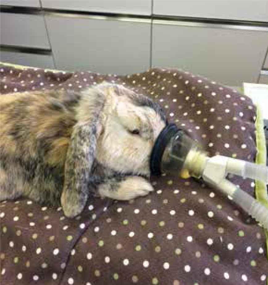
Before induction of anaesthesia, the rabbit should receive 100% oxygen via face mask (Figure 1), oxygen chamber or by allowing oxygen to flow by the nares. The aim of pre-oxygenation is to replace nitrogen in the functional residual lung capacity, with oxygen. This has a significant positive impact on body oxygen stores and, therefore, tolerance to apnoea is increased (Sirian and Wills, 2009). Excessive pre-oxygenation using a mask over the face can cause increased stress and anxiety, and a human study revealed a marked increase in oxygenation after only four breaths of 100% oxygen (Eatwell, 2014). Historically, a common technique used to induce anaesthesia in rabbits was to use gaseous volatile agents, such as isoflurane. The use of a single anaesthetising agent is by no means safer, provides no analgesia, is extremely stressful and can induce a breath holding response resulting in hypoxia. This approach has many flaws and unfortunately for the rabbit has probably contributed to the higher anaesthetic mortality rates seen in this species (Flecknell, 2006). Some gaseous agents (e.g isoflurane) are also irritant to the airways and eyes (Flaherty, 2009), and the noxious stimuli causes stress and anxiety immediately prior to anaesthesia, which can lead to a disturbed recovery in the author's opinion.
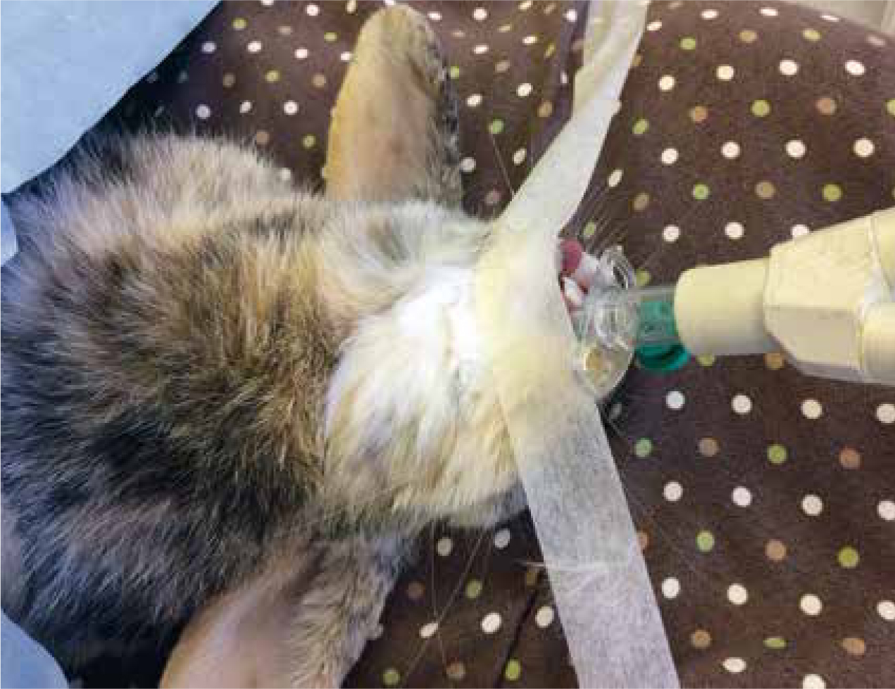
Injectable anaesthesia is preferred and can usually be administered intravenously or intramuscularly depending on the induction agent. Intravenous induction using agents such as propofol carries a higher risk of transient post-induction apnoea (Bourne, 2017). Therefore, the administrator should be confident that intubation can be achieved quickly so that intermittent positive-pressure ventilation (IPPV) can be commenced if required. Alternatively, supraglottic airway devices (V-gel®, Docsinnovent) are extremely useful (Figure 2). These devices can normally be placed more quickly than endotracheal tubes, and they provide protection of the airway and a means of delivering oxygen/volatile agent with less risk of gaseous leakage versus a face mask (Richardson, 2015). To ensure correct placement, the author recommends the use of capnography, however where this is unavailable, placement can be checked by close monitoring of vital signs, mucous membrane colour and once connected to the circuit, the reservoir bag can be observed moving. At the very minimum, a tight fitting oxygen mask should be placed over the nares and oxygen/volatile agent can be delivered.
Anaesthetic monitoring
The monitoring of anaesthesia often falls to the registered veterinary nurse (RVN) and as rabbit anaesthesia differs from that of cats and dogs, the RVN should familiarise themselves with the range of normal vital parameters, which can vary between different breeds. Due to the large relative size of the abdominal cavity compared with the thoracic cavity, abdominal contents can put pressure on the diaphragm resulting in increased respiratory rate and effort. Therefore, the thorax should always be elevated when a rabbit is anaesthetised. Once the rabbit is physically stabilised, anaesthetic monitors can be utilised. These may include capnographs, pulse oximeters, electrocardiograms, Doppler flow meters, however the availability of these pieces of equipment will vary from practice to practice so will not be discussed here.
To monitor the respiratory system without the use of specialised equipment, the anaesthetist should take note of the depth, rate and pattern of respiration. This can be achieved by watching the thorax rise and fall, auscultating the thorax with a paediatric stethoscope and/or monitoring the reservoir bag of the anaesthetic circuit moving synchronous to the rabbit's breaths. Very small rabbits may only cause subtle movements to the reservoir bag. Changes to respiration may occur throughout an anaesthetic and the anaesthetist should recognise potential cause for concern at the earliest opportunity (see Table 2).
| Change to respiration | Possible cause |
|---|---|
| Tachypnoea and/or increased depth | Noxious stimulation from inadequate analgesia, inadequate depth of anaesthesia, low tidal volume, hypercapnia, the use of a breathing system unsuitable for the species, excess dead space, one lung intubation, hyperthermia, hypotension |
| Bradypnoea and/or reduced depth | Certain medications, e.g opioids, too deep an anaesthetic plane, hypothermia, hypocapnia (this can occur when intermittent positive-pressure ventilation (IPPV) has been ‘over-used’, increased vagal tone |
| Apnoea | Intravenous (IV) induction, too deep level of anaesthesia, overdose of medication, breath holding, impending cardiac arrest |
Monitoring the heart rate can be achieved by auscultating over the cranial thorax with a paediatric stethoscope or by palpating the heart or pulse. The use of oesophageal stethoscopes can also be useful in larger rabbits. Small changes in heart rate may be more difficult to detect in rabbits than in dogs and cats due to the high resting heart rate of some breeds (see Table 3).
| Change to heart rate | Possible cause |
|---|---|
| Tachycardia | Hypercapnia, hypotension, cardiac arrhythmias, pain, too light a plane of anaesthesia, certain medications (ketamine), respiratory distress, hypovolaemia, anaphylaxis |
| Bradycardia | Certain medications (e.g. medetomidine), vagal nerve stimulation, severe hypoglycaemia, hypoxia, hypertension, hypothermia, metabolic acidosis, too deep an anaesthetic plane |
| Cardiac arrest | Too deep a plane of anaesthesia (apnoea usually observed first), overdose of medication, underlying cardiac disease, severe hypovolaemia, severe hypoxiad |
Along with monitoring the heart rate, pulse rate and quality should be assessed. Palpation of peripheral pulses is simple and should always feel synchronous to the rabbit's heart beat. Pulse deficits are an indication of cardiovascular compromise. There are limitations to using pulse palpation to assume the patient's overall cardiovascular health, as pulse quality is related to the difference between diastolic and systolic pressures, not the pressure itself. Therefore, very poor systemic blood pressure may present with strong pulses in certain scenarios (Baetge, 2007). The auricular artery located medially on the dorsal aspect of the ear pinna can be used in larger breeds of rabbit. The femoral artery can be used in all breeds and digital pulses usually only in very large rabbits.
Capillary refill time can be assessed in the way same as with cats and dogs, by blanching the tissues of the mucous membranes and waiting for the colour to return (normally within 2 seconds). Prolonged capillary refill time can indicate poor perfusion of tissues, however this is a limited indicator, but it is useful when taking into account the patient's current clinical signs/demeanour along with blood gas analysis/blood pressure/capnography (where appropriate) (Murrell and Ford-Fennah, 2011). The colour of the mucous membranes may be more reliable indicators of health status however as it is subjective, different assessors may come to different conclusions unless changes to mucous membrane colour are obvious or severe such as with cyanosis.
Reflexes
Reflexes are used to assess patient consciousness and central nervous system health. The most commonly used in small animal anaesthesia are listed below and these can be interpreted in the rabbit:
Anaesthetic emergencies — what to do
Apnoea The underlying cause should be established and this should include making sure the airway is patent. If the rabbit is not intubated this needs to be achieved to provide the best chance of resuscitation by performing IPPV. If intubation is not possible the V-gel®, or a tight fitting mask can be used to perform IPPV, but potential complications may arise if the stomach also inflates with oxygen as this can in itself cause respiratory compromise (Royal (Dick) School of Veterinary Studies (RDSVS), 2012). If the procedure allows, all anaesthetic gases should be turned off, 100% oxygen delivered and other vital signs assessed. If possible, reversible drugs (e.g medetomidine) should be reversed to provide the best chance of spontaneous respiration returning (RDSVS, 2012).
A more crude method of respiratory resuscitation is to rock the rabbit back and forth, moving the abdominal contents to and from the diaphragm. This method is only suitable for small herbivores as they rely on flattening of the diaphragm for breathing rather than outward movement of the ribcage like dogs and cats (Flecknell, 2006). If tracheal obstruction is suspected, a tracheostomy can be performed in the same way as for a cat or dog. It is important to note, some female rabbits may possess a large dewlap, which may hinder this procedure (Girling, 2013).
Cardiac arrest
Again the likely underlying cause should be established and all drugs that can be reversed should be. Anaesthetic gases should be turned off and 100% oxygen administered. Direct cardiac massage by compressing over the heart should be employed and, because of the rabbit's rapid resting heart rate, 100 beats per minute need to be achieved to maximise cardiac output (Girling and Fraser, 2009). Adrenaline at 0.2–1.0 mg/kg intravenously or via the trachea can be administered, this is particularly useful in fine ventricular fibrillation as adrenaline can be used to convert the electrical activity to coarse ventricular fibrillation, which is easier to convert to normal rhythms by cardiac massage (Girling and Fraser, 2009). It is important to note that up to 60% of domestic rabbits possess serum atropinesterases making atropine a relatively ineffective drug in the rabbit (Huynh et al, 2016). To support cardiovascular function, intravenous fluid therapy can be employed where the rabbit was dehydrated prior to arrest or is hypovolaemic. However, fluids should be avoided post cardiac resuscitation where there is no dehydration as fluids may decrease myocardial perfusion pressures and diminish nutrient delivery through coronary and cerebral vasculature (Girling and Fraser, 2009).
Recovery
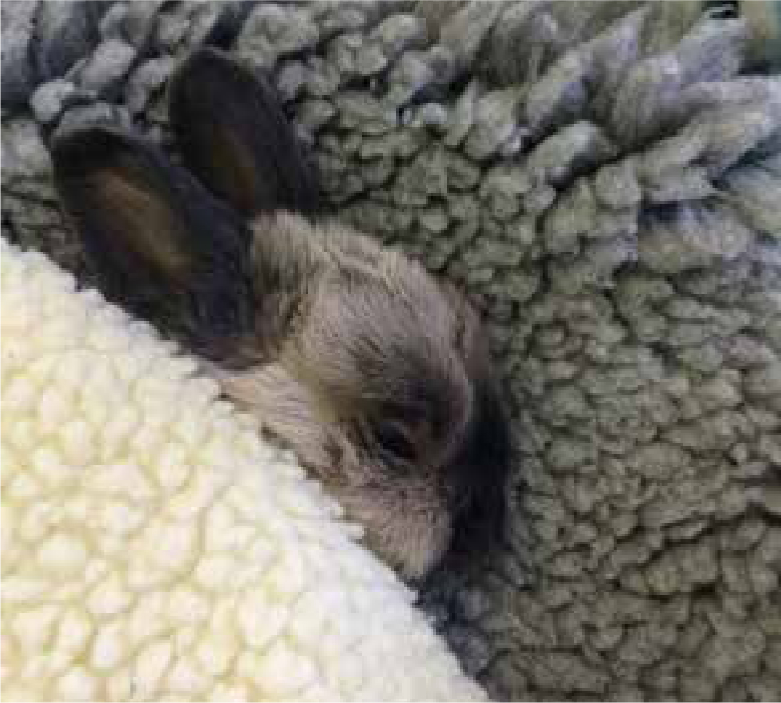
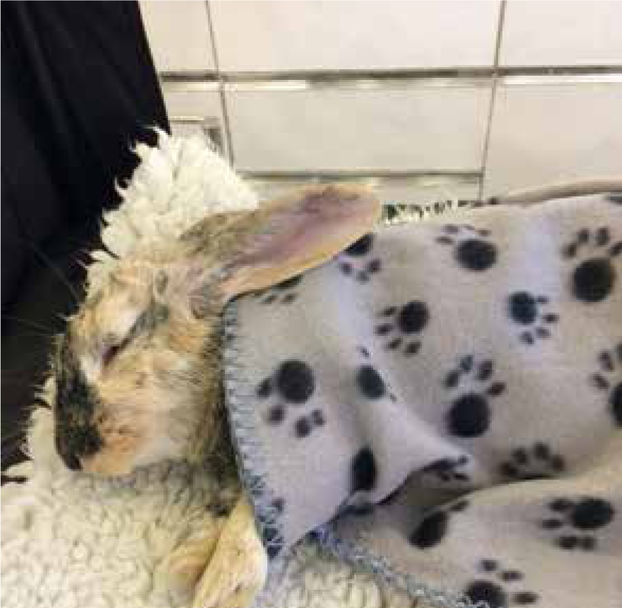
Recovery from anaesthesia should always occur away from predator species and environmental temperatures should be between 17–22°C (Mancinelli, 2015) (Figure 3 and 4). One of the main concerns post-operatively is anorexia which can lead to gastrointestinal stasis in the rabbit. Anorexia may be the result of post-operative pain and stress, however it should never be ‘expected’ and the underlying cause should be established and attempts made to resolve it. Deterioration after general anaesthesia warrants supportive care such as intravenous fluids and syringe feeding with a recovery diet, and diagnostic imaging/blood work where indicated. The rabbit should be offered familiar/palatable foods during recovery such as fresh herbs, which have a strong scent or the rabbit's usual food can be offered. Adequate analgesia is imperative to the rabbit's recovery and will improve the likelihood of the rabbit eating. Nurses should familiarise themselves with the subtle signs of pain in rabbits and a useful tool is the rabbit grimace scale. The key can be found at https://www.nc3rs.org.uk/rabbit-grimace-scale. The limitations of this scale should be acknowledged and other markers of pain in rabbits such as teeth grinding, hunched posture or abnormal behaviour should also be taken into account. The rabbit should be housed in a quiet, dark room and observation should not be invasive unless necessary. If the rabbit has a close bonded companion, it may be beneficial to the recovering rabbit to be in their presence as it will reduce stress and encourage appetite.
Conclusion
Although ultimate responsibility of the patient lies with the veterinary surgeon, the RVN plays an important role in the care of the rabbit through all steps of anaesthesia and a well prepared and knowledgeable RVN can mean the difference between life and death for the rabbit. As veterinary medicine advances, all members of the veterinary team should familiarise themselves with the current thought and best practice for their rabbit patients.