
Recumbency, normally due to processes such as pulmonary disease or thoracic wall disease, rib fractures, ventilated patients, trauma or surgical intervention, does not allow the patient to carry out normal daily activities. One of the main concerns in recumbent patients is respiratory function, the ability to maintain a healthy airway and provide good oxygenation while recovering. With limited research in the veterinary field involving recumbency, this article reviews both veterinary and human literature.
Gaseous exchange
The primary function and ability of the lung is to enable gaseous exchange, allowing air distribution within the lobes to match the perfusion of the body's tissues (Schilero et al, 2009). Altering body position of challenged patients with disease processes, alters biomechanical homeostasis (Vollman, 2004), subsequently requiring nursing interventions (NIs). Turning patients into variable positions and providing physical therapies are advised (Frownfelter and Dean, 2012). Clini and Ambrosino (2005) stated that early physiotherapy in human patients is cost effective, improves residual function, reduces the need for future admissions and improves quality of life. Physiotherapy is believed to restore respiratory function, ventilatory dependence, and reduce the risk of bed-rest complications. Therapy has been documented for strengthening the chest, lungs and stomach, initially by using deep breathing and postural exercises (Garrod and Lasserson, 2007). Change in posture is believed to decrease the incidence of further diseases developing, such as pneumonia, consolidation of lung lobes and atelectasis (Cook, 2003). Turning is a recognised technique, but current research into additional therapies has been controversial and spartan particularly in veterinary medicine, and postural exercises are difficult to replicate in veterinary patients.
Spinal cord injuries and neuromuscular disease processes commonly predispose patients to a reduced ability to cough effectively (Schilero et al, 2009), which can consequently cause mucus plug formation, sputum retention (Whitney et al, 2002), and predispose patients to infections, absorption ateclectasis and reduction in lung function (Guimarães and Zin, 2008; Treacy et al 2011). In addition, patients with spinal injuries are also recumbent or semi-recumbent, which increases their risk of developing secondary problems or of disease processes, such as pressure sores, occurring (Cook, 2003). Patients require the ability to establish an effective cough to clear fluid, and three determinants are fundamental.
An adequate vital capacity, the total amount of air that can be expelled after respiration (Aspinall and Capello, 2007), is required to establish an effective cough and to mobilise secretions from low lung volumes. A non-recumbent patient will use 85–90% of their inspiratory reserve volume for coughing. Expiratory muscle strength is required to clear secretions, along with intact bulbar function (IBF). IBF, in which the glottis closes while pressure builds up by contraction of the expiratory muscles, leads to the expiration of air (Whitney et al, 2002). In normal response to respiratory secretions, upwards transport is mediated by a mucocilliary escalator, the rate of which is determined by secretion rate, viscoelasticity of mucus and synchrony of cilia within the respiratory tract. Mucus is propelled by beating ciliary motions passing over the lower serous layer (Treacy et al, 2011). The parasympathetic nervous system enhances secretions; goblet cells increase amounts of mucus alongside the submucosal glands, initiating bronchoconstriction and subsequently producing a cough (Foster, 2002). During sleep it has been estimated by Hess (2005) that in humans body position will change every 12 minutes, which aids clearance in the normal patient.
When lower respiratory tract infection is present, volume and persistency of secretions can overwork cilia and reduce efficacy of the cough reflex, and induce incapability to clear secretions. In turn there is an inadequate perfusion of body tissues due to reduced gaseous exchange, which can instigate respiratory acidosis. If the chest wall is also non-compliant or other factors are present, such as, obesity, viscous secretions, abdominal surgery, the coughing reflex can also be duly compromised (Whitney et al, 2002). Patients exhibiting pneumonia reveal on radiography alveolar capacities and consolidated regions in the cranio-ventral and middle lung lobes (Eom et al, 2006).
Patient posture
There are many opposing views for correct positioning of the compromised patient. Vollman (2004) opposes upright positioning and stated that patients with large abdomens may struggle to inflate lungs effectively due to the abdominal contents exerting pressure onto the diaphragm, preventing even air distribution. Hess (2005) challenged this and commented that in supine there is a reduction in residual capacity due to alveolar closure in the lung area most compromised. There is a lack of specific literature on the effects of an animal's abdomen in supine position, but one would assume a large canine abdomen would cause more deleterious effects than a small abdomen indicating that more frequent posture changes need to be employed in veterinary care, as animals are normally maintained in the prone position if conscious. In the human patient, Vollman (2004) stated that a 45 degree angle assists lung expansion and capacity by relieving pressure onto the diaphragm, which could be adopted in the veterinary patient by artificially raising the chest. NIs such as using layered bedding or wedges to artificially raise the chest could be employed. Patient compliance could be a problem however as veterinary patients cannot understand the aims and may not, therefore, adhere to such requests.
March (2005) and Coyer et al (2007) have stated that prone positioning and continuous lateral rotation technique (CLRT) is advantageous in humans for correcting a mismatch of ventilation and tissue perfusion, however Collard (2003) previously found that there was no evidence for an optimal length of time to use a CRLT bed. Orpet and Welsh (2011) have advised that turning of veterinary patients should occur regularly every 3–4 hours, but Calvo (2012) stated that turning should occur every 4–6 hours with sternal recumbency being the position used most often, together with the addition of coupage at every turn. Park et al (2012) also stated that turning of veterinary patients should occur at least every 4 hours, preventing congestion and atelectasis suggesting that more frequent turning is required as Orpet and Welsh (2011) originally proposed. In human literature Coyer et al (2007) previously further shortened this time to every 2–4 hours in people, which provides personnel with a difficult decision for the ideal frequency of posture change.
Thomas et al (2006) documented that the altering the position of critically ill human patients has a critical effect on oxygenation and is very important in the reduction of cases of nosocomial pneumonia. Adopting the prone or semi-recumbent position may help to improve oxygenation if this could be provided to veterinary patients. The survey of 71 human physiotherapists and nurses from 38 Australian intensive care units reported that personnel were reluctant to alter patient posture as they were concerned about adverse effects such as desaturation of oxygen levels and interference with other equipment such as jugular catheters. The survey established that personnel would take extra care with patients that were on dialysis or had jugular or femoral catheters, and this may be mirrored in veterinary patients where gentle handling of these intensive care patients should be a priority. The study highlighted that there was an increase in risk of pressure sores developing if patients were static for more than 6 hours. The survey carried out revealed that 2 hourly repositioning is often not applied, and reasons have been documented as environmental, only achieving re-positioning every 4 hours.
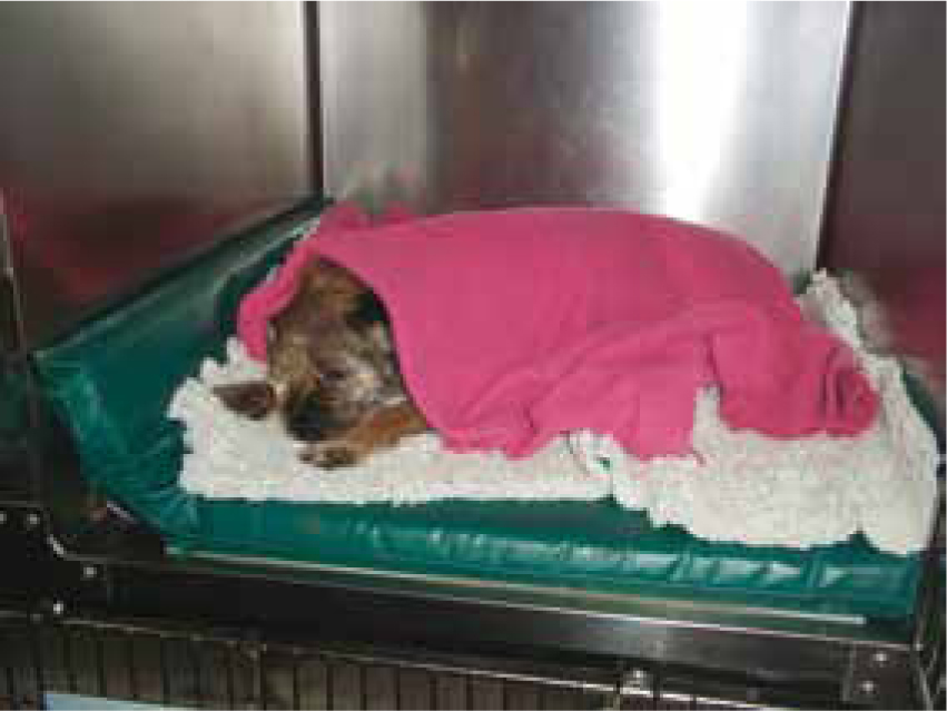
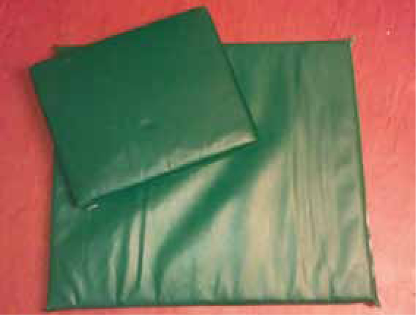
Ulcers, along with limb contraction, urine scalding and faecal soiling are all subsequent problems of recumbency in animals (Park et al, 2012), and in veterinary practice mattresses are often used to help reduce the incidence (Thompson, 2009) (Figures 1 and 2).
Investigation into supine position in recumbent animals is not available, so its use cannot be evaluated, and should be an area for further investigation. McMillan et al (2009) documented recumbent canine patients using the prone position and noted that arterial partial pressures of oxygen are significantly improved than when in lateral recumbency due to increased oxygen uptake from the lungs, rather than an increase in alveolar ventilation. The study looked at 21 conscious dogs and used arterial gas measurements to assess hypoxia. Measurements were recorded every 15 minutes to randomly assigned lateral or sternal participants. Each patient was turned every 15 minutes from sternal to lateral and vice versa. The study therefore suggests patients that are susceptible to hypoxia may subsequently benefit from sternal recumbency. Controversially, Thomas et al (2007) had previously studied oxygenation, haemodynamics and respiratory mechanics in human patients using the 90° lateral positioning and supine. The study revealed that in a range of patients with pulmonary pathology, no lung pathology, bilateral and unilateral pathology, overall there was not any change in the variables, such as arterial blood gasses, despite rotating patients' position from supine to lateral and return to supine. In 8% of the 34 subjects between 30 minutes and 2 hours there were minor adverse effects such as slight increase in cardiac output. Patients were turned every 30 minutes to 2 hours, and the study concluded that using lateral positioning in human ventilated patients who are not haemodynamically unstable is not detrimental.
Chest physiotherapy
In addition to body positioning, to aid the movement of secretions upwardly, physiotherapy methods can be employed, and are one of the most utilised techniques in human intensive care units (Clini and Ambrosino, 2005).
Percussion is striking the body, using alternate wrists, kept flexible, so movements are springy and invigorating (Sharp, 2008). The effectiveness of percussion has not been investigated fully in non-intubated patients (Clini and Ambrosino, 2005), but in a human study Rogers and Doull (2005) suggested that during deep breaths it is advantageous as intrapleural pressure changes allow loosening of mucus, and movement of secretions into the lumen to allow it to be expelled. They documented that percussion should be concentrated on the area of concern to maximise potential benefit.
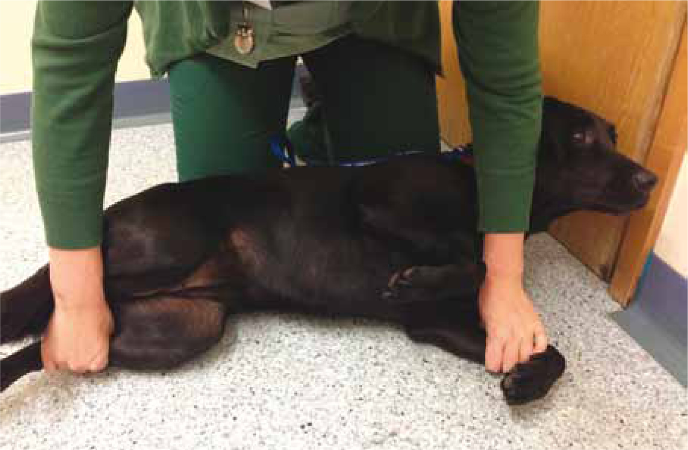
Coupage involves cupping the hands, and pronating the forearms gently to prevent lung damage (Orpet and Welsh, 2011). The elbows should be bent with alternate flexion and extension of the wrists, coming in contact with the chest wall. This resonates as a deep clapping sound over the ribs (Sharp, 2008), clearing secretions from the airway termed hypostatic congestion (Orpet and Welsh, 2011). Movements across the chest should be working caudal to cranial and always both sides of the thorax. Performance should be three to four times daily and sternal recumbency should be encouraged by using positioning aids such as sandbags and wedges (Orpet and Welsh, 2011).
Rhythmic shaking can be performed by holding the body part and moving side to side and up and down (Sharp, 2008), and in conjunction with vibrations can be used to convey a fine tremor through the hands and fingertips for a few seconds. Vibrations relax the nervous system, and help to reduce adhesions (Sharp, 2008). The technique increases the expiratory flow rate in central and peripheral airways (Wong et al, 2003). The three techniques in conjunction produce a positive mechanical forced energy to help dislodge mucus, creating a mist through the airways and aiding removal (Wong et al, 2003). These skills are all potentially able to be carried out by an RVN.
Wong et al (2003) conducted a study for human research using anaesthetised sheep to investigate physical therapy techniques and to establish if haemodynamic changes occur when techniques are used. Previous evidence prior to the trial tended to suggest cardiac arrhythmias may occur after coupage and with additional vibration would also increase heart rate, blood pressures, cardiac output and alter the volumes of anaesthesia or sedation required to establish unconsciousness. The animal model was used to enable invasive haemodynamic monitoring to occur. The study concluded that there were no adverse effects on haemodynamics of the sheep used within the study except for increased tidal volumes, which therefore implies that chest physiotherapy carried out by trained staff could be effective if carried out appropriately and therefore not detrimental to health, but further investigation into other species would need to occur. What this study does fail to detail is to take into account if patients are subject to any pain, coagulopathies or any recent surgery to their chest which could impact on whether physical therapies are detrimental or not.
A more recent study investigating chest wall vibrations in humans has been carried out by Shannon et al (2009) investigating repeatability of chest vibration technique between different physiotherapists various intervals. There were no significant differences in the force, duration, frequency of oscillation or amplitude of oscillation, but there were wide variations in vibrations between different physiotherapists. The study used a force mat placed onto the patient's chest wall and measured the forces exerted onto it. The study concluded that the great differences seen between staff may be due to the preferred posture of the physiotherapist while performing the procedure, wrist extension and hand size of the individual, and while all physiotherapists followed their undergraduate training, which taught them this technique, training varies. The study was limited to healthy subjects and eight physiotherapists, so a larger sample size may help to clarify the results. The force mat was developed by one of the authors, so conducting a similar study with a different research group that is not influenced by this individual would eliminate any bias from the study, despite the paper stating there were no conflicts of interest. This study could be repeated on veterinary subjects if the force pads are able to be secured easily, and could help veterinary physiotherapists and RVNs to apply effective physiotherapy to patients, improving repeatability of treatments between personnel.
Rogers and Doull (2005) suggested that physical activity providing systemic exercise in cystic fibrosis patients helps to mobilise secretions. This theory is supported by Coyer et al (2007) who have stated that limb exercising in all patients, both via passive and active physiotherapy, can improve soft tissue and muscle strength and help to restore the normal fluid distribution within the body. In recumbent veterinary patients mobilisation is achieved through sling and harness aided assistance (Thompson, 2009). If hypostatic pneumonia is suspected, radiography of the chest should be performed and thoracic auscultation should occur at least once daily. Keeping the patient hydrated will also help secretions maintain fluidity and stop consolidation forming, and therefore decreasing the risk of pulmonary infections (Carver,2012).
Conclusion
Recumbency in human medicine has not been investigated fully enough to enable an ideal protocol for maintenance of the health of recumbent patients, and this is mirrored in veterinary practice. It is, however, demonstrated through the literature, that alterations to a patient's position may be decided on, on a case by case basis, and therefore there may not be a specific protocol which can be applied to all patients. Physical therapy is suffering from a lack of scientific research and evidence, and work into this area of rehabilitation could standardise the type, rate, force and frequency of these techniques, or allow personnel to make adjustments to protocols on an individual basis. Physical therapy has no confirmed protocol in either human or veterinary patients, and this also needs to be addressed to enable RVNs to provide a comprehensive care plan to aid recovery from recumbency. RVNs under the direction of the veterinary surgeon could be more confident in providing a patient with the correct physical therapy, establishing a common rate, frequency and pressure to be employed. As evidence suggests, physical therapy can be adjusted on an individual case basis and coupled with posture changes, observation of clinical signs could enable recumbent patient care to be more clinically led by the RVN in the future, as the profession now holds its own accountability and disciplinary system.