
It is important to note that it is the horse's natural fight or flight instincts that can cause many of the wounds seen in veterinary practice, whether that be a barbed wire wound from a horse running through a fence or a wound on the distal limb caused by the horse kicking an object. Equine limb wounds often include an injury to the bone as this is relatively unprotected and therefore exposed to injury (Philips, 1995). Most traumatic wounds are either contaminated or dirty wounds (Southwood, 2008), which will increase the incidence of infection and this should be noted when dealing with such lesions. In addition, it is important to understand equine wound healing in order to nurse these patients successfully. It is also important to note the differences in wound healing between ponies and horses and how this may affect the final outcome of similar wounds in each species. Some tips are included on dressing the equine limb and these are based on the author's personal experience as an equine veterinary nurse. This article has concentrated on the ability of nurses to improve the outcome of the patient through thoughtful, critical and holistic dressing of the limb.
Wound healing
A wound is defined as an injury where there is a forcible break in the soft tissues, including open wounds and closed wounds where there is damage below the surface (Ousten, 2011).
The healing process takes place in three stages (Table 1). The length of each stage will depend on whether wound closure is a viable option; for example a wound healing via primary intention will take less time to resolve than a wound allowed to heal via secondary intention or after a delayed primary closure (Ousten, 2011).
| Stage of healing | When it occurs | What occurs |
|---|---|---|
| Haemorrhage, inflammation and debridement | In the first few hours after insult | Haemorrhage |
| Repair/granulation | 12 hours after insult | New cells produced over the surface of the wound (2 mm/day in a moist wound) |
| Maturation/scar formation | From insult until 2 years after injury | Open wounds can take weeks to months to heal as these tend to heal via second intention Surgically closed wounds, i.e. those that have undergone primary closure, heal via first intention which happens quicker with minimal granulation tissue Tissue regains strength for up to 2 years, only 80% after the first year |
Wound closure occurs via:
There are many factors that delay wound healing (Table 2), these factors need to be taken into account by all involved in wound management in order to achieve a successful outcome.
| Factor | Reason for delay |
|---|---|
| Infection | Good hygiene and cleaning of the wound initially can reduce the incidence of further infection. If a wound becomes infected the patient may show signs of systemic infection such as anorexia and pyrexia, the wound itself will look red, swollen, discharge may be seen with a odorous smell and the patient will be uncomfortable at the site |
| Movement | Movement at the site or of attached tissues delays healing, excessive mobility disrupts capillary beds and can cause a chronic inflammatory status within the wound. Lack of all movement can also be counter productive as it causes poor arrangement of the collagen fibres causing weaker healing |
| Foreign body | The most common reason for non-healing wounds; can include sand/grit particles, wood/plant matter, necrotic tissue, suture material and even hair from clipping around the wound |
| Necrotic tissue | Necrotic tissue retards healing. Tendon and bone are slow to exhibit non-viability and so careful debridement prior to closure is beneficial |
| Altered pH | Certain bacteria can alter the pH of the wound; the site should be at normal physiological pH or slightly acidic |
| Blood supply | Poor blood supply for example due to disruption of a blood vessel can cause reduced oxygen supply to healing tissues. Some areas such as the dorsal hock are thought to have a naturally poorer blood supply |
| Impaired oxygen supply | Lowered systemic oxygen concentration due to decreased blood flow or anaemia slows wound healing and increases inflammation |
| Poor nutrition or health | Older horses heal slower than the young; hypoalbuminaemia suppresses healing and encourages inflammation |
| Local factors | Wounds with a pouch of skin or excessive dead space can fail to heal. Accumulated fluids can be an ideal medium for bacteria. Self trauma localised to the wound will obviously slow or prevent healing |
| Iatrogenic factors | Incisions, swabbing, use of forceps and retractors can all cause localised injury to tissues, sutures can act as foreign bodies, excessive pressure can compromise blood supply and use of corticosteroids suppresses wound contraction |
| Genetic factors | Large horses heal less well than ponies, horses with congenitally weakened skin are more easily traumatised |
Factors that concern the veterinary nurse
The factors highlighted in Table 2 are those that nurses should be aware of and that can be minimised and acted on to achieve the best outcome. It will never be possible to remove every factor that delays wound healing but nurses should do everything they can to aid healing by reducing the effect of these factors. A wound involving a synovial structure that has not been identified may also affect wound healing and also the overall prognosis of the patient; it has been suggested that 17% of heel lacerations involve such a structure (Southwood, 2008), so initial examination by the clinician is very important.
Good hygiene
In order to ensure infection is not introduced into the wound good hygiene is of paramount importance. Gloves should always be worn when examining the patient and changed for each patient in order to help reduce infection, and this must be enforced for all those examining the wound. Passing an infection such as MRSA to a patient is an all too real possibility; it is known that humans contribute to the spread of infection (van Duijkeren et al, 2010).
Cleaning of the wound
A heavily contaminated wound should be flushed thoroughly using sterile saline, a syringe and an 18-gauge needle this combination produces the correct pressure for flushing contaminated wounds (Ousten, 2011). Disinfectant can be used but with caution: if it is too concentrated it becomes toxic to fibroblasts and other cells involved in wound healing (Phillips, 1995). It is advised to use disinfectants in concentrations that maintain antibacterial efficacy while minimising the risk of toxicity to cells, Phillips (1995) suggests 0.1% iodine and 0.05% chlorohexidine as adequate.
Hair from clipping around the wound
It is a very simple task to protect the wound before clipping to prevent further contamination; even if the wound is already contaminated it is not beneficial to add more contaminants. The use of hydrogel Intrasite Gel (Smith & Nephew) (Figure 1) or KY jelly applied into the wound will prevent hair from entering the wound during the clipping process. The contents of an unopened tube of both products is sterile and therefore will not introduce further contamination. However it is important to clean this gel away thoroughly before further investigation by the veterinary surgeon.
Self trauma
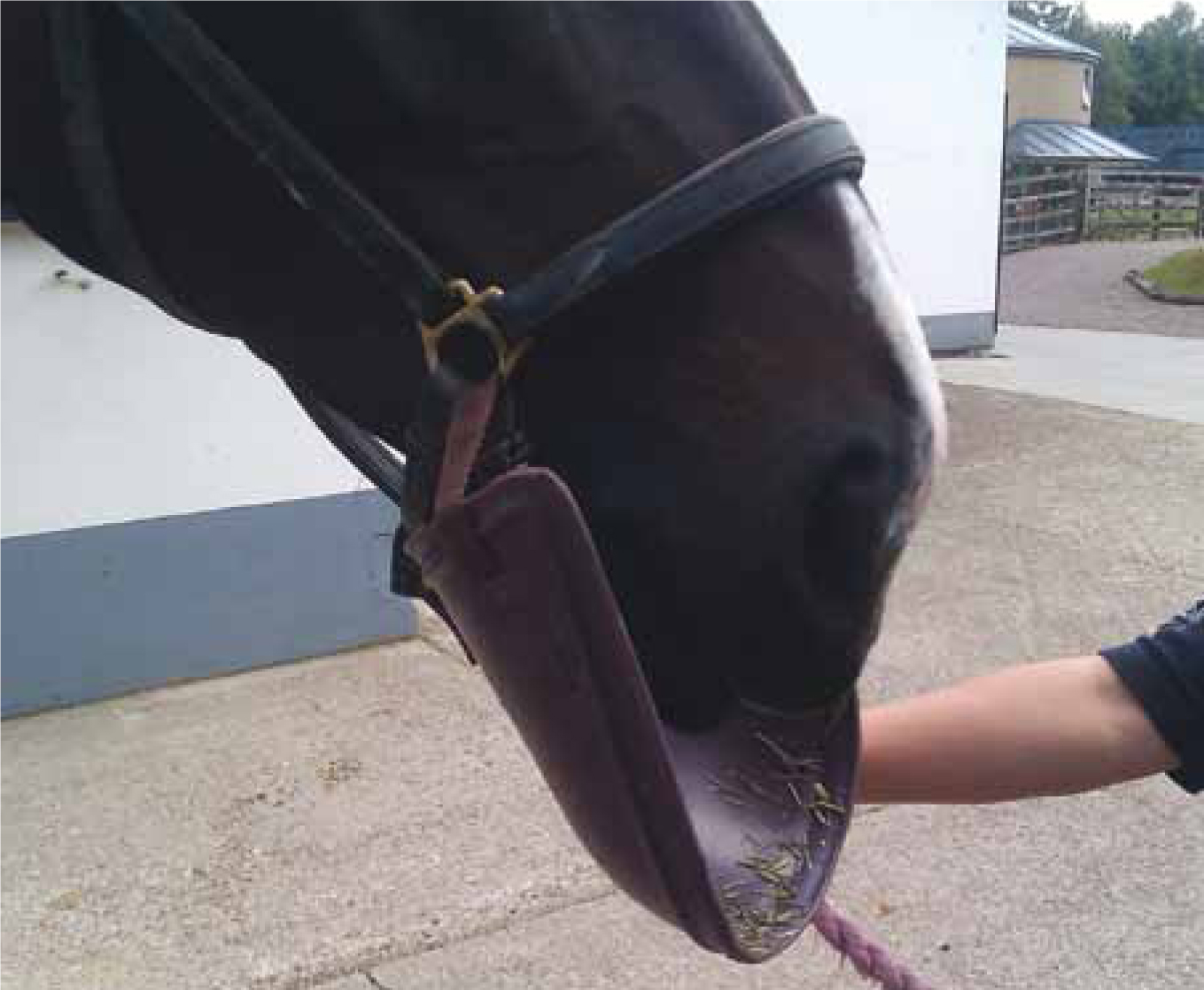
Self trauma may be more common in wounds to the body than the limbs, and an element of self trauma may still be evident when wounds are dressed. The temperament of some horses means they are not going to tolerate a dressing well. Youngstock in particular tend to struggle, they can be found pulling chunks out of their dressings while lying down (personal experience). Cribbox (hydrophane) can be used to aid in discouraging them from destroying the dressing; it is soul destroying to spend an hour placing a dressing only for the horse to destroy it in minutes. Distraction can also be used such as walking the horse when the dressing is new on (dependent on the wound or dressing type), snack balls, company (human or horse), tying them up for a period after the initial application and the use of bibs or a neck cradle (Figure 2). If all else fails, sedation, usually acepromazine so feeding can still continue, may play a part in discouraging this destructive behaviour until the new dressing has been ‘worn in’.
An underestimated area with respect to self trauma is consistency in the person placing the dressing. Everyone does dressings differently so if a patient has been unsettled in one person's dressing but much better with another person's then continuing with the preferred person will help to prevent stress in the patient and a large bill for the client.
Excessive pressure can compromise blood supply
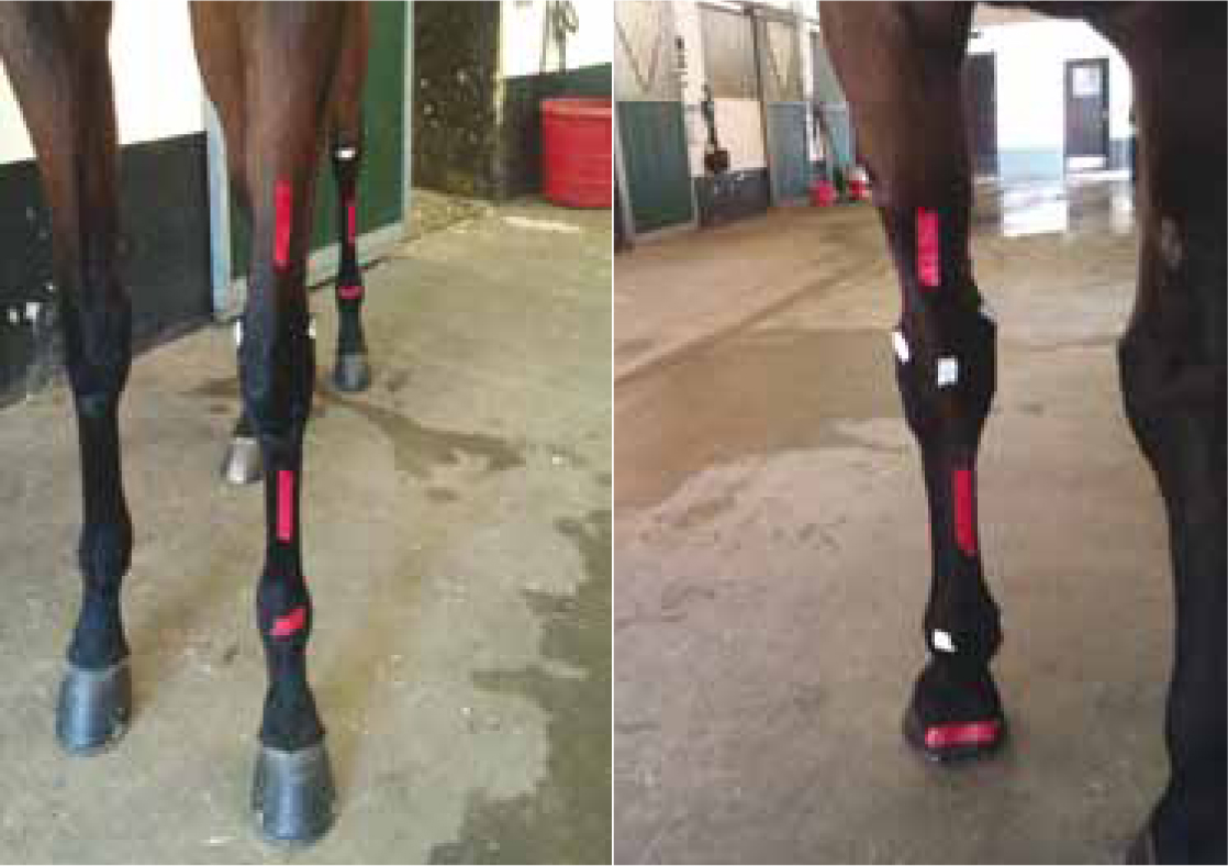
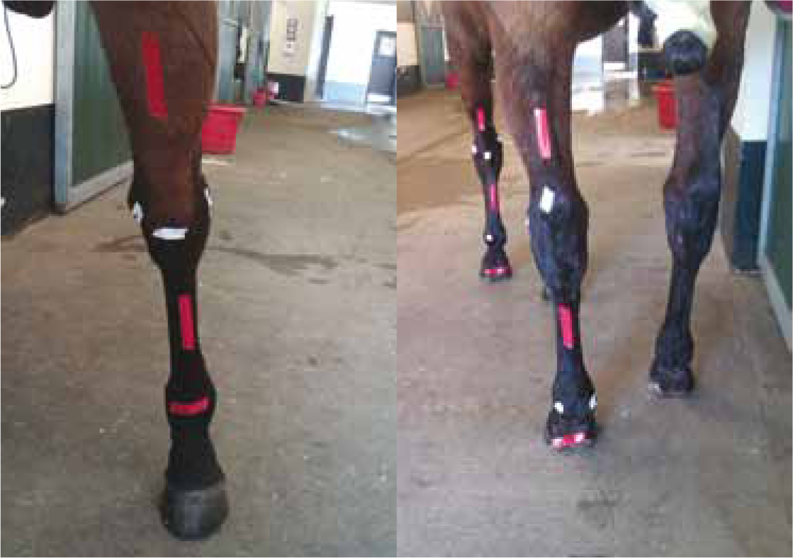
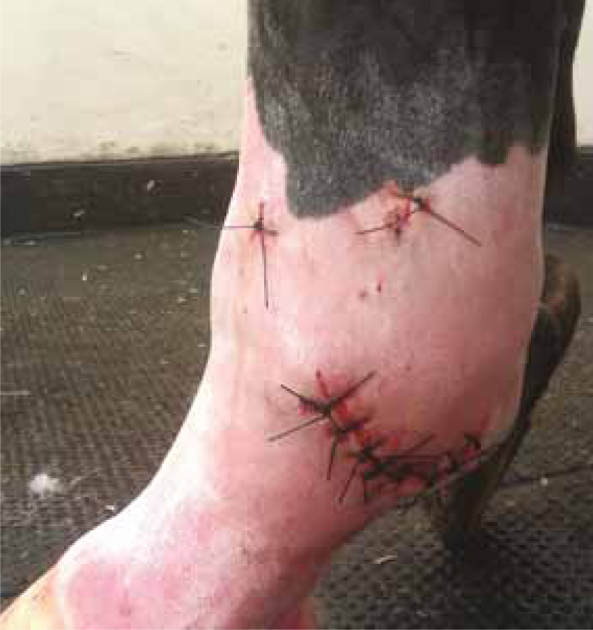
Excessive pressure can occur throughout the dressed area or in specific areas, the most common places that suffer from uneven pressure when dressed are shown in Figures 3 and 4. The red markers on the horses' legs show soft tissue areas that are affected by inappropriate pressure and the white marks are bony prominences that are commonly affected (personal experience). The areas the horse develops pressure sores depends on the conformation of the limb, some horses have very straight legs whereas others have relatively large, prominent joints. Veterinary nurses should be aware of the conformation of the patient's limbs and dress the limb in a holistic fashion; that is dress each leg as an individual, no two legs will be dressed the same. There are ways of minimising the damage for example: if a leg has a particularly prominent medial malleolus or if a horse has a pressure sore on its fetlock on admission, then additional padding can be added. If a pressure sore is evident on a dressing change it is important to adjust the dressing accordingly, not just replace the dressing as it was and hope for the best. Care should be taken not to make a dressing too tight as this could cause a horse to self traumatise — check the tension of the dressing numerous times throughout placement not just at the end.
Movement
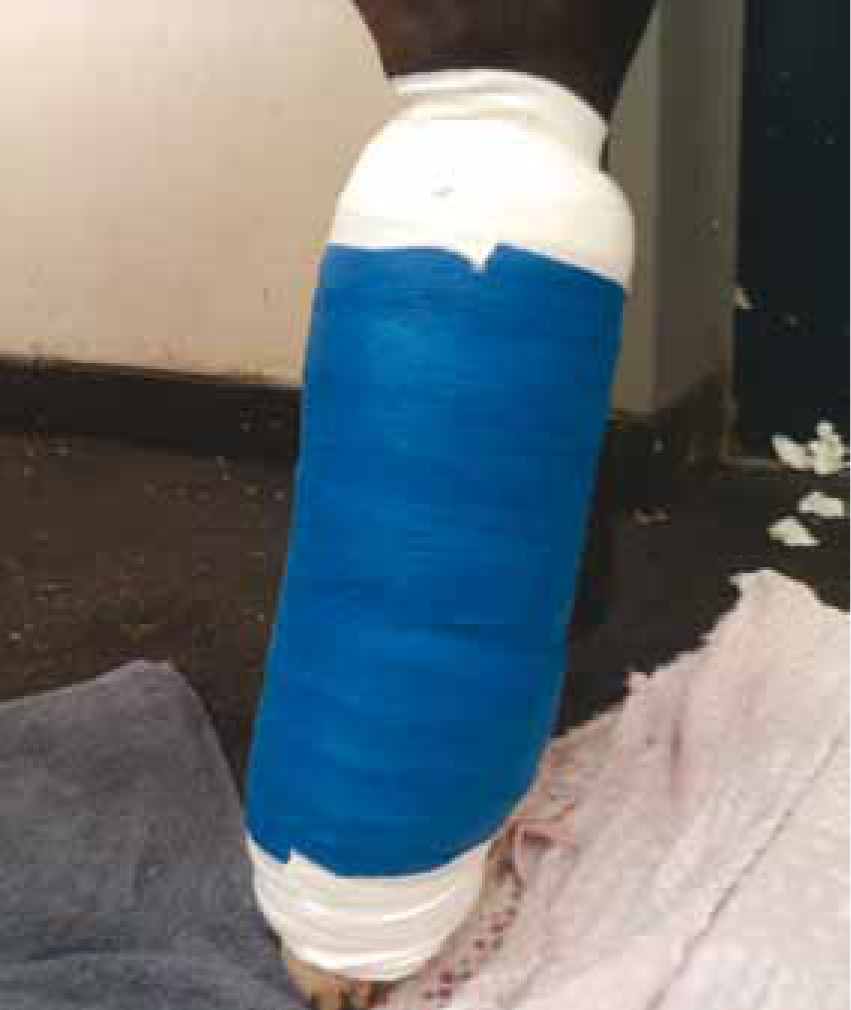
Movement is a problem in horses — it is difficult to get a horse to stay still, and any movement can result in the dressing sliping or the wound reopening — and dressings play a large part in minimising this movement. If the fetlock needs to have minimal flexion then the dressing needs to aim to stop this movement, if there is a dorsal hock wound then the aim is to reduce flexion of the hock. Ultimately the size of the dressing is down to the veterinary surgeon; the more layers they ask for then the more they wish to stop movement, i.e. a four layer full limb dressing will ensure the hock does not flex. The horse in Figures 5 and 6 had bone chips removed from her fetlock at surgery so a four layer dressing was applied to minimise movement of the joint.
Dressing the equine limb
The factors detailed above should be in the forefront of every nurse's mind when dressing a wound. The primary dressing will be decided by the clinician involved in the case but will usually be a non-adherent absorbent dressing, such as Melolin (Smith & Nephew) or a hydrocellular foam dressing such as Allevyn (Smith & Nephew), Tielle (Johnson and Johnson), depending on the amount of exudate. Some wounds may require other dressings, such as a manuka honey dressing, to aid in removal of contaminants from the wound (e.g. Kruuse Manuka honey dressing or Activon, Advancis Medical). Honey is known for its antimicrobial properties even after dilution and is effective against common wound pathogens, such as Escherichia coli, Pseudomonas spp. and meticillin-resistant Staphylococcus aureus (MRSA) (Carnwath et al, 2014).
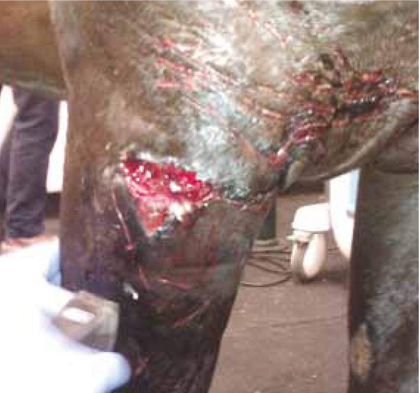
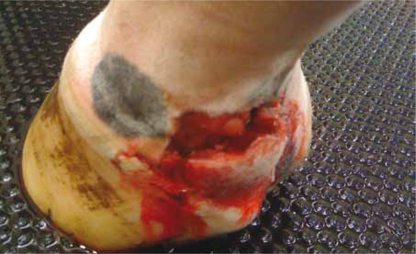
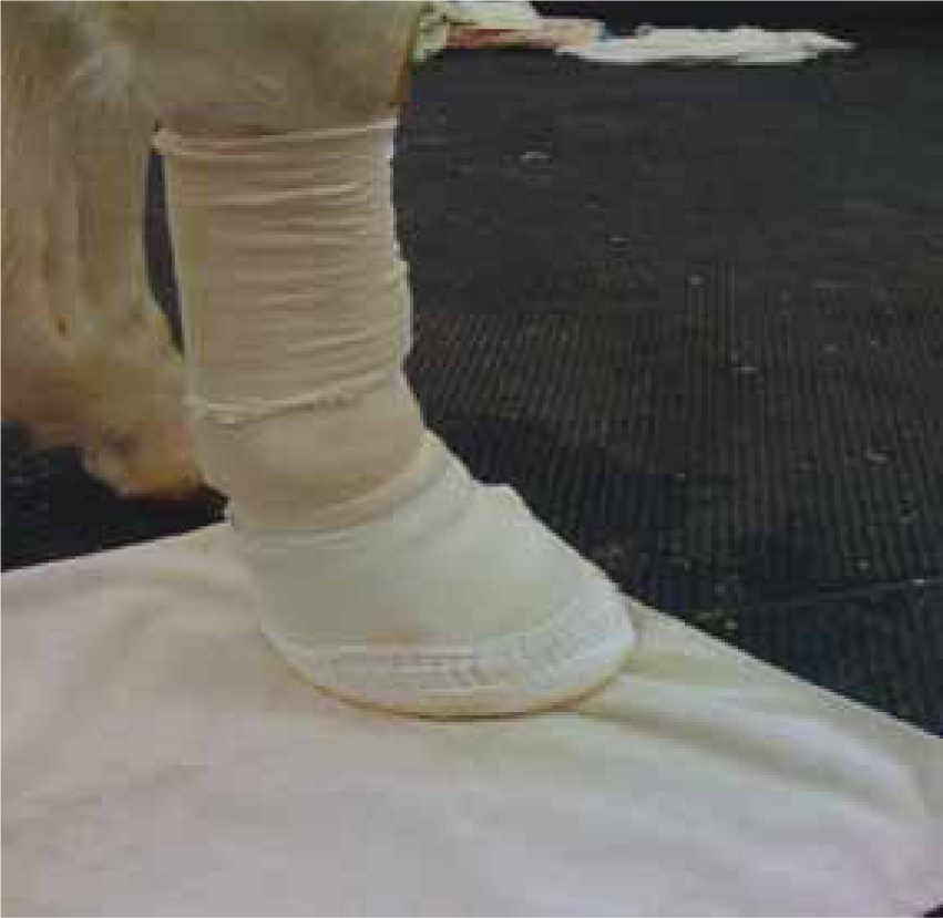
At the author's clinic honey is commonly used for contaminated heel bulb wounds to aid in reduction of bacterial load of the wound prior to a foot cast being placed, such as that seen in Figures 7 and 8.
How to protect the compromised areas
There are a number of ways to further protect areas of damaged/compromised skin and these can be as simple as applying a foam dressing to reddened skin; a foam dressing is ideal as it provides the covered area with a bit of padding to help to relieve soreness. Other materials that are commonly used to prevent or relieve pressure sores at the author's clinic are Soffban (BSN) and orthopaedic felt. Soffban has proved invaluable to the author's clinic when dressing limbs; ‘sausages’ are made out of it and used to the take pressure off compromised tendons and the orthopaedic felt is used if a patient has a particularly bad sore, such as ulcerated skin over bony prominences like the accessory carpal bone. The thickness of the felt allows the limb to be dressed for the original problem while at the same time preventing further damage to the bony prominence. Cutting a hole in the felt and placing a foam pad over the top to absorb any exudate from sores completely removes any pressure to the area and allows healing to occur. It is important to remember that both of these methods will change the shape of the leg that is to be re-dressed, so that extra padding will be needed. The author finds that padding out the narrow areas first (for example the pastern and the cannon) enables application of the first layer of cotton wool across the whole area to be dressed, achieving a functional and aesthetically pleasing dressing. Using 15 cm of conforming bandages may give a much more even pressure through the dressing than 10 cm, so the author uses the large 15 cm roles for all but the smallest of patients.
Robert Jones bandages (RJB) and casts
RJB and casts are used to completely immobilise an area, i.e. where there is a fracture or a large wound. The RJB at the author's clinic are only changed every 10 days unless something indicates a premature change is warranted, e.g. strike through or excessive lameness. Casts can be kept on for a number of weeks and can be a more cost-effective method of immobilisation than a RJB. As casts remain in place for a such a length of time it is exceptionally important to provide enough padding to delicate areas to prevent pressure sores. Bony prominences should be well padded and any compromised tendons should have pressure relieved where possible, it is not an easy or cheap job to change a cast if a problem occurs, so it is in everyone's best interest to do a good job the first time around.
Ponies versus horses
It is well documented that ponies heal better and more quickly than horses and there appear to be a number of reasons for this (Wilmink et al, 1999). There seems to be a higher rate of successful first intention healing in ponies, this is due to a more effective acute inflammatory response leading to reduced wound infection rates. Second intention healing also appears to occur faster in ponies due to a weak and slow onset inflammatory response in the horse. Horses are more prone to exuberant granulation tissue (also because of this weak inflammatory response) and this also persists over time causing a chronic inflammatory state (Lepage, 2011) delaying wound healing.
Conclusion
In summary, the veterinary nurse's role in wound healing is very much a supportive and preventative one. Veterinary nurses should be aware of how wounds heal but more importantly they should be aware of factors detrimental to healing and ways to minimise these factors. Good hygiene and prompt recognition of pressure sores are an extremely important part of the veterinary nurse's role and should always be in the forefront of their mind when dressing the equine limb. It should never be assumed that heavier breeds have tougher skin, as this is not always the case, and finally nurses should be critical about the dressings they have applied. The more critical a nurse is of the dressings applied the more they will strive to improve their work the next time.