References

Baines SJ. Thoracic Surgery. In: Martin C, Masters J Philadelphia: Elsevier; 2007

Holzman G, Raffel T. Surgical Patient Care for Veterinary Technicians and Nurses.Iowa: Wiley Blackwell; 2015

Jung J, Cho YK, Jung Y, Jeong J, Lee K. Diagnosis and Treatment of an aberrant Right Subclavian Artery with Persistent Right Ligamentum Arteriosum in a Kitten. Pak Vet J.. 2015; 35:(1)119-22

MacDonald KA. Congenital heart diseases of puppies and kittens. Vet Clin North Am Small Anim Pract.. 2006; 36:(3)503-31 https://doi.org/10.1016/j.cvsm.2005.12.006

Plesman R, Johnson M, Rurak S, Ambrose B, Shmon C. Thoracoscopic correction of a congenital persistent right aortic arch in a young cat. Can Vet J.. 2011; 52:(10)1123-8

Radlinsky MG. Surgery of the digestive tract, 4th ed. In: Fossum TW Missouri: Elsevier; 2007

Shannon D, Husnik R, Flectcher JM, Middleton G, Gaschen L Persistent right aortic arch with an aberrant left subclavian artery, Kommerell's diverticulum and bicarotid trunk in 3-year-old cat. JFMS Open Rep.. 2015; 1:(2) https://doi.org/10.1177/2055116915614590

Thrall DE. Principles of Radiographic Interpretation of the Thorax. Text-book of Veterinary Diagnostic Radiography, 6th Ed. Missouri: Elsevier Saunders; 2013

Tillson DM. Thoracic surgery; important considerations and practical steps. Vet Clin North Am Small Anim Pract.. 2015; 45:(3)489-506 https://doi.org/10.1016/j.cvsm.2015.01.007

White RN, Burton CA, Hale JSH. Vascular ring anomaly with coarctation of the aorta in a cat. J Small Anim Pract.. 2003; 44:(7)330-4 https://doi.org/10.1111/j.1748-5827.2003.tb00164.x

The regurgitating kitten

02 May 2019
12 mins read
Volume 10 · Issue 4

Abstract

Vascular ring anomalies present in young patients and will inhibit growth and development. Therefore it is essential that these patients have the anomaly corrected as soon as clinically stable enough to undergo surgical correction. In particular the congenital malformation of the persistent right aortic arch (PRAA) is uncommon in practice to see, but does require good knowledge and understanding by the veterinary team to provide a successful outcome.

The patient presented to the practice with a 1 month history of regurgitation of food, and upper respiratory noise at the point of the cranial chest and at the thoracic inlet.

Signalment

  • Species: Feline
  • Breed: Domestic Short Hair
  • Age: 2 months old
  • Gender: Male (entire)
  • Weight: 1.2 kg. Differential diagnosis

Differential diagnoses

Differential diagnoses included:

  • Vascular ring anomalies (VRA), specifically a persistent fourth right aortic arch (PRAA)
  • Megaoesophagus
  • Oesophageal foreign body
  • Oesophageal stricture
  • Oesophageal mass
  • Hiatal hernia.

Patient history and physical examination

The patient had been rescued from under a shed at approximately 4 weeks of age, and since then he had been hand reared. As growth occured, he developed regurgitation of his food, despite the owner feeding him from a height. He was vastly underweight for his age and was much smaller than his littermate. The referring veterinary surgeon (RVS) had radiographed the chest which revealed a megaoesophagus and that the aorta appeared to be on the right. On chest auscultation there was upper respiratory noise present at the thoracic inlet. The owner reported no coughing and the patient had been eating and drinking well. All other vital signs had no abnormalities. An intravenous (IV) 22 g catheter was placed in the right cephalic vein.

Tentative diagnosis by the veterinary surgeon

Based on the clinical examination and the RVS's radiographs (RADs) there was significant evidence that a PRAA may be present causing the clinical signs. PRAA is the most common of the vascular ring anomalies (VRA) seen as a congenital malformation of the great vessels and their branches (Plesman et al, 2011). VRAs are considered rarer in cats than dogs (Plesman et al, 2011; Shannon et al, 2015). It is documented that 95% of feline VRAs are PRAAs with or without other aberrant vessels involved and 44% of those have co-existing compressive arterial anomalies, however cardiac murmurs are rare (MacDonald, 2006). It has been documented that in a study where 16 cats with VRAs were diagnosed, the incidence of PRAA in the study was 81% (White et al, 2003). VRAs are thought to occur through multiple recessive genes which is complex and polygenic; normally the six pairs of aortic arches around the oesophagus and trachea are present in early fetal life (Tillson, 2015), but as maturation occurs, the arches should form the adult vasculature (MacDonald, 2006). The defects are present from birth and occur equally in males and females, but are usually diagnosed between 2 and 6 months when weaning occurs and semi-solid food is first fed (MacDonald, 2006; Plesman et al, 2011). VRAs normally also prelude to aspiration pneumonia as a secondary condition (Tillson, 2015), which needs to be addressed before surgery, and affected kittens are normally underweight, have an inability to thrive and require an increase in body condition before surgery. The kittens normally have a voracious appetite and some will eat regurgitated food.

Diagnostic imaging options

Options for diagnostics varied from plain RADs to the use of positive contrast RADs using barium sulphate liquid mixed with food. The latter approach requires a conscious patient and the ability for the patient to eat the liquid and be restrained for RADs, which does increase risk to personnel due to radiation exposure. Other oesophageal conditions can mimic VRAs and contrast studies need to be critiqued carefully, such as foreign bodies, strictures, masses, megaoesophagus and hiatal hernias (Radlinsky, 2007). Fluoroscopy is useful for assessing esophageal motility as a dilated oesophagus does not normally have adequate peristaltic contractions (Radlinsky, 2007). Angiography can be used to identify the type of VRA present and whether there are any other cardiac abnormalities which are of concern (Plesman et al, 2011), but are frequently are not conducted as they do not tend to add any further relevant information to change the surgical plan (White et al, 2003). Contrast computer tomography or magnetic resonance imaging tend to be used to identify abnormal vasculature (Jung et al, 2015; Shannon et al, 2015; Tillson, 2015). Endoscopy of the oesophagus is used to rule out other disease processes, including strictures or obstructions (Shannon et al, 2015) and can identify any ulcerations (Radlinsky, 2007). This modality confirms that there is a VRA present and that the oesophagus is not primarily weakened (Radlinsky, 2007).

The patient was anaesthetised with alfaxalone (5 mg/kg) after obtaining a blood glucose level and a premedication of acepromazine (0.005 mg/kg) and methadone (0.3 mg/kg) intramuscularly and remained in sternal recumbency once intubated. Due to client finances, thoracic plain RADs were initially taken, dorsal ventral view was taken first, to reduce lung atelectasis, followed by both lateral (LAT) views; all views were inflated to ensure any pathology present would be detected. The views confirmed a megaoesophagus due to a dilation of the cranial oesophagus (Jung et al, 2015) and that the aorta was located on the right side of the thorax. Following this modality, endoscopy of the oesophagus was undertaken which revealed the vena cava present on the left side of the thorax.

Surgical treatment options

Surgical treatment options were discussed in depth with the client. Minimally invasive thoracoscopy was discussed, and although not available at the author's practice, referral to another institution was offered, as this method can produce good results in small kittens of a similar size (Plesman et al, 2011). Early surgery was discussed as is required to improve the prognosis (MacDonald, 2006; Plesman et al, 2011). A left sided intercosatal thoracotomy was discussed indicated as a surgical approach to remove the PRAA as the treatment of choice (White et al, 2003; Shannon et al, 2015). Medical management was discussed but was not recommended (Plesman et al, 2011) as it is rendered a palliative option due to secondary effects of regurgitation and inability to thrive, despite use of gastrostomy feeding tubes to provide nutrition and bypass the defect (Radlinsky, 2007). It was also discussed that despite surgical intervention oesophageal dilation may have caused an irreversible nerve degeneration, causing hypomotility resulting in regurgitation (Shannon et al, 2015). The owners opted for surgical correction with the author's practice rather than being referred on again for surgery with further advanced imaging and surgical modalities, and surgery took place the following day.

Patient and equipment preparation

It is essential that the operating theatre is prepared thoroughly including stocking up with correct instrument packs and cleaning before the procedure begins (Holzman and Raffel, 2015). Ensuring equipment is present and ready before the procedure begins (Table 1) reduces the movement throughout the theatre and will allow the careful concentration that is required for invasive surgery like a thoracotomy (Baines, 2007). Prior to surgery the theatres are cleaned with Swift™ (Anistel) which is a sporicidal, mycobactericidal, virucidal, fungicidal and bactericidal surface cleaner to remove any harmful pathogens from the environment. An Anigene (Medimark Scientific Ltd) aerosol is also set off which has the same action as Swift™ (Anistel) to allow contact with all surfaces, including the ceilings. Anaesthetic concerns for the paediatric patient included hypoglycaemia, hypothermia due to a high surface area to bodyweight ratio, leading to bradycardia, decreased cardiac output and hypotension, reduction in ability to metabolise drugs administered and therefore prolonged recovery from general anaesthesia. Amoxicillin and clavulanate potassium was given intravenously (IV) through the IV fluid line to ensure this was given slowly to prevent an adverse reaction. Amoxicillin and clavulanate potassium is a broad spectrum antibiotic active against Gram-positive and Gram-negative aerobic organisms and many obligate anaerobes. It is used to reduce incidence of post-surgical infections by inhibiting bacterial cell wall synthesis and promoting destruction of bacterial cell walls. This reduces the strength and rigidity of the cell wall, increasing permeability and causing cell lysis (Radlinsky, 2007). The surgical site was clipped with #40 ethylene oxide (EtO) sterilised clipper blade using electric clippers (Holzman and Raffel, 2015). The clip was from the thoracic inlet cranially, to beyond the last rib caudally, past the midline dorsally and past the midline ventrally (Holzman and Raffel, 2015). A preliminary scrub was carried out using chlorhexadine gluconate 4% diluted with sterile NaCl (saline) 50:50 mixed in a sterile kidney dish with sterile swabs, wearing clean examination gloves and using a back and forth technique (Holzman and Raffel, 2015). Chlorhexadine gluconate requires 2–5 minutes' contact time with the skin to provide bactericidal and sporicidal properties, it also provides good residual action as it binds to the stratus corneum (Thrall, 2013). All personnel entering the operating room wore dedicated theatre scrubs, clogs, hats, masks (Plesman et al, 2011). The theatre table was prepared with the PetTherm (Inditherm Medical) heat mat prewarmed (Holzman and Raffel, 2015), and an absorbent pad was placed over the top end of the table approximately where the thorax would be placed. The patient was placed onto the table and a sandbag was placed under the patient at the level of the 5th intercostal space to elevate the thorax and aid visualisation of the surgical site for the surgeon. The patient was connected to the anaesthetic breathing system and monitoring equipment attached. A final preparation was carried using a Chloraprep® (Invicta Animal Health) 10 ml applicator by the circulating nurse, wearing sterile gloves put on with an open gloving technique, to prepare the area working in a back and forth motion over the surgical incision site for 30 seconds, and then moving outwards towards the hair edges with the remainder of the solution. The surgeon and scrub nurse performed a sterile scrub using Sterillium® (Bode Chemie GmbH) which is rubbed into dry hands undiluted, paying special attention to the fingertips, thumbs and wrists from an elbow-operated dispenser. Surgical hand disinfection is complete in 90 seconds, timed with a timer. Surgical gowns, and gloves were opened aseptically by the circulating nurse and then tied properly. The surgical packs and separately packed instruments were then opened aseptically by the circulating nurse and passed to the surgeon and scrub nurse for the inner sterile packets to be opened and instruments placed onto the instrument table organised ready for use.


Table 1. Instrument and equipment list
Drape pack — sterilised by manufacturer ethylene oxide
(4) Adhesive drapes size (1) Table drape
Thoracotomy kit — sterilised using steam sterilisation 135° double wrapped in self-seal pouches
45 degree curved forcepsDebakey atraumatic dissecting forcepsDebakey needleholdersDressing forcepsLahey chloecystecomy clampMetzanbaum curved scissors 14”Metzanbaum curved scissors 9” 90 degree curved forcepsMetzanbaum straight scissors 9”Rat toothed forcepsStatinsky forceps 20 mmStatinsky forceps 30 mm(2) Right angled clamps small(8) Spencer Wells forceps
General surgical pack — steam sterilisation at 134° wrapped in drape and in single self-seal pouch
(1) #3 blade handle(1) Rat tooth forceps(2) Dressing forceps(2) Allis tissue forceps(1) Olsen-Hegar needle holders (1) Straight Mayo scissors(1) Straight Metzanbaum scissors(4) Backhaus towel clamps(1) Brown Adson finger forceps(4) Mosquito forceps curved(4) Spencer Well forceps curved
Additional items — steam sterilisation at 121° double wrapped in self-seal pouches
Electrocautery  
Additional items — steam sterilisation at 134° double wrapped in self-seal pouches
Small Finochiettos rib retractorsCosmetic surgeons scissors straight Cosmetic surgeon scissors curved20 x 20 Laparotomy swabs x 2
Additional items — sterilised ethylene oxide by manufacturer
Suction tubingPlastic suction tip Mila chest drain tube 12 ga x 30 cm fenestrations 15 cmMila centesis valve and adapter 12.5 cm
Quality control methods
Class 1 external chemical indicator on self-seal bag for steam sterilisationClass 6 TST strips placed inside centre of packs prior to packaging for steam sterilisation

Operative report

Before surgery commenced a swab count was performed and the number of swabs was noted by the anaesthetic nurse to refer to at the end of surgery. A left sided 5th intercostal thoracotomy incision was made through the skin with a #15 blade on a #3 handle, which is the advised location for entry to the thoracic cavity for a PRAA (Baines, 2007). The subcutaneous tissue and cutaneous trunci muscle were also incised. The incision extended from just below the verterbral bodies to near the sternum. The incision was then deepened through the latissimus dorsi muscle with scissors and the correct intercostal space was confirmed by counting from the first rib. The scalenus and pectoral muscles were then transected with scissors perpendicular to the muscle fibres. The muscle fibres of the serratus ventralis were then separated at the intercostal space. Near the costochondral junction one scissor blade was placed under the external intercostal muscle fibres and the scissors were pushed dorsally in the centre of the intercostal space to incise the muscle. The internal intercostal muscle was then incised and the surgeon notified the anaesthetist of entry into the thoracic cavity and for intermittent positive pressure ventilation to begin.

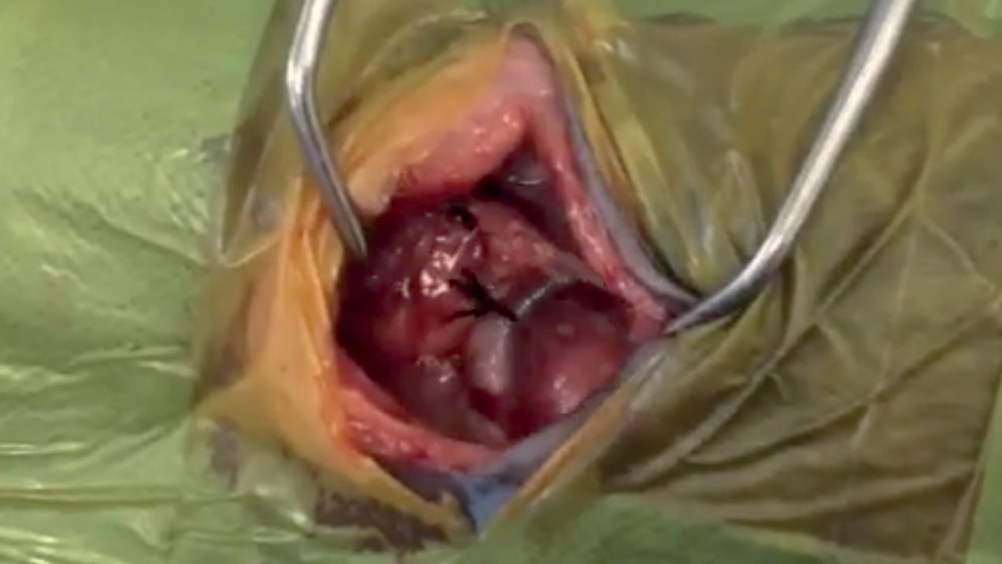
Once the lungs and pleura had been identified, using closed scissors, the pleura was then penetrated during maximum expiration. The excision was then extended dorsally and ventrally to provide visualisation (Figure 1; Video 1). Moistened laparotomy swabs were placed on the edges of the chest incision and a small Finochietto retractor was used to spread the ribs. The cranial lung lobes were packed with moistened swabs caudally to expose the mediastinum dorsal to the heart and to view the oesophageal dilation. Identification of all major structures including the aorta, pulmonary artery, ligamentum arteriosum and both the vagal and phrenic nerve was conducted. A 12 fr oesophageal stethoscope was attempted to be passed by the anaesthetic nurse but was unsuccessful. At this location there was a fibrous band location between the pulmonary artery and aorta. Dissection under the band was conducted carefully and two ligatures doubled were placed each side of the band with 2–0 Mersilk suture (braided silk). The fibrous tissue band was then cut and the fibrous tissue over the oesophagus was also dissected away. The 12 fr oesophageal stethoscope was then passed successfully and then replaced with an 18 fr oesophageal stethoscope which also passed successfully. The area was then flushed with 100 ml saline and all sutures checked for integrity. A Mila chest drain was then placed through the 7th intercostal space and attached using the connectors provided. The thoracotomy incision was closed by pre-placing six sutures of PDS Plus 3–0 non-absorbable monofilament sutures around the ribs adjacent to the incision. The sand bag beneath the patient was then removed. The sutures were then tied. The serratus ventralis, scalenus and pectoralis muscles were sutured in a continuous pattern with PDS Plus 3–0 non absorbable suture. The edges of the latissimus dorsi were opposed also with a continuous pattern. The residual air from the chest was removed from the thoracic cavity using a centesis valve attached to the chest drains in situ, which initiated spontaneous respiration and intermittent positive pressure ventilation could cease. The subcutaneous and skin were closed routinely with Monocryl Plus 3–0, followed by a bupivacaine intercostal block placed using a 2 ml syringe and 25 g needle, two intercostal spaces cranially and two caudally on the caudal aspect of the rib for analgesia at a maximum of 2 mg/kg (Baines, 2007). The patient was transferred into the radiography suite for postoperative RADs to confirm correct placement of the chest drain and to assess if any air was still present in the thoracic cavity. Once the removal of air was confirmed, bupivacaine (0.5 ml) was placed into the chest drain for further analgesia.

Figure 1. Visualisation of the surgical site. See Video 1 online.
Video

Visualisation of the surgical site

Postoperative care

On recovery the patient was placed into a pre-warmed recovery kennel with continuation of the IV fluid therapy (Plesman, 2011), placed in left lateral recumbency onto the chest drain to allow the right lung to expand adequately within the cavity without discomfort. Observation for dysponea was critical and pre-placement of a nasal oxygen catheter was conducted to assist in oxygenation in recovery (Radlinsky, 2007; Thrall, 2013). Blood glucose analysis was conducted every hour to ensure the patient did not become hypoglycaemic and this was reduced to once daily once he began to eat (Plesman et al, 2011). The chest drain was scheduled to be emptied every 15 minutes for the first hour initially to ensure there was not a dramatic increase in fluid or air on the chest which may compromise respiration, increasing to every 30 minutes in the second hour, and then every 2 hours. This was achieved using a Mila centesis valve adaption to reduce accidental air entry into the chest, using gloved hands when in contact with the drain and its contents. The amount of fluid and air removed from the chest was recorded on the hospitalisation sheet (Baines, 2007). Respiration rate and effort was also closely monitored, initially every 5 minutes, increasing to 15 minutes after the first 30 minutes as the patient remained stable. After 1 hour, respiratory checks decreased to every 30 minutes, and then hourly. Once air had ceased and fluid had reduced to 1 ml/kg/24 hours the following day the chest drain was removed. Opioids were continued into the postoperative period and antibiotics were continued due to the patient's debilitated condition and the risk of aspiration pneumonia. There is also a risk postoperatively of leakage or dehiscence of the oeosphagus post resection (Radlinsky, 2007). The patient was fed once normothermic a gruel consistency in an upright position with the head elevated and observed while eating (Figure 2). His position was continued for 5–10 minutes post feed to help with oesophageal muscle tone and reduce the incidence of reflux. Occasionally patients can be lame on the ipsilateral forelimb following thoracotomy due to the incisional trauma of the latissimi dorsi. and this should be evaluated in recovery (Plesman et al, 2011; Tillson, 2015).

Figure 2. Feeding elevated with a gruel like consistency.

Client education and prognosis

The clients were given both verbal and written discharge instructions. A medical pet T-shirt was provided along with a flexible Elizabethan collar to prevent patient interference with the surgical site and drain exit site. They were instructed to monitor the sites daily for erythema, discharge or signs of pain such as patient interference. Intradermal sutures would not require removal, however a postoperative check in 10 days at the practice would be required to take repeat RADs with barium sulphate to assess the oesophageal dilation. The clients were instructed to feed the patient soft food mixed with water and from a height. They were then asked to keep the head elevated for 5–10 minutes post feed to try and reduce incidence of reflux. They were also advised this may be a long-term situation and the patient may always require this method of feeding to avoid reflux despite surgery. The clients were then asked over a period of 2–4 weeks to decrease the amount of water mixed with the food until solid food could be tolerated. He was presented 10 days post discharge for barium sulphate RADs which concluded the dilation at the proximal oesophagus was much smaller than preoperatively, and the owners were intructed to continue feeding from a height until food has been gradually changed over to solids.

Conclusions

With a patient of such small size and a reduced ability to maintain body temperature, it is essential that the nursing team is aware of how the procedure described is executed. This is imperative foremost for the nursing care pre and post operatively, but also it is important that the scrub assistant is familiar with the anatomy and physiology and instrumentation required for the procedure to assist the veterinary surgeon effectively.

KEY POINTS

  • Knowledge of anatomy and physiology of vascular ring anomalies will aid the scrub veterinary nurse to assist the surgeon effectively to help with a successful outcome.
  • Advanced imaging and surgical options are available for treatment of vascular ring anomalies and should be discussed with the client to help achieve best outcomes.
  • Patients with vascular ring anomalies may require lifetime management despite surgical intervention.
  • Knowledge of equipment for theatre set up and how the instruments are used will reduce the amount of movement and thoroughfare through the surgical suite, minimising risk of poor asepsis.