The triage examination identifies patients with life-threatening conditions who require immediate attention from a veterinarian, and distinguishes them from patients that are not as ill (Aldrich, 2005; Drobatz, 2010). The systematic evaluation of patients in triage includes rapid assessment of the respiratory, cardiovascular, neurologic, and urogenital systems. Respiratory and cardiovascular triage were covered in part 1 of this series, and this manuscript will focus on neurologic and urogenital triage. Specific approaches to the history and owner communication during triage were also covered in part 1 of this series.
Neurologic triage
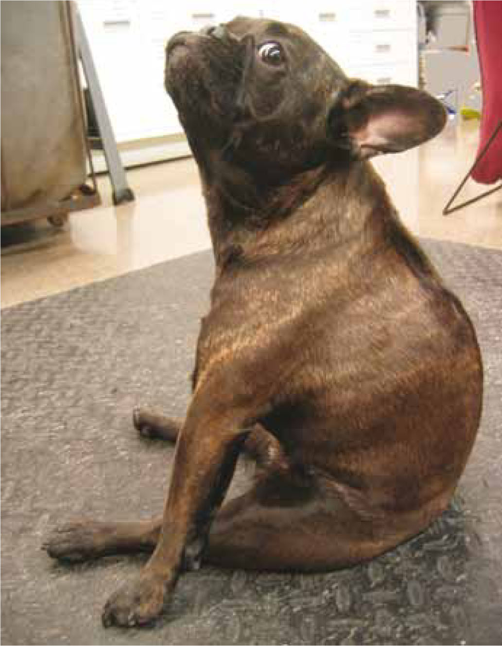
The neurologic triage examination begins as soon as the patient is seen. It is an abbreviated neurologic examination that observes components of the patient interacting with the environment. If abnormalities are identified on triage, a more in-depth neurologic examination should be conducted. Common neurologic diseases that cause an animal to present to the emergency room include seizures, head trauma, acute changes in mentation (e.g. as due to ingestion of toxins), and spinal cord disease (Figure 1) causing paresis or paralysis. Patients with neurologic consequences of systemic disease (e.g. hepatic encephalopathy or rapid changes in serum sodium concentration) may also present for or develop neurologic signs (Aldrich, 2005).
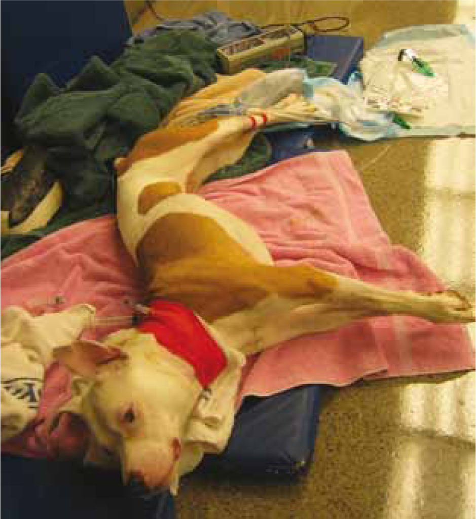
Patients should be aware of their surroundings, ambulatory on all four limbs, and otherwise alert (Syring, 2005). They should interact with the examiner on approach, and ataxia or knuckling of the paws should not be present. It is sometimes difficult to distinguish abnormalities in ambulation between orthopaedic or neurologic problems. Conditions such as rolling or circling, the inability to stand, or any paralysis should be further evaluated immediately. Patients that are non-responsive or laterally recumbent should be triaged immediately for further therapy and diagnostics. Opisthotonus describes a posture characterized by recumbency, neck hyperextension, and stiff extended limbs (Figure 2), and indicates impending herniation of the brain; patients must be treated immediately for increased intracranial pressure (Syring, 2005).
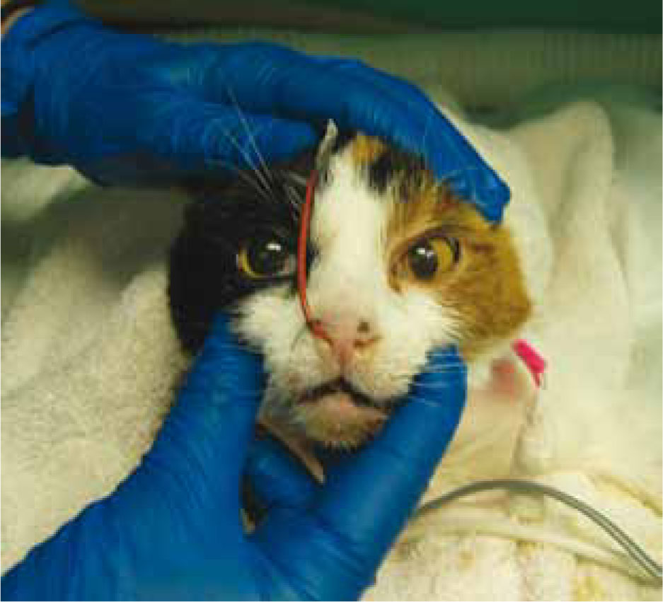
In addition to mentation and gait, the patient's pupils should be evaluated for asymmetry (anisocoria) (Figure 3) or for dilation (mydriasis) or constriction (miosis); all of these are abnormal, and the patient should be evaluated for intracranial disease immediately (Syring, 2005). Occasionally, topical medication (e.g. atropine) or systemic disease may cause these signs, but until proven otherwise, the possibility for neurologic disease should be assumed. Neurologic disease that affects the muscles of the face (e.g. tetanus) may also be seen on first approach. Facial droop due to decreased muscle tone may also be observed. Excessive drooling may be associated with paralysis of the tongue or an inability to swallow appropriately.
Seizures are often a reason for emergency room visits. If the patient is actively having a seizure, he or she should be immediately evaluated to decide if medication such as diazepam or midazolam should be administered to stop the seizure. Important historical facts include the frequency and duration of prior seizures, as well as the potential for toxin exposure and prior medical history. In addition to neurologic disease, low serum glucose concentrations can result in seizures. Hypoglycaemia can be seen in neonatal or malnourished patients as well as older patients with neoplasia or sepsis, or in those who have received an insulin overdose. Other systemic illness such as cardiac disease can result in syncopal episodes (collapse) that the owner may confuse with a seizure. The presence of a postictal period (a period of confusion and ataxia following a seizure event) may help to differentiate the two, as cardiogenic syncope is not always associated with a postictal period. Patients with cardiogenic syncope may also have an irregular heart rate.
Patients presenting for twitching may have neurologic disease, or may have other systemic disease. Twitching is associated with hypocalcaemia, pyrethrin flea treatment toxicity (especially in cats) and the ingestion of tremorigenic mycotoxins (Young et al, 2003; Boland and Angles, 2010). In patients with excessive muscle activity, it is important to measure and monitor body temperature, as the muscle activity can result in hyperthermia.
Urogenital tract triage
Emergencies involving the urogenital tract may not be as obvious as abnormalities of other systems. It is important to palpate for a bladder in certain animals. Feline urethral obstructions are a common emergency. They tend to affect young adult male cats, but older males and occasionally females may be affected (Lee and Drobatz, 2003; Rieser, 2005). The bladder should be palpated on every feline patient. If a bladder is firm and unable to be expressed, the patient may not be able to urinate due to obstruction by grit combined with mucus into a urethral plug (Lee and Drobatz, 2003). Urinary obstruction can also occur in dogs, although in these cases is more commonly associated with the presence of cystic calculi (urinary bladder stones), urethral strictures, or neoplasia such as transitional cell carcinoma. Animals with urethral obstructions may have varied clinical signs, including frequent trips to the litterbox, stranguria, or vocalizing while in the litter box (Lee and Drobatz, 2003). Owners frequently report that the cat has trouble defecating, or is eliminating inappropriately, and may report haematuria. Urethral obstruction is life threatening, and if the pet is not brought to a veterinarian immediately, he or she can become extremely ill from azotemia and severe hyperkalaemia (Rieser, 2005). At this point, a triage examination will note a cat with a firm to hard, non-expressable urinary bladder, bradycardia, and an obtunded to comatose mental status.
Intact female animals (especially dogs, but occasionally cats) are at risk for development of pyometra, which can progress to signs of septic shock if not treated in a timely manner. This is a condition that usually occurs in dogs approximately 1–2 months after the last heat cycle (Jutkowitz, 2005), and history about the prior heat cycle should be obtained during triage. Animals with pyometra may have vulvar discharge noted at triage. Both intact and castrated male animals are at risk for the development of paraphimosis (inability to replace the penis in the prepuce) or priapism (prolonged erection), both of which merit emergent evaluation and therapy (Root Kustritz, 2001).
Both cats and dogs may develop infections in the urinary tract; if an infection involves the kidney, pyeloneprhitis is present. Animals with pyelonephritis may show pain on palpation of the abdomen, and may have acute renal failure.
Other conditions
Other conditions noted in patients who present for emergency evaluation that merit immediate triage for further assessment and care are related to the underlying diseases. Animals presenting with haemorrhage should be evaluated immediately. Animals with a distended abdomen also merit an immediate secondary assessment; distention could be from air (e.g. a gastric dilatation-volvulus) or from fluid (e.g. acute haemoperitoneum, uroperitoneum, or peritonitis). Animals with known toxin ingestions that may require gastric decontamination should be seen immediately so that emesis may be induced if indicated, and those animals that have been bitten or stung by insects or snakes should be evaluated prior to worsening of clinical signs.
The role of the veterinary nurse in triage
All members of the veterinary care team should be trained in, and have a concept of, triage. The veterinary nurse, in particular, has experience and training to make educated assessments of patients arriving at the hospital. As noted previously, the nurse's role in triage is to use their experience of clinical presentations and client interactions to make a swift, informed decision. Recognizing an animal who requires immediate triage and intervention can mean the difference between life and death in some circumstances.
When communicating with the owners, the nurse should be direct and sympathetic. A physical examination only provides some of the needed pieces whereas a client who is asked the correct questions will help complete the puzzle. If necessary the conversation should be redirected back to the original questions. Confidence and compassion are extremely valuable and can calm and focus a distraught client. It should be remembered that the purpose of the triage examination is only to evaluate the animal, and not to offer diagnoses to the owner, although many will request your feelings on what might be wrong with their animal. In some areas, it is not legal for anyone other than a veterinarian to diagnose disease in animals, and questions about actual diagnosis should be deferred to the veterinarian.
If a patient is deemed critical and in need of further assessment, treatment, or stabilization, it is important to obtain permission from the owner prior to taking the patient away from the owner. This permission can vary from hospital to hospital, but usually verbal permission is acceptable. The owners at this point are not giving permission for hospitalization and a long list of diagnostics, only for initial stabilization and assessment. This may include permission for placement of an intravenous catheter (e.g. in an animal who may need intravenous fluid therapy or diazepam therapy to stop seizure activity) and basic blood work (such as packed cell volume (PCV) and total protein assessments, blood glucose, electrolytes, or lactate), or other more directed therapies (e.g. placement in an oxygen cage). In cases where an animal may need immediate and expensive therapies (e.g. a blood transfusion in a severely anaemic patient), it is also appropriate to give the owner a ballpark idea of the cost of this initial intervention.
Conclusions
Once the primary triage is completed and an action plan implemented to address identified abnormalities, patients must be continually reassessed. Especially in patients with neurologic disease, rapid changes in mentation or the progression of paresis to paralysis must be followed, as they may signal potentially life-threatening progression of disease. Animals that have a urinary obstruction may develop a post-obstructive diuresis, and the close monitoring of intravenous fluid volume administered (ins) compared with the amount of urine produced (outs) can alert the practitioner to the possibility of dehydration due to extreme diuresis (ins<outs) or removal or kinking of the urinary catheter (ins>outs).
As noted in part 1, a triage examination is a learned art, and practice makes perfect. The more an individual practices history taking and physical examination skills, the more comfortable he or she will be when these skills are put into practice.