Anaesthesia in reptiles progresses at a slower pace compared with other exotic species; however, anatomical differences make anaesthesia in reptiles more complex. Once understood, though, reptile anaesthesia can be highly rewarding. There are approximately 10 000 known species of reptiles, so this article will focus on the most commonly encountered groups in veterinary practice: snakes, lizards and chelonians.
This is part three of a series of articles designed to outline the principles of anaesthesia in exotic species and to instil confidence in the veterinary nurse as an advocate for exotic animal care.
Pre-anaesthetic preparation
Equipment
As with any anaesthetic procedure, equipment should be prepared and fully charged in advance to prevent avoidable failures during anaesthesia. As discussed in part 1 [Ayers, 2024), surgical loupes can be very useful for smaller patients to assist the veterinary surgeon in performing delicate procedures. Many reptiles become apnoeic after induction, which will be discussed in greater detail later; therefore, preparing a ventilator suitable for exotic reptile procedures is recommended.
Environmental factors
Reptiles are ectothermic, meaning they rely on environmental warmth to regulate their body temperature (Hedley, 2010). Each species has a preferred optimum temperature zone (POTZ), within which it can optimally regulate bodily systems, such as digestion, reproduction and immuno-competence. Sick reptiles will often seek the higher end of their POTZ, a behaviour known as ‘behavioural fever’, to enhance their immune response (Rakus et al, 2017). Very sick reptiles may also present as hypothermic as a result of an inability to mobilise; these animals should be gradually warmed to their POTZ before anaesthesia (Hedley, 2022).
As veterinary nurses, it is important to ensure that each environment the reptile patient encounters remains within its POTZ, including the hospital vivarium, theatre and prep room, to promote a smoother anaesthetic process and a more predictable, stable recovery (Hedley, 2022). Additionally, reptiles require UV light, including species such as snakes, which were historically thought not to require UV. Current research supports the provision of UV light to all domestic reptiles (Baines et al, 2016; Azevedo et al, 2021; Kane et al, 2023).
Patient evaluation
An accurate weight should be obtained before administering any drugs or fluids. Reptiles often display clinical signs of illness only once the disease process is advanced, which may take several years to develop; therefore, patient parameters will almost certainly be outdated from previous visits to the veterinary practice. Debilitated reptiles frequently present with chronic dehydration, which affects both physiological parameters and demeanour, making a full preoperative assessment challenging. As with any animal, it is important to stabilise reptiles beforeo anaesthesia.
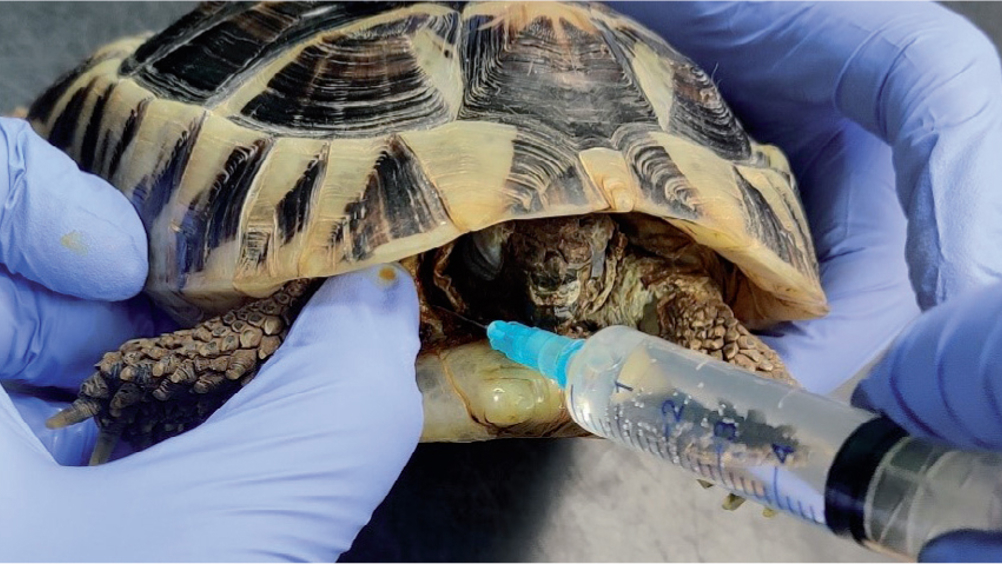
Rehydration in reptiles is relatively straightforward, with methods varying depending on the level of dehydration. If mildly dehydrated, fluids can be administered via stomach tube or through warm water baths, in which fluid is absorbed through the skin and cloaca (coprodeum) (Holz, 2020). For dehydration greater than 8%, fluids should ideally be given intravenously if possible, or otherwise via subcutaneous or epicoelomic injection (Figure 1) (Mitchell, 2009).

Fasting
Fasting is generally not necessary for herbivorous reptiles; however, species that receive live food or large whole prey should be adequately fasted. Insectivores should be fasted for at least 24 hours to allow digestion of live food before anaesthesia (Hedley, 2022). Small snakes should be fasted for at least two to three days, and larger snake species for longer, to avoid lung compression from a full stomach during anaesthesia (Hedley, 2022).
Handling
Like any exotic animal, reptiles are susceptible to high stress levels, which can be exacerbated by excessive handling. It is advisable to obtain a thorough history from the owner beforeinteracting with the animal, to assess the individual patient's comfort level with handling. Depending on temperament, a preoperative examination may not be possible; lack of regular handling may result in an animal displaying signs of anxiety and aggression, and many species are capable of making themselves unexaminable to the veterinary team while conscious (Hedley, 2022).
Anatomical differences
Heart
Most reptile species have a three-chambered heart, consisting of two atria and a single, incompletely partitioned ventricle. This structure allows mixing of oxygenated and deoxygenated blood, enabling blood shunting towards or away from the lungs under certain conditions. This variation in blood flow is hypothesised to enhance thermoregulation and extend dive times in aquatic species (Burggren et al, 2020); however, further research is needed in this area.
Reptiles can shunt blood away from the lungs during low-oxygen situations, such as when a snake has consumed a large prey item and is unable to use its lungs. During anaesthesia, reptiles may hold their breath and initiate cardiac shunting in response to inhaled volatile agents (Burggren et al, 2020). This can complicate the maintenance of adequate ventilation and anaesthetic depth, necessitating manual or mechanical ventilation. Once adequately anaesthetised, however, their capacity to bypass the lungs is reduced, which simplifies control over anaesthetic depth (Hedley, 2022).
The simplified illustrations in Figure 2 depict interspecies variations in ventricular septum size, which influence the degree of mixing between oxygenated and deoxygenated blood. In lizards, snakes and chelonians, the ventricular septum is incomplete, allowing some blood mixing between the ventricles. In crocodilians, the septum is approximately 80% complete but still permits partial blood mixing.

Lungs
Reptiles lack a diaphragm, so breathing is instead controlled by small movements of the intercostal, pectoral and abdominal muscles, or through limb movements in species that possess them. In lizards and snakes, lung ventilation is largely achieved by core axial muscles, which are also used for mobility. Consequently, many of these species must hold their breath during intense exercise (Doss et al, 2021). However, some lizard species are able to fully inflate their lungs during vigorous activity, allowing for prolonged aerobic respiration (Doss et al, 2021).
For veterinary nurses, this means that, much like birds, it is essential to avoid restricting chest movements during patient handling and restraint. This includes taking care with items such as heavy surgical drapes, towels and Bair Hugger systems.
Tortoises and turtles differ in that some species have a muscular sheet surrounding the lungs. When this muscle contracts, exhalation occurs; limb withdrawal into the shell forces air out of the lungs, while limb extension reduces pressure, allowing inhalation. As with other reptiles, exceptions exist; for instance, ventilation in box turtles does not correlate with limb movement (Doss et al, 2021). During anaesthetic induction, manual extension and flexion of the legs may be necessary to facilitate respiration in cases of apnoea, in addition to squeezing the reservoir bag (IPPV) or providing mechanical ventilation.
Larynx/glottis
Unlike in mammals, the reptile glottis remains closed except during inhalation (Figure 3). Reptiles lack an epiglottis, and their glottis is positioned rostrally, making intubation relatively straightforward in these species (Mans and Sladky, 2019). The tracheal rings are incomplete in snakes and lizards but complete in tortoises, turtles and crocodilians.

Renal portal system
In reptiles, the renal portal system supplies blood to the renal tubules within the kidneys when glomerular filtration is downregulated or absent, such as during dehydration. In these instances, blood from the caudal regions of the body flows through the kidneys to maintain renal tubule perfusion until glomerular blood flow can resume (Holz, 2020).
Hepatic first-pass
Venous blood from the caudal portion of the reptile body travels through the ventral abdominal vein(s) and is then transported either directly to the liver or indirectly via the hepatic portal vein before entering the systemic circulation; this process is known as hepatic first-pass (Mans et al, 2019). Consequently, any drug administered in the caudal portion of a reptile's body may be metabolised by the liver before it can be fully used by the body (Holz, 2020).
The clinical significance of the renal and hepatic portal systems will be discussed in further detail under Induction.
Catheterisation
Maintaining patency of intravenous catheters in reptiles is challenging and often unsuccessful once the animal is awake and mobile. Therefore, catheters can be maintained during anaesthesia to administer intravenous fluids for blood pressure support, and then removed upon recovery.
The jugular vein is typically preferred for chelonians, with transillumination used if needed (Figure 4). In snakes and lizards, the coccygeal vein is most commonly accessed. Alternatively, a butterfly catheter can be inserted into the ventral coccygeal vein and secured with tape, similar to blood collection procedures. In smaller lizards and snakes, the ventral approach can be used; however, in larger species with thicker tails, the lateral approach may be more effective (Figure 5).


Intraosseous catheters are rarely used and should not be relied upon without radiographic confirmation of correct placement. Placement can be time-consuming, and long-term use may impact normal behaviours and increase pain levels.
Skin preparation
Reptile skin harbours many commensal bacteria and viruses that typically cause no harm unless the immune system is compromised or a secondary disease is present (Ross et al, 2019). To prevent internal translocation of these commensals through the incision, it is important to reduce their presence before surgery.
In small animal surgery, research suggests that there is no significant difference in skin bacterial colonisation or postoperative surgical site infection rates when using either chlorhexidine or povidone-iodine for skin preparation (Marchionatti et al, 2022). However, in reptiles, chlorhexidine has been associated with neurological disease and even death, with chelonians appearing particularly susceptible. When used at a dilution of 0.024%, chlorhexidine appears to be safe (Platt, 2019). Povidone-iodine, which has a broader safety margin, is recommended by some authors for certain dermatological issues in reptiles, such as superficial infectious dermatitis (Platt, 2019; Aguilar and Mitchell, 2023).
Gentle removal of loose shed via warm water baths before surgery may be necessary, depending on the animal's condition. Scrubbing the skin with a sterile toothbrush allows for more effective debris removal and better penetration of the cleaning solution between the scales (Figure 6).

Induction of anaesthesia
Premedication
Premedication should be discussed among the veterinary team, taking into account the animal's signalment and history. A current weight should be obtained before drug administration, along with a note of the body condition score. Evidence indicates that obesity affects induction times in reptiles, as has also been observed in mammals (Kristensen et al, 2022).
Alfaxalone has been shown to be an effective induction agent for non-painful procedures in bearded dragons, with no significant difference in plasma concentration or time to loss of righting reflex between cranial and caudal injection sites (Shippy et al, 2023). Midazolam has been used successfully in pet royal pythons and leopard geckos (Doss et al, 2017; Yaw et al, 2020;); however, in wild-caught reptiles, it has been reported to cause dysphoria, complicating handling (Trenholme, 2023).
Ultimately, the choice of drug combination for each animal should be based on current research specific to the species. If the procedure is expected to be painful, the premedication protocol must include an analgesic, which will be discussed further in the recovery section.
Induction
Some very small snakes and lizards may be anaesthetised by sealing them in a transparent bag and administering anaesthetic gas until the righting reflex is lost, allowing for intubation. For larger reptiles and tortoises, injectable agents often result in smoother inductions (Hedley, 2022).
Some authors suggest that administering injectable medications into the caudal half of a reptile patient causes these drugs to be transported via the hepatic and renal portal systems to the liver and kidneys before reaching systemic circulation. This may lead to premature excretion, reduced bioavailability, and potential nephrotoxicity (Sykes and Greenacre, 2006; Yaw et al, 2018; Mans et al, 2019). However, other studies have not supported this claim and have reported no effect of injection site on blood plasma levels (Holz et al, 2002; Lai et al, 2020; Shippy et al, 2023). Since results vary across species, further research is needed.
The metabolic rate of reptiles can be as low as one-tenth that of mammals (Trenholme, 2023); combined with the first-pass effect, this affects drug metabolism. This consideration is particularly important for analgesia, as adequate time must be allowed for these drugs to take effect before starting painful procedures. Consequently, analgesics may need to be administered on patient admission. Additionally, the site and route of injection should align with the most current evidence.
Because of the lack of concrete evidence at present, intramuscular injections should generally be administered in the proximal third of the body. Interestingly, intravenous administration does not appear to undergo hepatic firstpass metabolism (Ferreira and Mans, 2022).
For anaesthetic induction, the coccygeal vein, accessed via either a ventral or lateral approach (Figure 5), is generally appropriate in most species, except in chelonians, where the subcarapacial sinus, jugular vein (Figure 4) or brachial plexus (Figure 7) is more commonly used. When using the subcarapacial sinus, practitioners should note the moderate risk of lymphatic contamination (Hedley, 2022). Veterinary nurses should also be aware that cardiac shunting and the ectothermic nature of reptiles can slow the speed of induction (Williams et al, 2023).

Intubation
In most snakes and lizards, the glottis is positioned rostrally, making visualisation and intubation relatively straightforward. In chelonians, however, the glottis is located at the base of a thick, fleshy tongue, which complicates intubation (Mans et al, 2019). In crocodilians, the glottis sits behind the epiglottal (gular) flap, which must be displaced ventrally to allow intubation (Mans et al, 2019).
Additionally, the trachea in many reptiles bifurcates cranially, so short endotracheal tubes are important to avoid unilateral ventilation. Specialised endotracheal tubes known as Cole tubes are available; these have a ‘shoulder’ and no cuff, creating a seal around the laryngotracheal opening to minimise tracheal damage.
Maintenance of anaesthesia
Once successfully intubated, reptiles can be maintained on a gaseous anaesthetic agent, typically isoflurane or sevoflurane. Kane et al (2020) investigated the use of isoflurane, sevoflurane and desflurane in rattlesnakes and found that, although desflurane resulted in the quickest loss of righting reflex, it failed to achieve a sufficiently deep anaesthetic plane for intubation in 4 out of 12 subjects and also demonstrated the longest recovery time to extubation. Sevoflurane slightly reduced the overall recovery time in rattlesnakes; however, other studies have shown that this is not consistently the case in other reptile species (Bertelsen et al, 2005; Morrison et al, 2016; Kane et al, 2020).
Many reptiles are prone to prolonged periods of apnoea under general anaesthesia. This can be managed through ventilation, either manually via intermittent positive pressure ventilation (IPPV) or with a mechanical ventilator (Figure 8).

Heart rate can be monitored using a Doppler. For chelonians, the Doppler probe should be positioned in the distal cervical area, directed caudally toward the heart (Figure 9c). In snakes, the probe should be placed approximately onethird of the way down the body (Figure 9a) and in lizards, it should be positioned between the scapulae with the animal in dorsal recumbency (Figure 9b).

A very recent study has documented the use of the temporoorbital pulse in ball pythons as a reliable measure of pulse rate, which was shown to correspond accurately with heart rate; this theory has also been tested in other species of reptile including bearded dragons, leopard geckos and tortoises with the same success (Ito et al, 2024).
As a result of a reptile's unique metabolism and ability to shunt blood, it is advisable to maintain a high level of gaseous anaesthesia until the peak of surgical stimulation has passed. At the suturing stage, it is usually appropriate to turn off the volatile agent entirely to prevent prolonged recovery.
Reflexes in reptiles may be more challenging to assess because of anatomical barriers (eg the shell in chelonians; lack of eyelids in snakes). However, there are reflexes that can be used, such as toe/tail pinch, jaw/cloacal tone, and physiological parameters like changes in heart and respiratory rate, to monitor anaesthetic depth. Trends in these parameters should be assessed rather than individual measurements.
Environmental temperature is a critical consideration when nursing reptiles. If the environment is outside the animal's preferred optimal temperature zone, anaesthesia is likely to be less predictable, and recovery may be slower (Hedley, 2022).
Recovery
Reptiles do not benefit from 100% oxygen during recovery because their stimulus to breathe is a lowered PaO2 (hypoxia). Providing 100% oxygen can lead to apnoea (Hedley, 2022). To stimulate spontaneous breathing in reptiles, they should be ventilated with room air. This can be achieved using an Ambu bag attached to the endotracheal tube, with a manual breath given to match the reptile's resting respiratory rate. As a result of their slower metabolism compared to mammals and birds, reptiles have a prolonged recovery, and extubation should only occur once jaw tone and spontaneous respiration have returned.
Nutrition, while important for all species, is less of a priority in the initial recovery period for reptiles because of their slow metabolism and the ability of many species to fast for extended periods. If anorexia persists, feeding via a stomach tube should be considered. Nonetheless, it is essential to ensure fluid balance is maintained and supplemented as necessary.
It is also crucial to remember that anaesthetic agents alone do not provide lasting analgesia; therefore, any anaesthetic protocol must include an analgesic for potentially painful procedures. Sladky (2023) reviewed analgesia in reptiles, concluding that morphine and hydromorphone have the most reliable evidence for effectiveness, while opinions on the effectiveness of butorphanol are varied. Meloxicam remains a commonly used anti-inflammatory drug in reptiles, with species-dependent variability in results. Tramadol and local anaesthesia also have recent evidence supporting their effective use (Sladky and Mans, 2019). Since the physiology of pain and analgesic use in reptiles is not fully understood, consulting the latest literature is recommended to determine the best protocol for each species.
Species-specific considerations
- The vasovagal response (VVR) involves stimulation of the vagal nerve, which can present similarly to tonic immobility (TI), discussed in part one of this series as an immobile, hypnotic state. Research has highlighted the differences in physiological responses between TI and VVR in mammals, with TI presenting as sudden bradycardia and stable blood pressure, and VVR presenting initially as tachycardia and tachypnoea, followed by bradycardia and hypotension (Carli and Farabollini, 2021)
- The VVR can be used to induce a hands-off, drug-free restraint in lizards and chelonians by applying continuous pressure until movement ceases; this may be full-body (Figure 10a/b) or targeted, such as over the eyelids using cotton wool and bandages (Figure 10c). This technique can facilitate procedures like X-rays, with movement returning once the animal is stimulated (Schilliger et al, 2021). There are limited data on the impact of this procedure on reptile stress and physiology, so further research is warranted in this area
- In species that spend much of their time underwater, such as turtles, it is important to be mindful of the dive response. This response is believed to limit the negative effects of hypoxia on oxygen-demanding organs like the heart and brain (Mans and Sladky, 2019). Aquatic and semi-aquatic reptiles are highly tolerant of hypoxia and, as a result, will not take spontaneous breaths during anaesthesia, so ventilation is necessary (Mans and Sladky, 2019). Breaths should be administered approximately every 10 seconds, with pressures not exceeding 20 mmHg during induction; the frequency of breaths can be reduced once an appropriate plane of anaesthesia is achieved (Mans and Sladky, 2019)
- Some lizards will shed or ‘drop’ their tails as a flight response to a perceived threat (Figure 11). Tail regrowth may occur depending on the species but is often smaller and misshapen if it does. For veterinary nurses, it is essential to be aware of this phenomenon to prepare in advance, minimise handling time and avoid procedures that could trigger tail autotomy in at-risk species.


Conclusions
Reptile anaesthesia progresses at a slower pace than in birds and mammals; however, the anatomical differences and unique physiological responses to anaesthetic drugs require the veterinary nurse to have prior knowledge in order to respond appropriately to changes during anaesthesia. Veterinary nurses should keep in mind the impact of the hepatic first-pass effect and the renal portal system when administering medication, as well as the anatomical differences and the importance of warmth and the POTZ for ectotherms. By understanding the key differences between reptiles and mammals, veterinary nurses can provide the same high standard of anaesthetic care for reptiles as they would for traditional companion animal patients.
KEY POINTS
- Preparation is essential for a smooth, well-planned anaesthetic; this should involve the entire team.
- Reptile anaesthesia can be challenging because of anatomical differences. Familiarity with these differences will give the veterinary nurse greater confidence in managing reptile anaesthetics.
- The ectothermic nature of reptiles places a greater emphasis on environmental temperature control.
- Monitoring trends in values, rather than focusing solely on what is considered ‘normal’ for the species, is crucial for identifying and addressing changes during exotic animal anaesthesia.
- Reptile recovery differs from that of birds and mammals because of respiratory and cardiovascular differences; the veterinary nurse plays a pivotal role in monitoring the reptile patient during this period.