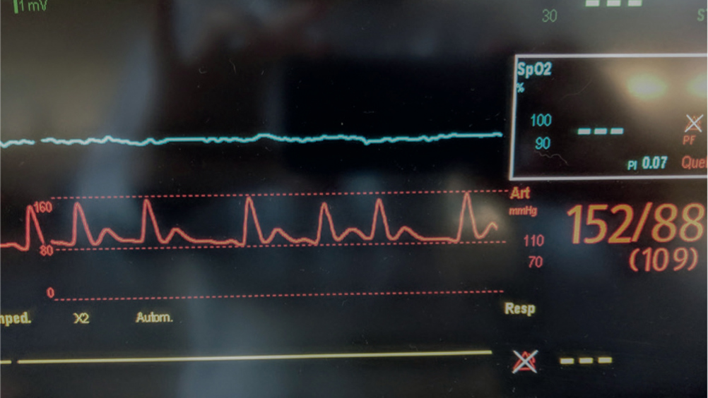
Placing an arterial line is an invasive procedure, it can be painful and uncomfortable for the patient and presents other risks, as discussed in this article. It is, however, a valuable tool in intensive medicine and provides benefits such as continuous blood pressure (BP) measurement and arterial blood gas sampling. Benefits and risks of using non-invasive blood pressure (NIBP) versus invasive blood pressure (IBP) monitoring techniques need to be considered with each patient. This article reviews IBP monitoring, including: how to place an arterial line; waveform analysis; complications; troubleshooting and comparison to NIBP (Figure 1).

What is IBP?
IBP is an invasive and direct measurement technique. An arterial line is a catheter that is placed into an artery. This is attached to compatible tubing which is connected to a monitor via a transducer. This allows the measurement of IBP, observation of the arterial waveform and sampling of arterial blood gases.
BP measurement, whether direct or indirect (via oscillometric, high definition oscillometric (HDO) or ultrasonic Doppler flow), is defined by the measurement of pressure of blood flow on the vessel wall. The unit of measurement is millimetres of mercury (mmHg). Systolic arterial pressure (SAP) is the measurement of pressure during cardiac systole (contraction). Diastolic arterial pressure (DAP) is the measurement of pressure during cardiac diastole (relaxation). The mean arterial pressure (MAP) is calculated from the SAP and DAP.
During a cardiac cycle, the heart spends twice as much time in relaxation (diastole) as it does in contraction (systole), accordingly the calculation for MAP reflects this. The MAP is calculated from systolic and diastolic pressures with the following calculation:
MAP = ([SAP-DAP]/3) + DAP
Normal values for blood pressure are species-specific and are shown in Table 1.
Table 1. Normal arterial blood pressure values in dogs and cats
Normal values | Dogs | Cats |
---|---|---|
Systolic arterial pressure | 90–140 | 80–140 mmHg |
Diastolic arterial pressure | 50–80 mmHg | 55–75 mmHg |
Mean arterial pressure | 60–100 mmHg | 60–100 mmHg |
Indications for arterial line placement — why?
Arterial lines allow reliable BP measurement in critically ill patients and high-risk anaesthesia/surgery patients. These patients may be more likely to experience rapid changes in pressures for various reasons and having a continuous IBP is advantageous. Arterial lines also enable blood to be taken conveniently as blood can be taken from the catheter, for example for blood gas analysis.
Key points:
- Continuous BP monitoring allows for constant assessment of the patient's perfusion status
- Using continuous IBP allows for better titration of vasopressor/catecholamine (e.g. dopamine/noradrenaline), vasodilator (e.g.nitroprusside) or inotropic (e.g.dobutamine) medication
- When BP falls, it can be very difficult to measure with a non-invasive device. Having an arterial line means that even when the BP drops, an accurate measurement is possible
- It is important to place an arterial catheter earlier rather than later before pulses are non-palpable making placement more difficult (see Box 1, and Tables 2 and 3).
Box 1.Example cases benefiting from arterial lines
- The septic abdomen patient. Depending on consciousness and awareness levels a catheter will be placed before or just after the patient is anaesthetised. These patients are often unstable and need intensive monitoring/nursing, fluid resuscitation and vasopressor/catecholamine therapy. Having an accurate invasive blood pressure (IBP) will guide this treatment. After recovery, the arterial line will often be left indwelling for continued monitoring and blood sampling.
- Patients at risk of haemorrhage during surgery, and subsequently needing a transfusion. For example (but not limited to) splenectomy/liver lobectomy surgery, laminectomy/hemilaminectomy or ventral slot. One of the signs of acute blood loss is a rapid drop in blood pressure. These patients are often stable after surgery (where no significant bleeding has occurred), in those patients arterial lines can be removed soon after waking up.
Table 2. Diseases associated with secondary hypertension in cats and dogs
Dogs | Cats |
---|---|
Acute kidney disease | Chronic kidney disease |
Chronic kidney disease | Diabetes mellitus |
Diabetes mellitus | Hyperthyroidism |
Hyperadrenocorticism | Obesity |
Hypothyroidism | Pheochromocytoma |
Obesity | Primary hyperaldosteronism |
Pheochromocytoma | |
Primary hyperaldosteronism |
Table 3. Possible causes of hypotension in cats and dogs
Hypovolaemic and distributive shock1 |
Lowered systemic vascular resistance through; sepsis, anaphylaxis, drug-induced1 |
Vasodilation through anaesthesia1,2, |
Decreased myocardial contractility3 |
Cardiac arrhythmias and cardiogenic shock3 |
Obstruction of venous return3 |
How to place an arterial catheter
Unlike intravenous catheters (IVC), arterial catheters are placed based on palpation only; there is no visual aid. Making pulse palpation (including pedal pulse) a part of the veterinary nurse's daily routine will help in the first step to placing an arterial catheter.
Common arterial catheter placement sites in veterinary patients are the dorsal pedal artery and the coccygeal artery; less common but also suitable are the lingual, radial and auricular arteries. The dorsal pedal artery can be easily palpated in medium to large dogs, but in smaller dogs and cats this becomes increasingly more difficult. In cats, the coccygeal artery can sometimes be more accessible, but the catheter will need to be removed sooner due to the risk of contamination from soiling.
Placement begins with palpation; once the artery is located, the following steps are taken:
- The area is clipped and cleaned with an antimicrobial detergent such as a chlorhexidine 2% solution and surgical spirit. The placer must disinfect their hands before re-palpation of the artery after prepping. Sterile or non-sterile gloves can be worn.
- A skin stab incision can be made over the placement site with a large bore needle. This avoids blunting of the catheter before entering the artery lumen and may allow for smoother catheter placement, but may come with a greater risk of infection (Band and Maki, 1979). Using a 20G or 22G (24G for small dogs and cats may be suitable) the larger the catheter lumen the less likely it is to occlude later (Scheer et al, 2002). The catheter is placed with short, confident movements at a 10–30 degree angle.
- Once blood is seen in the stylet hub the catheter is flattened down and advanced. The stylet is removed, the catheter is capped and taped in securely, the bandage placed over the catheter should be marked with ‘Arterial Catheter’ to avoid accidental administration of drugs via this catheter (Figures 2–4).


At this point, a blood sample for arterial blood gas analysis is often taken (Box 2). Next, the catheter and the transducer need to be connected via plastic low compliance tubing to minimise dampening of the arterial signal. After that, the transducer can be connected to the monitor and the system must undergo the zeroing process described below.
Box 2.How to measure blood gases – three syringe sampling techniqueBlood may be sampled directly at the catheter or at the flushing port/transducer.Discard: The author uses a five ml syringe to draw back the two ml of heparinised saline in the extension line leading the to patient plus one to three ml of blood, this is to ensure no dilution of the blood sample (Cole et al, 2007)Sampling: The sample is collected using a one ml blood gas syringe.Flush: The line is flushed with another two ml of heparinised or non-heparinised saline.Cole et al, 2007
Zeroing the transducer
The transducer needs to be zeroed every time it is manipulated. Examples include: after patient movement; after disconnecting; and after blood sampling. Zeroing should also be performed when the measurements and waveforms are unreliable, incorrect and when artefacts are present. The transducer must always be positioned at the level of the heart for a correct reading.
- Close the three-way stopcock to the patient.
- Remove cap from the stopcock, exposing it to the atmosphere.
- Zero the system via the monitor.
- Once zeroing is successful, a new cap is placed on the stopcock and it is reopened to the patient.
Points to note: if the patient moves from lying to sitting or standing then readings will no longer be reliable. The transducer must always follow the patient to stay at the level of the heart (left atrium) in order to measure correctly. If the transducer is too low, over measuring will occur and if too high under measuring will take place.
Arterial waveforms, what do they tell us?
The waveform can give further information and can also indicate if the numbers are reliable. The waveform is made up of three phases:
- The systolic phase (anacrotic limb)
- The dicrotic notch (the dicrotic notch represents the closing of the aortic valve just after systole)
- The diastolic phase (dicrotic phase).
These are all outlined in Figure 5.

A steep incline represents a greater pressure over time indicating strong contractility. A shallow slower rise would represent weaker contractility. The dicrotic notch or delay thereof may be associated with vascular resistance and therefore helpful in treatment for vasodilatory shock (Nirmalan and Dark, 2014).
Dampening
Dampening is the absorption of the arterial signal, this can occur through various mechanisms, and can be under or over amplified. This is known as over or under-damping. Over-damped waveforms are waveforms which look like they have been flattened out. The systolic may be underestimated while the diastolic may be overestimated. An underdamped waveform will cause the opposite effect, an overly heightened or spikey waveform with excessive oscillations. Under-damping may overestimate systolic pressure and underestimate diastolic pressure.
Both under and over-damping are usually resolved by the following: the measuring system should be checked for leaks, kinks, clots or air bubbles, switching from longer to shorter and high to low compliance tubing. See Table 4 for causes of over and under-damped waveforms.
Table 4. Causes of over and under-damped waveforms
Causes of over-damped waveforms | Causes of under-damped waveforms |
---|---|
Air bubbles | Stiff tubing |
Loose connections | Long tubing |
Embolisms/clots | Increased vascular resistance |
Low flush bag pressure |
Maintenance and management
Maintenance of the arterial catheter is similar to a venous catheter — it must be checked daily and flushed at least every 2 hours or ideally continuously. Care should be taken so the bandage remains clean and not contaminated with blood/urine/faeces. The tape surrounding the catheter should also remain as clean as possible to prevent catheter-related infection.
Key points:
- The catheter must be secured using sterile tape (e.g. Micropore™, 3M) and protected with a sterile padding layer (e.g. Soffban™, BSN medical) and a conforming tertiary layer (e.g. Coban™, 3M; Elastomull (BSN medical) (Figure 6).
- The placement site must be inspected at least daily for signs of inflammation or infection using non-sterile gloves.
- The bandage must be changed daily or more frequently if necessary.
- The bandage must be clearly labelled along with the tubing. Ideally, a different colour tertiary bandage should be used, to reduce the risk of errors.
- The catheter must be flushed regularly to avoid occlusion or thromboembolisms either manually or via a pressurised saline bag (delivering fluid at a rate of 3–4 ml/hour).

How and when to remove the catheter
The time in which to remove the catheter must be discussed with the veterinary surgeon. Usually, removal is carried out when the patient is deemed stable, it is no longer required or the catheter is occluded. The catheter is removed much like an IVC — first the protective layers and tape, then the catheter itself. When removing an arterial catheter, one must have a pressure bandage prepared to place during the removal of the catheter itself to avoid excessive bleeding and soiling of the patient.
Tip: make a note on the patient's chart to remove the pressure bandage in 30–60 minutes to avoid swelling and discomfort from the bandage.
Care needs to be taken in coagulopathic patients, and with these patients, the catheter should be removed as soon as the patient is stable. Consideration needs to placed on patients waking up from general anaesthesia since they may make more erratic movements than usual. Seizuring patients or patients in status epilepticus recovering can be a challenge too — the decision of when to remove the catheter needs to be made on a case by case basis.
To reduce the risk of complication to the patient the Centers for Disease Control and Prevention recommends removal of arterial catheters as soon as they are no longer needed (O'Grady et al, 2011). It is unclear what the ‘safe’ amount of time is to keep an indwelling catheter in place, but what is clear is that the risk of complication goes up with time left indwelling (Khalifa et al, 2008). As it is especially difficult to keep veterinary patients clean and hygienic, to reduce risk, in the author's practice, arterial catheters are removed at a maximum of 48 hours post placement.
Complications
Arterial lines, like any indwelling catheter, come with complications. This section touches on the main complications and how to avoid them.
Infection
Infection is a complication present in indwelling catheters (Lemaster et al, 2010). Infection may be avoided by:
- Disinfecting the site and hands before placement (Coopersmith et al, 2002; Vallés et al, 2007)
- Use of Teflon™ catheters (Lopez-Lopez et al, 1991)
- Cleaning away blood spillage (an excellent medium for bacteria) prior to securing the catheter and cleaning the patient regularly to avoid bandage soiling
- Bandage changes daily
- Daily catheter site inspection
- Replacing the flush bag and administration set every 96 hours (Ullman et al, 2013; Guillaumin et al, 2017)
- Aseptic technique while zeroing the pressure and blood sampling
- Removal of the catheter as soon as possible (Khalifa et al, 2008).
Bleeding
Bleeding may occur when a patient pulls the catheter themselves either by excessive movement (e.g. recovering post anaesthesia) or by chewing. Patients should be monitored by the nursing team 24/7 and not left unattended. Caution should be taken placing arterial catheters in patients with coagulopathies. If coagulopathy is suspected, a platelet count and coagulation profile should be checked prior to placement and the risk versus benefit decided by the veterinary surgeon.
Thromboembolism and ischaemic limbs
Thromboembolisms may occur during or post placement of an arterial catheter; this is believed to occur because of changes to vessel wall integrity after a catheter is placed (Bedford and Wollman, 1973). Clots may block/partially block the catheter or may get flushed into the circulation. Regular flushing with heparinised or normal saline may reduce these risks. Studies have shown no improved catheter patency by flushing with heparinised saline vs normal saline (Del Cotillo et al, 2008; López-Briz et al, 2014). If an embolism is present causing a temporary occlusion, a rare complication known as limb necrosis may occur (Bowlt et al, 2013). Due to these risks, especially in smaller patients where vessels are smaller, and embolisms are more prevalent (Scheer et al, 2002), the catheter should be removed as soon as it is reasonable to do so. The occurrence of permanent ischaemic damage in human patients is <1%, and to the author's knowledge the figure is unknown for veterinary patients (Scheer et al, 2002).
Accidental drug injection
Accidental drug injection can cause grave deleterious effects and must be avoided at all costs. In order to help prevent this, the catheter must be well labelled. Ideally, any ports should be covered with sterile tape to hinder accidental injection into an extension port, and the extension lines should also be labelled clearly. Injection of medication into an artery can lead to paraesthesia, severe pain, motor dysfunction, compartment syndrome, tissue necrosis, and limb loss (Dasgupta et al, 2012)
Troubleshooting
Keep the setup simple. Remove any unnecessary lines, ports and three-way stopcocks. Doing this will avoid interference with the waveform damping as well as avoiding introducing air bubbles into the circuit.
- Use the correct infusion lines: use tubing suitable for arterial BP monitoring. The tubing must not be overly compliant or too large in diameter. It should also not be excessively long.
- Keep the bag pressurised: the saline bag should be kept in a pressure bag at a pressure over 200 mmHg.
- Remove air bubbles: air bubbles not only have the potential to cause harm by producing an embolism but they are also a culprit in waveform artefacts.
- Zero the transducer: regular zeroing of the transducer should take place after patient movement, blood sampling and any interruption otherwise.
- Assess the transducer height regularly: the transducer must stay at the level of the heart to produce a reliable reading (Figure 7).

Comparisons with non-invasive methods
NIBP methods include oscillometric including high definition oscillometry (HDO) and ultrasonic Doppler flow. Oscillometric methods rely on recording motions under a pressurised cuff, these movement/oscillations are caused by arterial pulse pressure. Using oscillometric methods one can measure systolic blood pressure (SBP) and diastolic blood pressure (DBP) and from that calculate the MAP (Table 5).
Table 5. Advantages and disadvantages of invasive blood pressure (IBP) monitoring
Advantages | Disadvantages |
---|---|
IBP affords constant measurement and therefore a constant value, real-time changes are seen on screen; this allows for quicker treatment | More risk to the patient |
No errors due to incorrect cuff fitting | Painful to place. Arterial lines are often placed while the patient is already under general anaesthetic (GA) or sedated |
Most reliable measurement | Skill and training is necessary for placing arterial catheters |
Continuous blood pressure (BP) despite hypotension | An expense of material and equipment |
Considerations for oscillometric measurements
If the patient moves, pants, shivers or has arrhythmias this can create disturbances in the measurements. Measurements can take varying amounts of time to obtain, this may cause an elevation in BP due to stress during measurement making the value inaccurate.
Blood pressure monitoring using Doppler
The Doppler method is similar to the auscultatory (Korotkoff) method used in human medicine. BP is measured listening for pulse sounds with a Doppler crystal placed over an artery while reducing the pressure on the cuff placed proximal to the artery. The first audible sound is taken as the SAP; this has been shown to be a reliable SAP measurement in dogs (Chalifoux et al, 1985), but in cats, this may be better correlated to the MAP (Caulkett et al, 1998).
Doppler has been proven in one study to be more reliable than oscillometric measurement (Binns et al, 1995). However, in a more recent study oscillometric came out as being ‘the best prediction of systolic measurement’, but also stated that all indirect forms of BP monitoring underestimated the systolic BP (Caulkett et al, 1998). NIBP comes with less risk to the patient and delivers good results, but ultimately IBP is still deemed as the ‘gold standard’ of BP measurement techniques in veterinary patients (Bosiack et al, 2010).
Conclusion
In conclusion, BP monitoring is key in the intensive care patient. IBP monitoring is the most accurate form but does require skill and time to place, which is not always afforded in the emergency situation. Invasive methods come at an extra cost of materials along with potential risk, but the author believes the benefits outway the risks in the majority of critical patients. Non-invasive methods are useful, but may become unreliable in the severely hypotensive patient (Bosiack et al, 2010). Placement needs to be assessed on a case by case basis.
In the author's opinion, as the front-line carer for the veterinary patient, it is often the veterinary nurse's job to suggest or recommend certain interventions for their patients. Knowing not only how to perform such a procedure but also the reasoning, benefits and disadvantages, maintenance and aftercare helps nurses assist veterinary surgeons and above all better care for patients.
Key Points
- Invasive blood pressure (IBP) involves the placement of a catheter in the arterial lumen.
- IBP monitoring is considered the ‘gold standard’ in veterinary blood pressure (BP) monitoring.
- There are risks associated with an indwelling arterial catheters.
- Aseptic technique and good hygiene must be practised while handling/placing an arterial catheter.
- Patients with arterial catheters should not be left unattended.