
Kirby (2014) explained that the terminology keeps changing regarding resorptive lesions because they are a relatively poorly understood phenomenon; the aetiology has not been definitively proven. They have been called cervical neck lesions, feline odontoclastic resorptive lesions (FORL) and feline dental resorptive lesions (Zivkovich et al, 2010; Zivkovich et al, 2011), however they are now simply referred to as tooth resorption (TR), and they are commonly documented on dental charts using RL to indicate a tooth with a resorptive lesion.
What is a RL?
A RL is a hard tissue defect which most veterinary practitioners first identify when it reaches the cementoenamel junction (CEJ), and at this stage it can be identified by visual and/or tactile examination (Gorrel 2015). Gorrel (2015) advised that clinical examination will only facilitate detection of coronal defects, so concavities in the tooth structure at the CEJ or above (Zivkovich et al, 2010), as opposed to those developing on the root surfaces. They can appear anywhere on the tooth surface, however they tend to be more commonly located on the buccal and labial surfaces (Zivkovich et al, 2011).
Gorrel (2015) stated that TR is a common occurrence; various factors have been implicated, but not definitively proven regarding TR pathogenesis including: acidic pH of food, excess vitamin D, the presence of periodontal disease, dietary factors, mechanical stresses/abnormal forces, developmental defects, the breed of the patient, and viral diseases (Knaake, 2013; Niemiec, 2014). Zivkovich et al (2011) postulated that changes in the local environment around susceptible teeth may play a role in TR where the transfer of ions and organic molecules from the teeth into the saliva, and vice versa, may result in the dissolution of dental tissues, and thus concavities form.
There are some additional key facts and findings from research and expert clinical experience that veterinary practitioners should be mindful of when considering the identification and management of TR in their feline patients:
RL development
The initial resorption takes place on the roots of the teeth. Either the hard tissues of the root surface are destroyed by odontoclasts and are replaced by cementum, or the resorption starts in the cementum before extending into the dentine, the dentinal tubules and eventually the crown, so the crown and root are ultimately both affected by the resorption in many cases. Gorrel (2015) advised that the peripulpal dentine is relatively resistant to resorption so typically the pulp is only involved later in the disease process.
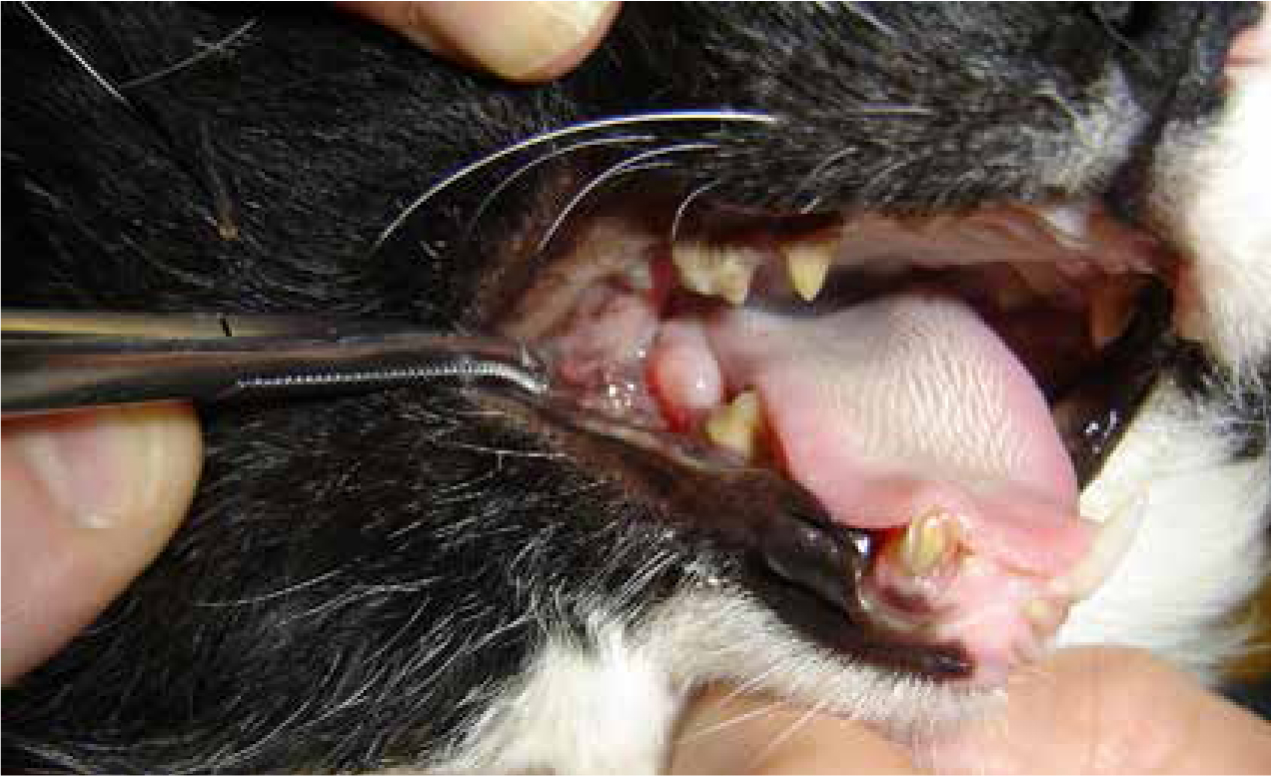
If the resorption extends through all of the crown's dentine the enamel can start to resorb, which will result in the creation of an enamel defect and the crown may fracture off completely due to its integrity being undermined (Gorrel, 2015). Without radiography, TR will only become evident clinically when an RL causes a defect at the CEJ or when the crown fractures off. When the crown is lost, root remnants are left behind which are likely to be resorbing and they become covered with intact gingiva, which may or may not be inflamed. So, during consultations when the veterinary practitioner is examining the oral cavity of a ‘gummy’ cat and the client says the patient's teeth have just disappeared over time, it is likely the crowns have fractured off due to RLs and there may well be root remnants subgingivally, which may or may not cause the patient problems (Clarke and Caiafa, 2014) (Figure 1). This cannot be confirmed without the use of intraoral radiography.

Internal and external root resorption
Gorrel (2015) described two types of resorptive processes, internal and external, which have been summarised in Table 1.
| Internal | The integrity of odontoblast layer of pulp is broached, so the resorption starts in the pulp and extends towards external aspects of the tooth |
|
| External | External resorption typically follows damage to the periodontal ligament (PDL) and cementoblast layers (CBL). |
Surface resorption This seems to be self-limiting and reversible and occurs in response to minor trauma, such as occlusal trauma, chewing, bruxism etc, or due to local damage to the PDL and CBL |
| Inflammatory resorption This is believed to begin when there is inflammation in tissues adjacent to the root, resulting in peripheral inflammatory root resorption (PIRR) and/or external inflammatory root resorption (EIRR) |
||
| Replacement resorption |
||
Diagnosis and radiographic appearance
It is clear that TR cannot be identified clinically until the lesions have reached the level of the CEJ and gingival margin where they can be visualised, however this tends to be during the later stages of the disease process (Gorrel, 2015). RLs can sometimes be hidden underneath calculus so they go unnoticed (Kirby, 2014) (Figure 2), or they may be identified as an erosive lesion filled with granulation tissue or hidden beneath haemorrhaging or hyperplastic gingiva (Southerden, 2010; Clarke and Caiafa, 2013). This is why examination under general anaesthesia is essential so a dental explorer or resorptive lesion probe can be used at a 90°angle to the tooth's surface, using small strokes, which will catch on the edge of any enamel defect and provide the operator with tactile feedback about the integrity of the tooth's surface (Kirby, 2014).
Clinical signs associated with RLs include: dysphagia, ptyalism, face rubbing, jaw chattering, inappetance, weight loss, a change of food preference, a decrease in food intake (Southerden, 2010; Gorrel, 2015); however the clinical signs vary dramatically between individuals and the extent of their RLs. If a RL has reached the CEJ/gingival margin and there is communication between the internal structures of the tooth and the oral environment then there is likely to be more sensitivity, or even pain experienced by the cat, potentially leading onto some of the other signs mentioned. Cats with RL confined to their roots are likely to be asymptomatic (Southerden, 2010).
Intraoral radiography is an invaluable tool in the diagnostic process as the images will allow the veterinary surgeon to identify lesions that are localised to the root surfaces in the alveolar bone (Mihaljevic et al, 2011; Kirby, 2014; Gorrel, 2015). The loss of tooth substance, the loss or irregularity of adjacent bone, and any changes in the radiodensity of the tissues can be properly assessed, which facilitates identification of the type and stage of the resorption present (Southerden, 2010; Gorrel, 2015) (Figure 3). Only when the veterinary surgeon has this information can they confirm with the owner the number of teeth/roots that need to be removed (Kirby, 2014); it is simply guesswork without radiography. Gorrel (2015) advised a full mouth series of radiographs in these patients; this will enable the veterinary surgeon to fully classify the RL and also identify root remnants which may or may not require extraction (Clarke and Caiafa, 2013). If cost is an issue and the owner has not consented to a full mouth series then radiography of at least 307 and 407 is advised as a screening protocol as these teeth are typically affected first by TR (Mihaljevic et al, 2011; Zivkovich et al, 2011; Gorrel, 2015); if they have RL then one can assume that it is highly likely other teeth will also be affected.
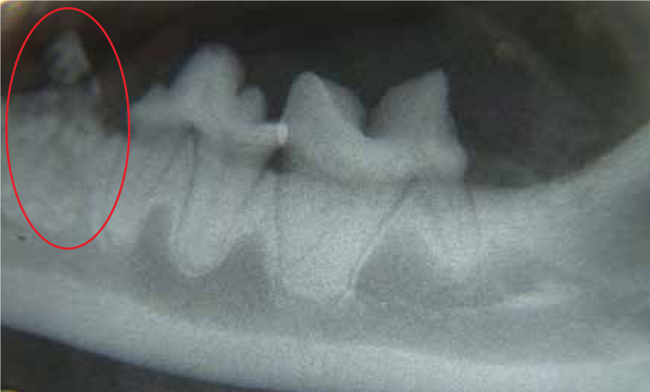
Table 2 outlines the types of root resorption according to radiographic features, their suspected cause and recommended treatments. Once the veterinary surgeon has ascertained the type of lesion present on an individual tooth this can be recorded on the dental chart.
| Type | Appearance/features | Cause | Treatment |
|---|---|---|---|
| 1 |
Otherwise normal root radiodensity however there are focal or multifocal radiolucencies evident within the tooth structure |
Possibly inflammatory and associated with periodontal disease | Standard extraction is warranted — a surgical (open) approach would be prudent |
| 2 |
Radiolucent roots or part of roots evident |
Truly idiopathic (there are suggested models for this type of resorption) | Crown amputation can be performed along with flap closure |
| 3 |
Features of both types 1 and 2 lesions described above in the same tooth | Combination of the above | A standard extraction of any root/part root is required which has a normal PDL space |
Knaake (2013) devised a 5 stage grading system for TR in cats, which is very user-friendly for all veterinary practitioners, and the stages outlined here can be used to help record what is found following radiographs on a dental chart (as suggested in the brackets following each descriptor):
Treatment
Kirby (2014) warned that feline roots are likely to crack during extraction and the presence of TR as well increases the likelihood of this happening. Gorrel (2015) outlined that treatment should aim to relieve pain if this is evident, prevent the progression of pathology, and restore function if this has been affected. Gorrel (2015) also clarified that there is no known, specific treatment with proven efficacy available for the prevention, development and progression of idiopathic RLs because it is very difficult to treat something effectively when the true cause remains unknown.
Restoration has been advocated in the past (filling the defect with restorative materials); however as the resorption is likely to continue the restoration will simply be lost so this is not a recommended treatment option (Gorrel, 2015). Zivkovich et al (2010) highlighted reported failure rates of 65% in literature, which was associated with any one restoration at any one site on a tooth within 6 months of it being placed.
Where crowns have cracked off and left behind root remnants, some veterinary practitioners have performed root atomisation, or root pulverisation, which is absolutely not an acceptable way to treat these patients. Southerden (2010) advised that this approach risks incomplete extraction of the tooth material and potential damage to the near- by neurovascular structures, which could result in serious consequences.
There are three main potential management techniques for patients with RLs, which are outlined in Table 3.
| Conservative management | This involves monitoring the patient's teeth clinically and radiographically. It is advisable to get the owner to bring the cat in at least every 3 months for a visual inspection; if a resorptive lesion (RL) does ‘surface’ then at least the cat is not left for too long without the lesion being identified |
| Tooth extraction | This is considered the gold standard treatment, removal of the whole tooth, especially with type 1 RL. |
| Coronal amputation | This can be performed when the root has proven, radiographically, to be extensively resorbed and it would therefore be impossible to extract all of the tooth material. Type 2 RLs |
Conclusion
It is always challenging trying to treat a problem for which the aetiology is unknown, however there is enough evidence from literature for all veterinary practitioners to use to create a protocol for optimal treatment of TR cases in practice. Perhaps, with ongoing research, experts in the field will identify the true aetiology of TR, which may lead on to the development of different treatment modalities. However, based on what is known, restoration of RLs as a result of TR is inadvisable due to the ongoing resorptive processes, therefore the optimal treatment remains intraoral radiography to identify the lesion type and stage followed by tooth extraction or coronal amputation.