References

Anderson A. Treatment of hip dysplasia. J Small Anim Pract.. 2011; 52:(4)182-9 https://doi.org/10.1111/j.1748-5827.2011.01049.x

Dyce J, Wisner ER, Wang Q, Olmstead ML. Evaluation of risk factors for luxation after total hip replacement in dogs. Vet Surg.. 2000; 29:(6)524-32 https://doi.org/10.1053/jvet.2000.17858

Fossum T. Textbook of small animal surgery.Missouri: Elsevier; 2007

Fries CL, Remedios AM. The pathogenesis and diagnosis of canine hip dysplasia: a review. Can Vet J.. 1995; 36:(8)494-502

Guevara F, Franklin SP. Triple pelvic osteotomy and Double pelvic osteotomy. Vet Clin North Am Small Anim Pract. 2017; 47:(4)865-84 https://doi.org/10.1016/j.cvsm.2017.02.005

Harper TAM. Conservative management of hip dysplasia. Vet Clin North Am Small Anim Pract. 2017; 47:(4)807-21 https://doi.org/10.1016/j.cvsm.2017.02.007

Henrigson B, Norberg I, Olssons S-E. On the etiology and pathogenesis of hip dysplasia: a comparative review. J Small Anim Pract. 1966; 7:(11)673-88 https://doi.org/10.1111/j.1748-5827.1966.tb04393.x

King MD. Etiopathogenesis of Canine Hip Dysplasia, Prevalence, and Genetics. Vet Clin North Am Small Anim Pract. 2017; 47:(4)753-67 https://doi.org/10.1016/j.cvsm.2017.03.00

Nelson LL Surgical site infections in Small Animal Surgery. Vet Clin North Am Small Anim Pract. 2011; 41:(5)1041-56 https://doi.org/10.1016/j.cvsm.2011.05.010

Punke JP, Fox DB, Tomlinson JL, Davis JW, Mann FA. Acetabular ventro-version with double pelvic osteotomy versus triple pelvic osteotomy: a cadaveric study in dogs. Vet Surg. 2011; 40:(5)555-62 https://doi.org/10.1111/j.1532-950X.2011.00802.x

Rasmussen LM, Kramek BA, Lipowitz AJ. Preoperative variables affecting long-term outcome of triple pelvic osteotomy for treatment of naturally developing hip dysplasia in dogs. J Am Vet Med Assoc.. 1998; 213:(1)80-5

Richardson DC. The role of nutrition in canine hip dysplasia. Vet Clin North Am Small Anim Pract. 1992; 22:(3)529-40 https://doi.org/10.1016/S0195-5616(92)50053-5

Smith GK, Karbe GT, Agnello KA Pathogenesis, diagnosis, and control of canine hip dysplasia’, 1st edition. St Louis: Elsevier; 2012

Surgical treatment options for hip dysplasia
02 December 2019
7 mins read
Volume 10 · Issue 10

Abstract

Hip dysplasia is thought to be the most commonly diagnosed orthopaedic condition in dogs. There are both conservative and surgical treatment options available to the owner and there will be a number of factors which will be involved in their decision making. This article will focus on the surgical treatment options, giving the veterinary nurse (VN) the knowledge of the options available and what is involved, in order that the VN can help the owner in their decision and, if applicable, the VN has the knowledge to be able to work within the surgical team when performing these surgical procedures

Hip dysplasia (HD) is thought to be the most commonly diagnosed orthopaedic condition in the dog and, although primarily genetic, environmental factors can have a great bearing on the severity of the disease (Smith et al, 2012).

HD was defined by Henrigson et al (1966) as ‘a varying degree of joint laxity of the hip joint, permitting subluxation during early life, giving rise to varying degrees of shallow acetabulum and flattening of the femoral head, finally, inevitably leading to osteoarthritis’.

Joint laxity refers to the instability which occurs during weight bearing which then leads to subluxation of the femoral head (Smith et al, 2012). In order to create a normal hip joint there needs to be normal load present between the femoral head and the acetabulum. In the dyplastic hip, the constant subluxation of the femoral head from the acetabulum leads to more mechanical load concentrated on the dorsal acetabular rim which in turn will then effect development and can lead to problems such as microfractures of the acetabulum (King, 2017). Osteoarthritis (OA) follows secondary to hip dysplasia due to the ongoing effects of joint laxity if left untreated. In the young dog, in-flammation of the hip joint capsule, abnormal wearing of cartilage and microfractures of the acetabular rim (from the repetitive abnormal load) contribute to the signs of OA (Fries and Remedios, 1995). Although OA cannot be cured, HD can be treated before it progresses to OA.

There are conservative treatment methods for OA in-cluding weight control, non-steroidal anti-inflammatory drugs, hydrotherapy and neutraceuticals, however, the focus of this article will be on surgical treatment options for HD. These include double (DPO)/triple pelvic osteotomy (TPO), femoral head and neck excision (FHNE) and total hip replacement (THR). Treatment options will depend on factors such as the age of the patient, client finances, expertise available and what the future performance of the patient is required to be (Harper, 2017).

Femoral head and neck excision

As the name suggests, this is removal of the head and neck of the affected femur with the outcome being immediate relief from the pain of OA or the prevention of symptoms of OA (Harper, 2017). The head of the femur and the acetabulum will no longer be in contact and the removal of the femoral head will result in the formation of a ‘psudoarthrosis’ composed of fibrous tissue (Fossum, 2007).

In smaller patients the outcome of this surgery is thought to be more successful, although there is not a specific size limit to this procedure. It should be remembered, however, that very young and skeletally immature animals may improve clinical signs with maturity so the surgery should be avoided in these patients. There are few complications associated with this surgery but patients may develop a decreased range of motion in the affected limb and some limb shortening may occur (Fossum, 2007).

FHNE can also be carried out for fractures of the femoral head and neck and also acetabular fractures

This procedure is probably the least technically demanding on the veterinary surgeon (VS) and requires only a small amount of surgical kit with no implants required (see Box 1). A scrub nurse may be required to help with retraction of soft tissues and lavage when the power saw is in use.

Box 1.Surgical kit required for femoral head and neck excision

  • Basic surgical kit
  • Scalpel handle and blade
  • Mayo and Metzenbaum scissors
  • Artery forceps
  • Towel clips
  • Dissecting forceps
  • Needle holders
  • Saline lavage and 20 ml syringe
  • Suction
  • Power saw
  • Gelpi self retaining retractor
  • Langenbeck retractors
  • Diathermy

Pelvic osteotomy

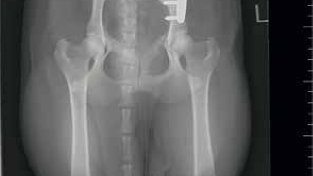
This procedure is carried out in young dogs, normally before 6 months of age before some of the effects of hip sub-luxation become apparent, such as bone formation in the acetabulum or damage to the femoral head (Fossum, 2007). If surgery is successful, the outcome is to improve dorsal coverage of the femoral head by the acetabulum in order to reduce the risk of luxation (Anderson, 2011). This is achieved by rotation of the acetabulum through cuts made to different parts of the pelvis (ischium, pubis and ilium for triple pelvic osteotomy and pubis and ilium for double pelvic osteotomy) (Guevara and Franklin, 2017). The osteotomy of the ilial wing is held in place with a specially designed plate which is selected based on the degree of rotation required (Figure 1).

Figure 1. A postoperative radiograph following a triple pelvic osteotomy.

DPOs, which omit the cut to the ischium, are thought to be associated with fewer complications than TPOs, however, due to the age of patients undergoing this procedure there may be a higher risk for implant failure due to the immature skeletal anatomy and bone density (Punke et al, 2011).

The age of the dog at the time of surgery can have an impact on how successful it is, which can make the window of opportunity for this surgery small. Rasmussen et al (1998) found that dogs with the average age of 7 months old at the time of surgery were seven times less likely to develop OA than if the dog was an average of 12 months old at the time of surgery.

As with any surgery, there is the risk of infection, which could be increased due to the use of implants. Other complications associated with TPO/DPOs include incorrect surgical technique, implant failure and narrowing of the pelvic canal (Vezzoni, 2002).

Total hip replacement

The most common reason for this surgery is HD and secondary arthritis, although there are other indications for the surgery such as femoral head fractures and revision of FHNE and pelvic osteotomies (Dyce et al, 2000). This surgery involves remodelling of the acetabulum with an acetabular cup and inserting a femoral stem component followed by femoral head prosthesis. There are two main systems used for this — cemented (acetabular cup and femoral stem) or cementless (biological fixation) which relies on ingrowth of bone around the femoral stem. There are no clear advantages to using any system for a particular age of patient, however it is thought that the biological system may be more advantageous for use in younger more athletic animals and the cemented system is suitable for older animals where bone quality may be more questionable (Anderson, 2011). There are implants available for a range of sizes including micro and nano sizes for cats which are only available as a cemented system.

The surgery is technically challenging and the decision to carry out the surgery should not be taken lightly. All treatment options should be thoroughly discussed with the owners. Surgeons require to have carried out a relevant certification course and perform a number of surgeries on cadavers and also under supervision of experienced surgeons. There is also the need for a scrub team, including a nurse, in order to keep surgery time as quick and efficient as possible in order to reduce infection risks. The scrub nurse should have knowledge of the surgical procedure and understood the order in which the stages take place in order to prepare equipment; in order to achieve this there is a certain amount of training which needs to take place with support from more experienced members of the team.

The VS needs to take preoperative radiographs of the patient in order to plan the size of implant needed (Figure 2).

Figure 2. Pre-operative ‘templating’ of the acetabular cup and femoral stem in order to ascertain the size of implant required.

Risks associated with this surgery include luxation of the joint postoperatively, femoral fracture (which can occur perior postoperatively) and infection (Dyce et al, 2000).

Sterility is of the utmost importance in this surgery. Infection has large consequences for the patient such as the need for revision surgery, amputation and even mortality.

At the author's workplace, the theatre in which the surgery takes place remains ‘closed’ throughout with no entry into or out of theatre (unless in an emergency). The scrub and circulating team all wear full surgical hoods and the patient has their anal glands emptied and a purse string suture placed pre-operatively to reduce the risk of faecal contamination.

Prior to surgery the patient is assessed fully including checking for any signs of remote infection, e.g. skin, ears, as any sort of infection such as this may have an effect on the risk of surgical infection. If there is any evidence of infection the surgery is cancelled until the infection is resolved.

Postoperatively the patient is taken to radiography to check that the implants are in the correct place (Figure 3). The surgical kit should be covered with a sterile drape and kept sterile during this time in case a return to theatre is required. Although it is not always avoidable, a return to theatre to revise any aspect of the surgery is thought to increase the infection risk in these surgeries (Nelson, 2011), and so it is a good idea to make a note of this for future record.

Figure 3. Postoperative radiographs showing the total hip replacement implants in the correct position.

The VN's role

The VN in general practice is often the person who spends the most time with the client, either in consultation or over the phone. Although these procedures may not take place at every practice, and the patient may be referred for surgical treatment, it is important that the VN has knowledge of the surgical treatment options for HD. Having this knowledge can help to counsel the owner in their decision for treatment. There may be factors affecting the decision for surgical treatment such as financial or owner constraints. The ability to help the owner make a decision is something which VNs should have the confidence to be able to assist with.

If the practice does carry out these surgical procedures, the VN may be involved in the surgery as a circulating or scrub nurse. Knowledge of the procedures will help them to run efficiently, which is important in all procedures, especially in the case of THR.

The VN may also be involved in the postoperative care of these patients. Considerations include ensuring that the patient is comfortable and adequately analgesed postoperatively, keeping the wound dry and covered, and keeping activity levels to a minimum.

After THR, it is vitally important that the animal remains calm (temperament should be taken into account when making the decision for surgery) and is sling walked with a minimum of two people for toileting purposes only for the first 24–48 hours, depending on the dog's temperament. If the VN suspects that there may be any chance of luxation or any other complication they should inform the VS.

The use of cryotherapy such as cold compresses around the surgical site in the immediate postoperative phase can aid with inflammation and pain relief, however, this should only be carried out under the instruction of the VS in charge of the case. The cold compress should be adequately covered and not applied directly to the skin of the patient.

Conclusion

There are a number of surgical treatment options available for HD in order to slow down the onset of OA, each with their own technicalities and potential complications. By having the knowledge of these, the VN is able to (alongside the VS) help the owner in making an informed choice for their pet.

The VN may also be involved with these patients in the pre-, perior postoperative phases and so the knowledge and skills to enable them to help with care of the patient and efficiency of the surgical procedure is advantageous.

KEY POINTS

  • There are a number of different surgical treatment options for hip dysplasia.
  • There are many factors which will need to be taken into account by the VS when deciding on the best course of action.
  • VNs should have a knowledge of all the options so that they can aid the client in making a decision.
  • VNs will have a very active role in the care of dogs receiving a total hip replacement, whether that is pre and post operative or as a part of the theatre team.
  • A knowledge of any of the discussed surgical treatments is required to aid with efficiency of the surgical procedure.