
Urethral obstruction is a problem that occurs predominantly in male cats of any age, breed and gender. This is because the urethra of a male cat is much longer and narrower than that of a female cat, thus is more susceptible to becoming blocked. Other factors that may influence urethral obstruction are being middle-aged, overweight, neutered and an indoor cat (International Cat Care, 2016). The blocked male cat is a familiar case in both general and emergency veterinary practice.
Signs and causes
Cats with urethral obstruction will typically show signs of urinary tract inflammation and discomfort. Signs may vary from mild to severe. Systemic signs are often not present until 24 hours after the urethra becomes blocked. The patient may present with the following signs:
Urethral obstruction can be a life-threatening emergency that requires immediate veterinary attention. If left untreated, it can cause acute kidney failure, electrolyte imbalance and potentially death within 3–6 days (Brooks, 2017).
Small uroliths or an accrual of crystals, characteristically struvite or calcium oxalate, may cause a urinary obstruction. These uroliths and crystals form in the kidney and move down to the bladder, or form in the bladder but can become lodged in the urethra. Swelling and spasm of the urethra may also contribute to a blockage if the cat is unable to relax the muscle due to inflammation. Typically, a mucoid plug rather than uroliths obstructs the urethra at the most narrow point and prevents urinary flow (International Cat Care, 2016). This causes filling of the bladder. Once the bladder has reached maximum capacity, the urinary pressure will increase up the ureter to the level of the kidneys. Due to an increase in pressure within the renal tubules, glomerular filtration ceases. This leads to azotaemia, hyperkalaemia, and metabolic acidosis.
Diagnostics
On initial presentation, clinical examination will reveal a distended and painful bladder when a complete obstruction has occurred. Limiting diagnostics at first allows time to focus on the most life-threatening abnormalities of hyperkalaemia, poor perfusion and acidosis. Initial diagnostics should include minimum database: blood for baseline haematology, biochemistry and electrolytes; evaluating these is vital to check for potential life-threatening disturbances.
Additional diagnostics are patient dependent, and may be carried out after urethral catheterisation has been successful. These may include:
If possible, collecting urine samples during the placement of a urethral catheter to prepare urine sediment for slide evaluation is advantageous. Urinary sediment may reveal a urinary tract infection, crystalluria, cystitis or abnormal cells. Identification of crystals is vital; this guides further treatment and prevention (Sabino et al, 2016). Radiographs and ultrasound investigations are unwarranted in the initial diagnosis for identification of urethral obstruction. Once the urethra is unblocked, ultrasonography and radiographic examinations may be undertaken for further diagnostic evaluation.
Obtaining a blood sample is advantageous to check haematology, biochemistry and electrolyte status. Haematology may reveal a raised leucocyte and neutrophil count, or no abnormalities at all. Biochemistry may indicate post-renal azotaemia hyperkalaemia and metabolic acidosis. Despite the potentially life-threatening consequences of hyperkalaemia, symptoms are often absent or mild. The increasing potassium levels have an effect on the myocardium and can cause arrhythmias. For cats presenting with bradyarrythmias, performing an electrocardiogram (ECG) is beneficial to check for any abnormalities. Quite frequently, and depending on the severity and speed of onset, hyperkalaemia may be associated with substantial ECG changes that can lead to death. Appropriate interventions may be required. Table 1 demonstrate significant ECG changes.
ECG changes do not become obvious until plasma potassium concentration is >7 mmol/litre, and become progressively more obvious when potassium concentrations reach >9 mmol/litre. Therefore, the visible ECG abnormalities shown in Table 1 indicate urgent intervention to reduce potassium levels before anaesthesia to unblock the patient (Norkus, 2006).
The cardiotoxic effects of hyperkalaemia manifest when potassium concentrations exceed 7.5 mEg/litre, which can significantly increase anaesthetic risks. Patients with serum potassium levels >5.5 mEq/litre should not be anaesthetised until the potassium levels have been reduced (Pachtinger, 2014). Therefore, instigating supportive treatment to decrease serum potassium levels is required. Fractious cats that appear painful may benefit from sedation prior to relieving the obstruction.
Treatment of hyperkalaemia
In cats with moderate to severe serum potassium levels, intravenous fluid therapy should commence to correct hypovolaemia, hyperkalaemia and metabolic acidosis. Assuming there are no confounding factors, e.g. cardiac disease, a bolus of 10–20 ml/kg of a balanced isotonic electrolyte solution is a suitable first bolus. There has been a lot of discussion on which choice of fluids to use for supporting patients with urethral obstruction. Dobromylskyj (2014) states dilution with potassium-free fluids such as saline 0.9% will achieve this more rapidly than the use of Hartmann's, but the administration of fluid containing 5 mmol/litre potassium will still reduce potassium concentrations of >9 mmol/litre. However, because many patients are mildly hyponatraemic, dextrose-supplemented normal saline will correct this while promoting endogenous insulin secretion to drive potassium back into intracellular domain. Conversely, Hetzel (2010) states that patients with urethral obstruction are often in a metabolic acidotic state and an acidifying solution, such as saline, may worsen the hyperkalaemia as hydrogen ions move into the cells in exchange for potassium ions.
Correcting hypoperfusion and establishing urine drainage will diminish potassium levels, but often-supplementary treatment is required. Drug options for hyperkalaemia include dextrose or insulin and sodium bicarbonate to reduce serum potassium concentrations, or calcium gluconate to reduce the effects of hyperkalaemia on the heart (Thomovsky, 2011).
If using dextrose-supplemented saline as a fluid choice there is no need for administration of dextrose or insulin. However, if Hartmann's is used, patients will benefit from administration of dextrose to stimulate endogenous insulin production and drive potassium from extracellular to intracellular.
The benefit of administering sodium bicarbonate is that it reduces the pH in the extracellular spaces by stimulating the exchange of intracellular hydrogen ions for extracellular potassium ions (Thomovsky, 2011).
Calcium gluconate 10% solution administered slowly by intravenous injection (0.5–1.5 ml/kg) is especially useful. While it has no effect on serum potassium levels, it will restore the difference between resting and threshold potentials and allow normal cardiac conduction to occur (Matthew, 1999). The effect lasts for 20–30 minutes. Administration of calcium gluconate must be gradual; too rapid injection can lead to further ECG abnormalities and even cardiac arrest. Intravenous dextrose (0.5 g/kg) and regular insulin (0.2 IU/kg) will directly reduce serum potassium levels by promoting intracellular translocation of potassium. Sodium bicarbonate was historically the first line of treatment for hyperkalaemia, but is now infrequently used due to better alternatives and because it can decrease the ionised calcium concentration further.
Premedication and anaesthesia
Sedation or general anaesthesia is required in order to catheterise and remove the urethral obstruction. The use of alpha-2 agonists should be avoided in cases of hyperkalaemia due to their effects on the cardiovascular system, and because they inhibit the release of endogenous insulin for approximately 2 hours, thus causing hyperglycaemia (Dobromylskyj, 2014). This insulin suppression will interfere with the use of glucose in the management of hyperkalaemia, as insulin is required for treatment to work (Dobromylskyj, 2014). Phenothiazines, such as acepromazine, may be safe at a low dose rate; they aid in reducing spasm associated with urethral obstruction, which can make it easier to dislodge calculi. One of the safest combinations for induction of anaesthesia in haemodynamically unstable patients is a benzodiazepine combined with an opioid (Norkus, 2006). Opioids perioperatively and postoperatively are essential in managing pain and providing comfort. Propofol is a useful injectable anaesthetic agent because doses can be administered slowly and to effect.
Relieving the obstruction
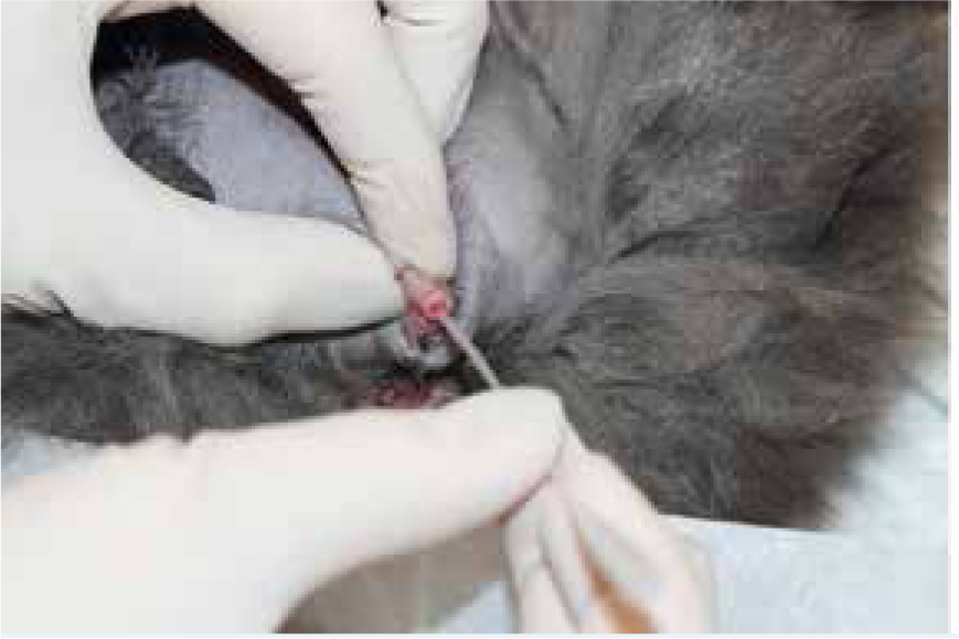
Relief of obstruction in male cats involves urethral catheterisation (Figure 2). Some cases may have an intra-penile obstruction; the penis can look cyanotic, and it may be possible to break down the obstruction physically with massage.
Equipment required for catheterisation:
With the patient positioned in lateral recumbency, using the thumb and index finger push the prepuce cranially exposing the glans penis. Introduce the tip of a lubricated catheter aseptically into the penile urethra. Once the catheter is in, clasp and pull the prepuce caudally to straighten the urethra and allow safe passage of the catheter. The catheter requires gentle advancing until urine appears at the catheter hub. Chandler (2007) advises to take care when advancing urinary catheters, particularly when passing the ischial curve. Corgozinho (2007) explains that damage can manifest from mild inflammation as a small amount of bleeding, to more severe urethral rupture and uroabdomen.
In cats with a blockage caused by a urethral plug, it may be necessary to use a catheter with a very small gauge and an aperture at the tip, such as the cannula of a 22 gauge intravenous catheter, or a lachrymal catheter. The catheter is advanced while flushing with copious amounts of saline. In some cases where catheterisation proves unsuccessful, emptying the bladder via cystocentesis can facilitate catheter placement, as the full bladder is no longer pushing caudally on the urethra. Cystocentesis should only be employed as a last resort as temporary decompression. Lane (2009) advises not to carry out cystocentesis on a prolonged obstruction or if the urine is extremely bloody in colour, which suggests a devitalised bladder wall. To avoid excessive bladder wall trauma a small gauge needle attached to an extension tube should be used. Once the catheter is in place and the blockage has been relieved, the catheter should be fixed in place. A closed urinary system should be attached; this averts any urine scalds to the patient and allows for accurate monitoring of urine output.
It may be necessary to use an alternative technique known as retrograde urohydropropulsion if catheterisation proves difficult. This method involves two people; the first person occludes the proximal urethra against the ventral pelvic bone, using a finger with rectal palpation. To occlude the urethra opening compress the tip of the penis, around the catheter. The second person is responsible for flushing saline into the catheter (this dilates the section of urethra between the two sites) until resistance is felt by the first person, who quickly releases the tension in the pelvic urethra. Saline flushed into the catheter, will flush the urolith into the bladder. Depending on individual cases, there are two options to treat the uroliths; these include surgery or medical therapy.
Post-obstructive diuresis (POD) is a complication that may occur in animals that have had a urethral obstruction for a long period (Thomovsky, 2011). Patients will produce a large amount of urine that exceeds the volume of fluids being administered, because of these further complications such as dehydration, hypovolaemia and shock may arise. Managing POD involves frequent monitoring of urine production and matching this to the rate of intravenous fluid administration. The gold standard method of monitoring urine output involves using a closed urinary collection system and measuring the volume of urine produced. Thomovsky (2011) recommends measuring urine every 4 hours, to work out urine output: divide the total volume of urine by the time over which it was produced to determine the total milliliters of urine per hour. To ensure intake and outputs match accurately, the fluid pump should then be set to the same milliliters of urine per hour until the next urine measurement is due. This allows for accurate measurements and alteration of fluid rate in accordance with the previous amount of urine production.
Nursing care
After removing the obstruction, the cat requires close and attentive monitoring. Analgesia is essential, and methadone is often the preferred choice, as an initial dose; in the author's experience buprenorphine may be administered 4 hours after the methadone, and continued every 8 hours. It is beneficial for the veterinary team to carry out pain scores on patients with a urethral obstruction to improve patient welfare. Pain is subjective, therefore to make pain management more effective, the same person should carry out the pain scores where possible. In addition to this, regular assessment of the patient's pain will allow for early detection and intervention if required. Opioids should be continued and used at the lowest dose needed to achieve comfort and help relax the urethral sphincter.
Other possible treatments include the use of dantrolene and prazosin. Studies indicate that they may be effective in relaxing intraurethral skeletal and smooth musculature in male cats. However, the studies revealed that it is not certain that administration of muscle relaxants would facilitate urethral catheterisation and removal of the obstruction in male cats with blockage of the lower urinary tract (Straeter-Knowlen, 1995).
Non-steroidal anti-inflammatory drugs should be avoided until hypovolaemia and azotaemia are fully resolved. The use of these can be beneficial in reducing inflammation and any disturbance in muscular coordination post obstruction. A suitably sized Elizabethan collar needs to be worn at all times to avoid interference with the catheter; self-removal of the catheter is unwanted due to the potential for re-obstruction, trauma to the urethra and the risks associated with re-catheterisation.
Blockage of the catheter may be a problem for cats that have obstructed initially due to urethral plugs or uroliths. To detect any problems, an ultrasound examination will demonstrate that the tip of the catheter is in the correct position in the bladder. It is crucial to monitor urine output thoroughly by emptying the bag and accurately measuring the urine using a measuring jug. Monitoring the quality and quantity or urine is essential in ensuring the patient is not re-obstructing or becoming dehydrated. Initially urine will be darker than anticipated in a healthy patient due to haematuria from the stretching and inflammation of the bladder, as well as catheterisation attempts (Segev et al, 2011). The aim is to maintain at least 1–2 ml/kg/hour. However, in the presence of renal tubular injury or loss of concentrating ability for other reasons, urine output can be extremely high (25–40 ml/kg/hour). In this situation, it is important to measure urine output and use this to guide adequate fluid replacement (Matthews, 1999).
Electrolyte assessment, specifically potassium, should be made every 4–24 hours; this depends on the severity of initial potassium levels and clinical assessment of the patient. Serum potassium is often high to start with. However, levels may decline during fluid diuresis, and may even need supplementing. If neutral insulin has been a line of treating hyperkalaemia, blood glucose checks should be taken regularly as the patient may become hypoglycaemic; the effect of intracellular translocation of potassium levels lasts approximately 4–6 hours. In addition, urea and creatinine levels should be measured every 12–24 hours to ensure they are decreasing as expected.
It is imperative to monitor the cat's weight daily. Cats with urethral obstruction may be inappetant; therefore, assisted feeding may be implemented. It is useful to try different bowls and food, but care must be taken not to overwhelm the cat. It may be necessary to place a naso-oesophageal feeding tube to initiate feeding, the cat will still be able to eat on its own if it wishes. Alternatively, mirtazapine 3.75 mg/cat (1/4 of a 15 mg tablet) per os (PO), every 3 days may be necessary to stimulate appetite. Syringe feeding should be avoided due to potential complications such as risk of aspiration, causing food aversions and increasing stress levels.
Reducing stress in the hospital environment is imperative and achievable. Outlined below is a methodical approach to reducing stress in feline patients:
Due to frequent blood samples being required in order to check electrolyte parameters, it would be advantageous to consider the following:
Potential complications
Risks and complications need to be considered in relation to catheterisation. With proper catheterisation technique, urethral damage is unlikely. Partial tears or ruptures may occur when there have been forceful and repeated catheterisation attempts. If this is suspected ultrasonography examination can be used to investigate tears and ruptures
Soft, non-irritating catheters are beneficial, these are less likely to cause bladder trauma when left indwelling.
It is fundamental for the patient's wellbeing that indwelling urinary catheters are managed correctly, and nurses are aware of the possible problems that may occur. Urinary tract infections can be caused at the time of catheterisation, a result of introducing bacteria into the bladder. The risk increases with repeated catheterisation attempts. Therefore, daily urinalysis can help lead to early recognition of urinary tract infections. Should the prepuce become soiled, it should be cleaned with chlorhexidine solution and warm water. Antibiotics are to be avoided unless required to treat an existing infection. The urine can be cultured after 72 hours or sooner if indicated and the catheter should be removed as soon as possible (Matthews, 1999).
Management at home
Preventing any further episodes of obstruction is the main long-term goal. Management of feline urethral obstruction needs to be continued once the patient is at home. It is imperative that an effective nurse consultation is carried out that highlights key areas that need to be addressed.
Dietary manipulation can aid in reducing the risk factors of urolith formation. There are various prescription diets available on the market which all help to reduce and effectively dissolve struvite uroliths. It is advised by Royal Canin that 5 to 12 weeks of dietary support is required to dissolve struvite uroliths (personal communication). To avoid recurrence, the diet should be fed for a further 6 months and regular urinalysis is advised. Once a patients is being successfully managed it can be maintained on a urinary diet. In general most urinary foods contain reduced magnesium, (as this is a component of struvite crystals), but low urinary magnesium concentrations have the potential to increase the risk of the formation of calcium-containing uroliths. This highlights the importance of regular urinalysis for a cat on a therapeutic urinary diet (Ackerman, 2012).
In addition to dietary changes, methods should be advocated to increase water intake, these are outlined below:
Client education plays a vital role in managing patients with urethral obstruction, where possible potential risk factors should be discussed at first vaccination or routine check-up appointments to help educate clients. For example, weight management should be discussed at routine appointments as most cats with urethral obstruction are overweight. This could lead to further discussions into diet paying particular attention to low carbohydrate, but high fibre diets. In addition to this, clients need to be made aware of the clinical signs of feline urethral obstruction to allow them to act quickly and seek veterinary attention.
Conclusion
Recognition of potential complications associated with feline urethral obstruction and the nursing care involved is pivotal to successful treatment of these cases. When handling the urinary collection system, strict aseptic techniques should be adhered to at all times. Close and vigilant monitoring of hydration status is crucial. Failure to detect electrolyte derangements, prolonging relief of urethral obstruction, and failure to match intake and output of fluids can have detrimental effects on patient welfare and successful recovery. To prevent further issues, veterinary nurses can have a huge impact on home management of these cases by carrying out effective nursing consultations and creating a home care plan for clients to follow.