
Mammary masses are one of the most common reasons for presentation to practice in female rats. A high incidence (80–95%) of these masses are benign fibroadenomas (Percy and Barthold, 1993) with this diagnosis coinciding with a high incidence of concurrent pituitary tumours. More rarely, adenocarcinomas and interstitial cell tumours can present, which carry a poorer prognosis. Because of the high incidence of fibroadenomas, these will be the focus of this article along with their management and prognosis. Male rats can also be affected, but this is less common as mammary growths are primarily stimulated by prolactin secretion and indirectly affected by oestrogen.
Hormonal influence
Fibroadenomas are known to be under hormonal influence, more specifically it is theorised that high levels of prolactin from prolactin secreting pituitary adenomas influence the development of these neoplasms (Bennett, 2012). A study by Hotchkiss (1995) concluded that oestrogen does not appear to increase the incidence of tumour development itself, but may be a contributing factor in the development of pituitary tumours, which in turn increases the incidence of mammary tumours. Ovariectomised rats (at 90 days of age) had a significantly lower incidence of pituitary adenomas than intact females (Hotchkiss, 1995); Hoefer and Latney (2011) reported a 90% incidence of fibroadenomas with concurrent pituitary tumours. In a study by Planas-Silva et al (2008) ovariectomy performed between 5–7 months reduced the incidence of spontaneous mammary neoplasms by 95%. More positively ovariectomy also increased the 110-week survival rate and overall lifespan, which could potentially be a result of a lesser incidence of pituitary tumours, however this was not analysed. Currently ovariectomy in general practice is not routinely carried out in female rats. This is likely for a variety of reasons. Owners rarely present rats to practice unless there is a perceived problem and, in the author's experience, new pet checks and discussion of preventative medicine is rare in rodent species. Financial implications for the client and the perception and reality of the risk of small mammal anaesthesia and surgery create barriers to performing such procedures. It may often be concluded that removal of a demarcated mass later in life is a simpler procedure than ovariectomy, however the majority of the former patients are geriatric and, in reality, likely pose a higher anaesthetic risk. Further evidence-based studies involving pet rats are likely required to confidently encourage prophylactic ovariectomy in this species.
Diagnosis
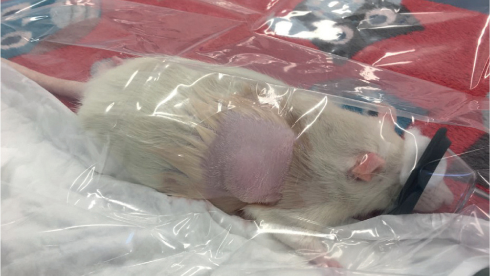
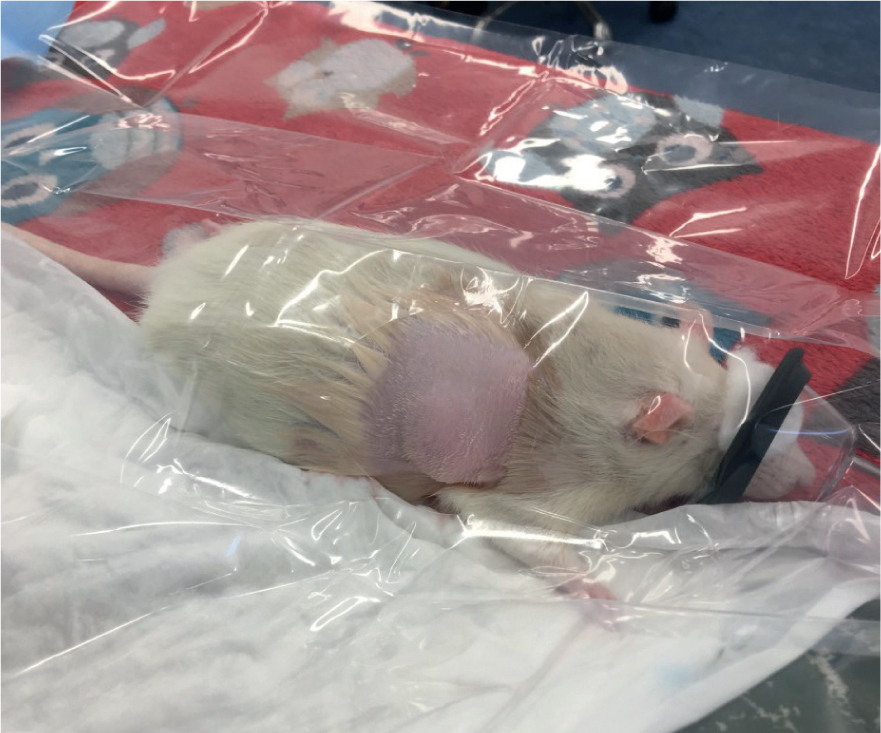
Mammary tissue in rats extends from the cervical to inguinal region ventrally and as high as the scapula dorsally. Initial presentation at an early stage may reveal a small pea sized lump situated anywhere along these mammary chains. These are usually non painful and well demarcated. Differential diagnosis by the veterinary surgeon at this point may include a subcutaneous abscess so a fine needle aspirate is a simple and easy technique to perform (under veterinary direction) to rule out the presence of infection. Often abscesses are associated with a bite or scratch that may be visible on the skin and/or fighting is reported by owners. Clipping over the area and applying a local anaesthetic cream before sampling is advised to improve patient tolerance. Other differentials may include granulomas, mastitis or mammary hyperplasia or adenocarcinoma. In the later stages where masses are large (Figure 1), a presumptive diagnosis is made by the veterinary surgeon based on the onset of a rapidly growing mass in the mammary regions. Definitive diagnosis can be achieved via fine needle aspirate, biopsy or histopathology.
Treatment
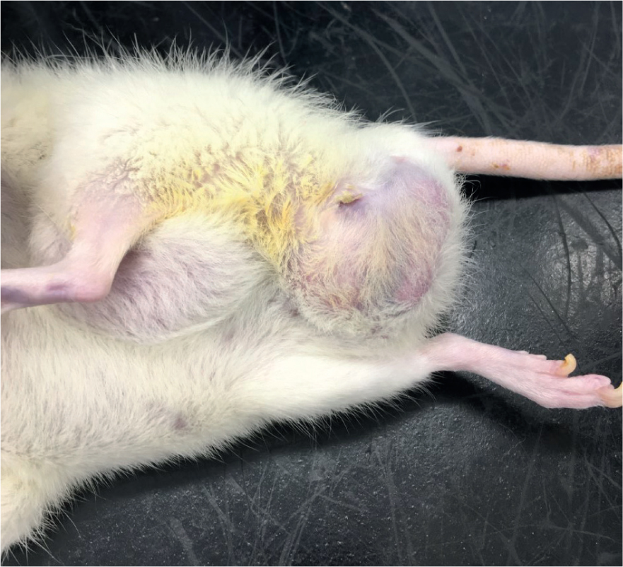
Surgical removal of the mass is the treatment of choice to improve quality of life where the mass is negatively affecting the rat or to prevent it from doing so. Bennett (2012) described surgical treatment as removal of the tumour and associated mammary gland along with ovariectomy in appropriate surgical candidates. If the mass is ulcerated or known to be malignant then removal of the overlying skin is important. In the case of fibroadenomas the skin can be preserved, facilitating easier closure of the wound. There are usually a few significant blood vessels supplying the tumour that need to be ligated while bluntly dissecting the tumour and then closure of the wound in two layers to limit dead-space (Bennett, 2012). Issues can arise in cases where inguinal masses are associated with the vulva/urethra (Figure 2). There is risk of iatrogenic damage to urogenital structures and the veterinary surgeon must perform a risk versus benefit analysis for surgical treatment of these cases. Assuming the patient is stable under anaesthesia, surgery can then progress to an ovariectomy, however it may be more appropriate to stage the procedures and perform the ovariectomy once the rat has recovered from the initial surgery. This mitigates the increased risk of haemorrhage, hypothermia and surgical/anaesthetic time that will occur when both surgeries are performed at once. In a lot of cases sole removal of the mass will be undertaken, however, it is important to inform clients that recurrence is common and several surgeries may be required (Frohlich, 2021).
Anaesthetic considerations for surgery
Rats do not need to be starved before anaesthesia because of their inability to vomit. The owner should be instructed to admit the rat in a familiar carrier with a hide and provide familiar foods. As with other small mammals, a companion will provide support to the surgical candidate and help to relieve stress and encourage eating postoperatively. As prey mammals, rats should be housed in a quiet ward away from predators, and acclimatisation to the environment is encouraged before restraint and/or drug administration. Although prescription of anaesthetic drugs can only be performed by the veterinary surgeon, the veterinary nurse should be encouraged to have a clinical discussion surrounding anaesthetic plans in this species in order to maximise patient comfort and the provision of an appropriate anaesthetic protocol. Premedication has several benefits and if the patient is tolerant of pre-operative injections then implementing the triad of anaesthesia (hypnosis, analgesia and muscle relaxation) as in other species is certainly beneficial. There are several drug protocols appropriate for surgery in rats and ultimately there is no ‘one size fits all’ for anaesthesia in small mammals. However, as with canine and feline anaesthesia certain drug protocols are more appropriate than others in certain scenarios. As with other prey mammals, rats are adept at disguising signs of illness and pain, and unfortunately it is likely less pre-anaesthetic screening (for example bloods) will be carried out, and monitoring of critical parameters such as blood pressure may be difficult because of patient size. Therefore, adopting protocols used for ‘critical’ patients may be more appropriate. In the author's practice a common premedication includes a subcutaneous injection of midazolam at 1 mg/kg plus buprenorphine at 0.05 mg/kg (Hawkins and Pascoe, 2012), or methadone at 0.5 mg/kg (methadone dose extrapolated from rabbit anaesthesia protocols). This often sufficiently calms the animal for inhalational induction with isoflurane or sevoflurane. In resistant patients, ketamine at 5 mg/kg can be added to the protocol (again extrapolated from rabbit anaesthesia protocols). Constant rate infusions should not be overlooked in these patients, and ketamine as a constant infusion provides excellent somatic analgesia, which is very useful for the likes of mass removal (Allweiler, 2013). Non-steroidal anti-inflammatory drugs (NSAIDs) should be provided as standard for surgery, provided there are no contraindications. Rats require much higher doses than canine and feline companions, and a study by Roughan and Flecknell (2003) concluded that a dose of 1 mg/kg of meloxicam significantly reduced post-operative laparotomy pain. Local anaesthesia should also not be overlooked, and incisional blocks of lidocaine with or without bupivacaine provide true analgesia at the site and should help to reduce the incidence of postoperative patient interference, which can be a problem in this species. In the author's experience, postoperative interference is more likely in patients that have received inadequate analgesia, where infection has occurred or where skin sutures (versus intradermal sutures) have been used. Rodents are generally intolerant of bandages and dressings and if skin sutures are used these can create a focus for the animal and divert attention to a healing wound. Gentle surgical technique from the veterinary surgeon, and excellent peri-operative analgesia, helps to reduce patient interference. The use of small Elizabethan collars in this species is not ideal and likely results in significant stress, especially for a prey animal as sight and sound may be altered. Postoperative analgesia may include the use of NSAIDs and tramadol at 5–20 mg/kg per os (Hawkins and Pascoe, 2012). Seroma formation, local infection, cellulitis, tumour recurrence and self-mutilation are all potential complications associated with this surgery, and must be discussed with the client before obtaining informed consent.
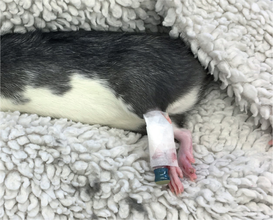
Fluid therapy is an essential part of anaesthetic management of these patients and intravenous catheters can be placed in the cephalic, lateral saphenous or lateral tail vein (Figure 3). A 26 g catheter is often needed, however, in larger individuals a 24 g catheter can be placed. If this is unable to be achieved then the subcutaneous, intraosseous and intraperitoneal (by a veterinary surgeon only) routes are available, however, there are limitations to these sites. Subcutaneous routes are most commonly used and relatively large volumes (5–10 mls) of warmed isotonic fluids can be delivered very easily in the scruff region (de Matos, 2011). There are very few contraindications to subcutaneous fluid therapy, especially in small mammals, however uptake can be variable and slow in compromised, hypothermic patients. The intraosseous route requires more skill and will usually be achieved when the patient is anaesthetised because of the pain associated with this procedure. The proximal femur is the most common site used and most drugs and fluids can be administered via this route, apart from hypertonic or alkaline solutions, which should be avoided (de Matos, 2011). The intraperitoneal route is often reserved for emergencies and is not routinely used in pet rats because of the risks of inadvertent puncture of internal organs, the risk of introduction of infection, the reliability of the technique (fluids may enter the subcutaneous space, muscle or lumen of an organ), and because drugs and fluids must first pass through the portal circulation, which may alter metabolism and biotransformation (de Matos, 2011). Often masses are extremely large, and significant proportions of tissues and their associated blood supply are removed. This may result in shock and significant hypovolaemia, especially if haemorrhage occurs, necessitating fluid therapy. Maintenance rates in rats have been estimated at 90–100 ml/kg/day (Girling, 2013). Thermal support is critical in these patients and hot hands, Bair Huggers, heat pads and snuggle safes can all be used. Incubators provide an ideal place for pre-operative warming after pre-medication and in recovery, and the author usually sets this to 26–28°C initially, then reduces this to 21–23°C once the rat is mobile and eating. Maintenance under gaseous anaesthesia is usually achieved via a face mask because of the difficulties in safely intubating this species in general practice. Fresh gas flow contributes to patient cooling and the drying of mucous membranes, therefore heat pads placed around the inspiratory tubing as close to the patient as possible may help to warm inspired gases.
Medical management
The influence of hormones on tumour development has resulted in numerous studies looking at medical trials for management of mammary neoplasia. A theoretical contender for treatment is gonadotropin-releasing hormone agonist (deslorelin impant), which suppresses the reproductive endocrine system and prevents the production of pituitary hormones (follicle stimulating hormone and luteinising hormone), and hormones produced by the gonads (oestradiol and progesterone in females and testosterone in males). Unfortunately, a recent study by Vergneau-Grosset et al (2019) failed to demonstrate a decreased risk of development of further mammary neoplasia in rats that had previously undergone mass removal (within 2 months) that were implanted with a 4.7 mg deslorein implant. Disappointingly these rats also did not have an increased survival time. It may be possible that, as discussed above, these rats had concurrent pituitary tumours secreting prolactin, which contributed to the development of subsequent tumours, however, more work in this area is required.
Some work has been carried out in relation to the drug cabergoline in rats and humans with prolactin secreting pituitary tumours. In humans cabergoline caused a significant decrease in mean adenoma size and the tumour disappeared in four macroadenomas and in 11 microadenomas after 12 months of treatment (Cannavo et al, 1999). In a rat study by Mayer et al (2011) cabergoline was administered at 0.6 mg/kg PO every 72 hours to a male rat that was diagnosed using magnetic resonance imaging (MRI) to have a large pituitary mass. A follow-up MRI 2 months later after starting treatment revealed the mass to have significantly decreased in size and the rat had no clinical signs of disease. Unfortunately, 8.5 months after the start of treatment the rat deteriorated and was euthanased. Post-mortem histopathology confirmed the diagnosis of pituitary adenoma. It would appear from these studies that cabergoline is promising in the development of treatment for pituitary disease in rats, but its use for the management of fibroadenomas is currently unknown.
In some cases owners may opt for palliative care with no surgical intervention. Often this is in aged animals. In these cases the focus is on the comfort of the animal and provision of analgesia where appropriate. Often large masses may become ulcerated and secondarily infected, and antibiotic therapy may be appropriate as an adjunct to analgesia. Provision of single-tiered cages with dry soft bedding is paramount when mobility is affected.
Conclusion
Rats are intelligent, curious and lovable pets and are favoured by many clients and veterinary professionals alike. Advancing medicine and surgery in this species affords clients alternative options for their pets and improves ethical decision making in veterinary practice. The development of fibroadenomas and concurrent pituitary tumours can grossly affect a rat's quality of life and necessitates the nurse and clinician to adopt a logical and structured treatment plan that benefits the patient and owner alike.
KEY POINTS
- Unneutered female rats have a high incidence of mammary fibroadenomas and concurrent pituitary adenomas.
- Mammary fibroadenomas are known to be under hormonal influence — mainly prolactin and to a lesser extent oestrogen.
- Surgical removal of masses can be curative but recurrence rates are high.
- Rats require species-specific surgical and anaesthetic care.
- Adequate analgesia is required for surgical candidates to help prevent post-operative interference with surgical wounds.