The purpose of this article is to give detailed instruction on how to place a jugular catheter. It will discuss appropriate occasions for placement and care and maintenance of the catheter.
Indications for placement of a catheter into a jugular vein include:
The introduction of foreign material or infectious agents into the central circulation can have far more serious consequences than peripheral vessel contamination; maintenance of aseptic technique is vital when placing jugular catheters.
It is worth noting that there are other sites for central venous access, e.g. the femoral vein but for the purpose of this article only the jugular vein will be discussed.
Contraindications
There are a number of contraindications to using a jugular catheter. These include:
Choosing the right type of catheter
Considerations
The compound used to make different types of catheters affects rigidity, reactivity and thrombogenicity. For example, catheters made of silicone and polyurethane are less thrombogenic than other materials such as polypropylene.
The length and gauge of catheter required is dependent on the purpose of the catheter and the patient size. Critically ill patients often require large volumes of fluid and therefore the largest gauge catheter should be placed. Length is important if the patient needs central venous pressure monitoring
Two techniques of catheter placement are available, the peel-away sheath needle technique and the modified Seldinger jugular catheter technique (BSAVA, 2007).
Types of jugular catheters available include single, double and triple lumen and it is important to decide prior to placement which is the most appropriate for your patient. This decision will be based on the functional requirements of the catheter, e.g. is frequent blood sampling and IV fluids required in which case a double lumen would be suitable. If the patient also requires total parenteral nutrition then a triple lumen may be more appropriate. It is important also to bear in mind that the more lines there are the narrower the lines therefore the largest catheter practicable should be considered.
Technique for catheter placement
Peel away sheath needle technique
There are a number of advantages and disadvantages to using the peel-away sheath needle technique for introducing catheters.
Advantages — different sizes of sheath needle and catheters can be purchased separately, the user can choose a combination of sheath needle and catheter that best suits the individual being catheterized.
Disadvantages — blood spillage and/or air embolization are possible during the placement procedure.
Modified Seldinger technique
There are also a number of advantages and disadvantages to using the modified Seldinger technique for introducing catheters.
Advantages — this technique allows entry into the vein without cutdown and because small gauge needles are used there is minimum trauma to the surrounding tissue and reduced risk of blood spillage.
Disadvantage — increased cost associated with purchasing the kit.
Comparison of the two techniques has shown there to be little significant difference between the techniques in ease of placement, haematoma formation and time to place (Portillo et al, 2006), but for the purpose of this article we will focus on the modified Seldinger technique.
Before beginning the procedure the required distance for catheter insertion is premeasured, the reason for this is that if the catheter is too long it may pose a risk of inducing cardiac arrhythmia (Crowe, 2006). The aim for a jugular catheter is to have a tip lying within the thoracic cavity, just cranial to the right atrium; this can be measured from the insertion site to the caudal edge of the triceps muscle or the first rib.
Patient positioning for catheter placement
Correct patient positioning and immobilization of the vein are vital to achieve successful catheterization. It is important that the patient remains still during jugular catheter placement to avoid trauma to the jugular vein, therefore sedation or general anaesthesia is often required. On occasions where the patient’s condition is so compromised that this proves impractical, local anaesthesia may be used, e.g. Emla cream or lidocaine placed at and around the insertion site. Jugular catheters are placed ante grade with the tip of the catheter always directed toward the heart (Davies, 2009).
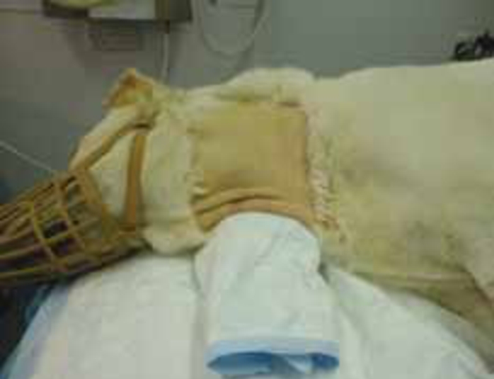
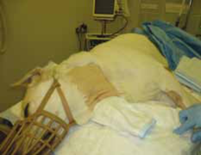
The patient is placed in lateral recumbence with a support placed under the neck (e.g. a sandbag) to help visualize the jugular vein. The patient’s head is extended and its forelimbs positioned caudally, this helps to both immobilize and make the vessel more accessible.
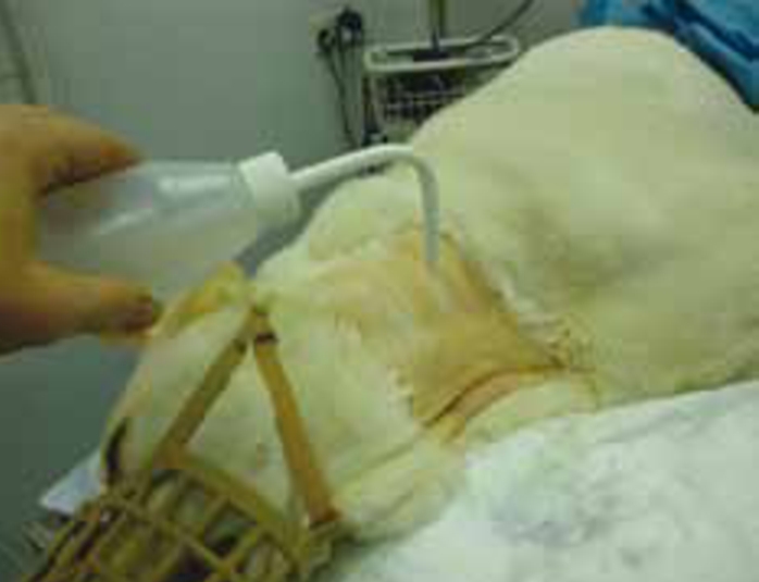
Preparation of the catheter insertion site
The site of catheter insertion should be identified by raising the jugular vein at the thoracic inlet (Crowe, 2006). An area is clipped around the site from the jaw line to the thoracic inlet giving at least 5 to 10 cm either side of incision in order to allow the multiple lumen of the catheter to be fixed appropriately, a larger area may be appropriate in cases where the patient has an excessively long coat. The area should be prepared as a surgical site. In order to achieve this it is important that the person preparing the site should first carry out effective hand hygiene (Vincent, 2012) and disposable gloves should be worn. The area should be prepared as a surgical site using any suitable surgical scrub solution; the author prefers a 50% chlorhexidine solution applied with clean (non-sterile) swabs, the area is then prepared with isopropyl alcohol.
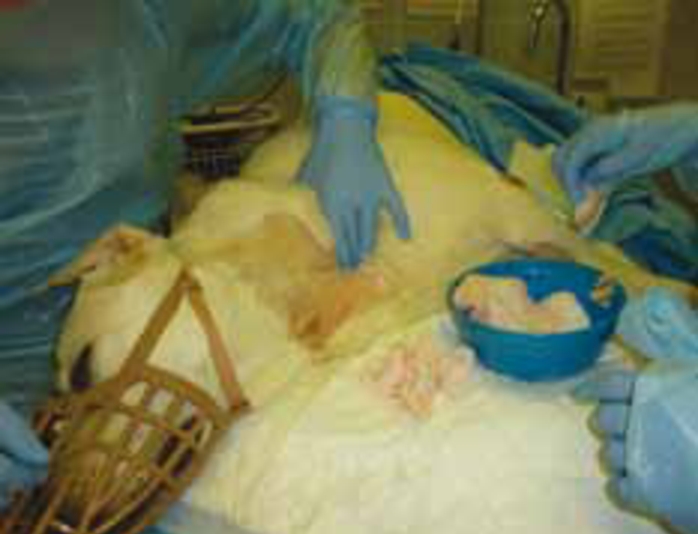
The procedure is a sterile surgical procedure and sterile gloves should be worn and asepsis maintained throughout. In some circumstances, if for example the patient is immunocompromised, it may be appropriate to also wear hats, sterile drape and mask. It may also be helpful to have an assistant to raise the jugular vein at the thoracic inlet and to assist in placing the catheter.
Equipment required for jugular catheter placement
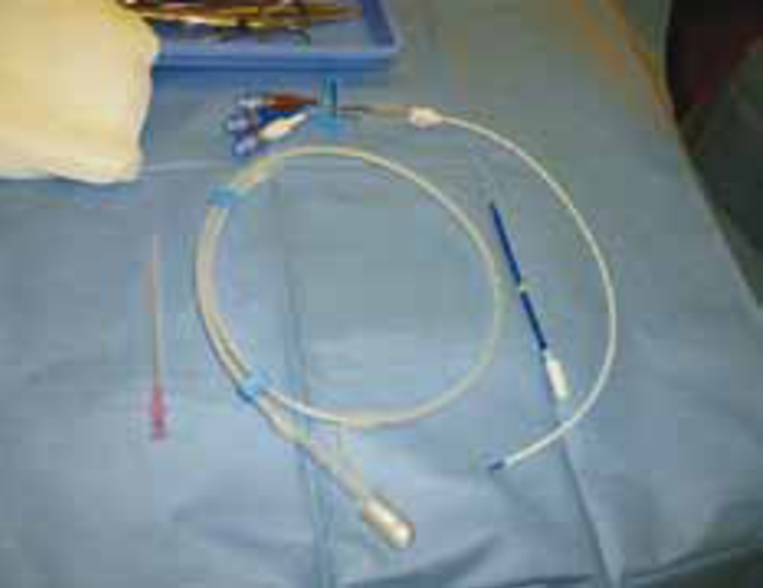
The jugular catheter kit must be opened aseptically within the surgical field and equipment arranged in the order it is to be used on a sterile drape. Equipment required should include the following:
Commercially available central venous catheter set for Seldinger includes:
Catheter placement
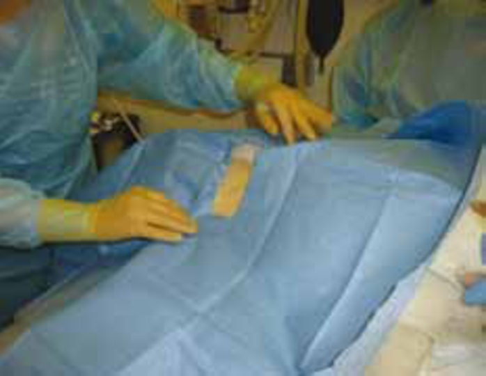
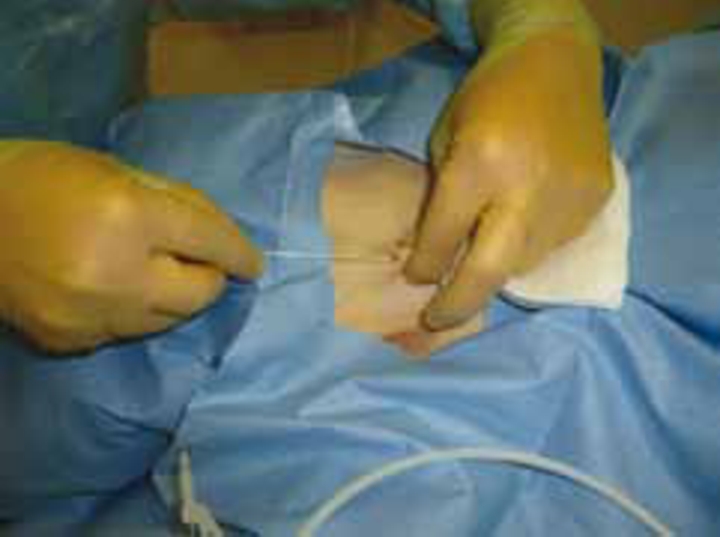
A fenestrated sterile drape is placed over the jugular site to maintain asepsis. The jugular vein is raised using digital pressure at the thoracic inlet. If the vein cannot be visualized it can normally be palpated and if necessary a sterile No. 11 blade used to make a small incision at the sight of catheter insertion (personal experience).
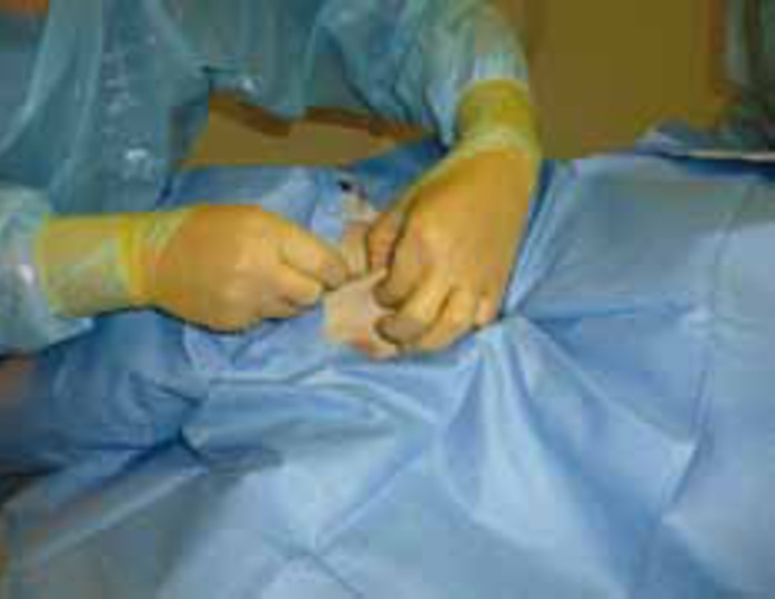
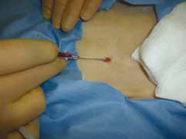
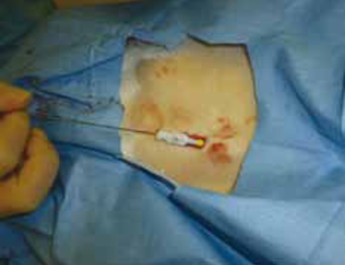
Once visualized the introducer needle is passed through the skin and into the vessel. Beal and Hughes (2000) suggest a ‘quick, controlled, stabbing motion can facilitate entry into the vein lumen’ due to the relatively mobile nature of the vein’. A flash of blood into the catheter indicates that it is placed correctly into the vein.
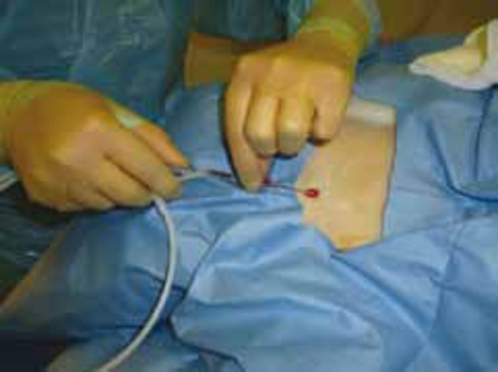
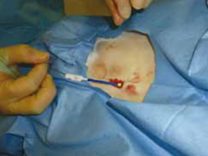
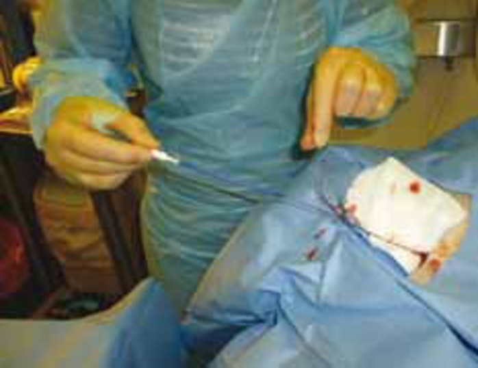
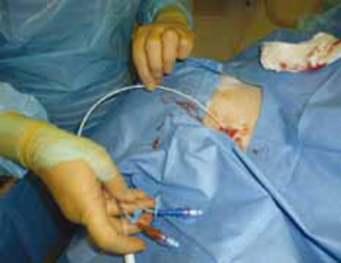
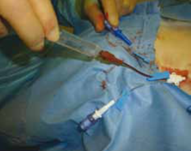
The J end guide wire is then passed through the introducer needle and into the vein; approximately two thirds should be passed into the vein. The J tip prevents damage to the vessel wall. The guide wire should then be held in place while the introducer needle is withdrawn and the vessel dilator passed over the wire. The dilator is grasped at the distal end and advanced with a forward twisting motion into the vessel. As the dilator is withdrawn a sterile swab should be placed over the site to stem any bleeding, the guide wire remains in place and the multi lumen catheter is threaded over the wire until the proximal end of the guide wire protrudes from the hub of the catheter. The proximal end of the guide wire is held while the catheter is advanced into the vessel the desired distance as determined by previous measurement. The wire is removed.
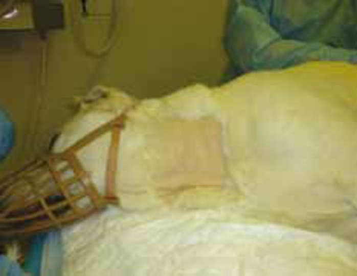
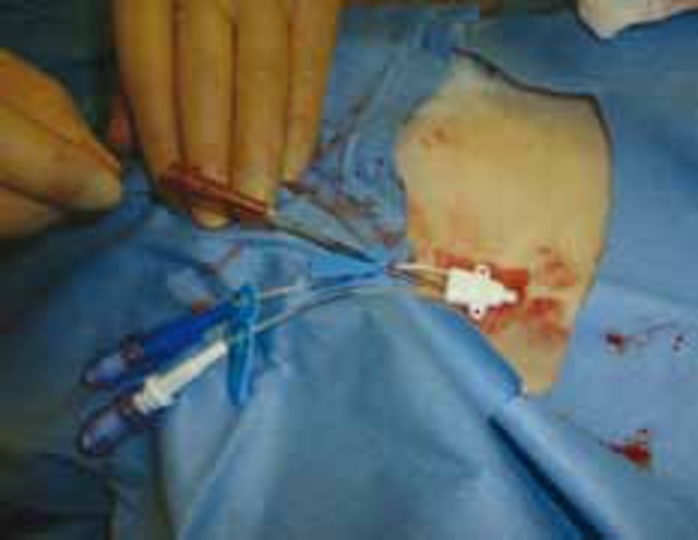
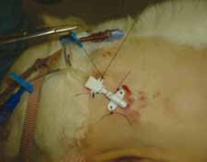
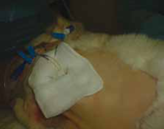
All ports are then aspirated to remove any air and to ensure that blood is easily drawn through the catheter. If necessary the catheter may be repositioned to allow aspiration of the blood. All ports are flushed with heparinized saline. The catheter is then sutured in place and the insertion site covered with sterile gauze and bandaged appropriately.
Jugular catheter maintenance
Jugular catheters are placed for long-term access, factors that make them particularly suitable include: their location; they are less vulnerable to patient interference when compared with peripheral catheters; they are more comfortable for the patient; they are easier for the nursing staff to use as they are more easily accessible compared with femoral or saphenous veins; and they do not require frequent replacement as is the case with peripheral catheters.
General recommendations for central venous catheter maintenance
It is vital that jugular catheters are maintained appropriately to avoid risk of contamination and that patients are closely monitored while the catheter is in place. The veterinary nurse plays a very important role in ensuring that this is the case.
The nurse should ensure that while handling the patient appropriate hand hygiene is observed and disposable gloves should always be worn when handling the catheter. In the case of dogs, harnesses should be used for exercise to prevent additional stress to the jugular site
Veterinary nurses should check the catheter insertion site twice daily for evidence of infection, e.g. pain, inflammation or abscessation of the skin. If this occurs the clinician must be notified as the catheter will need to be removed. Sterile gloves should be worn to redress the site and sterile swabs used to place over the site of insertion prior to redressing.
If the patient develops an unexplained fever (temperature >39°C) it should be noted and the clinician in charge advised.
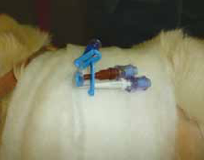
All injection ports should be wiped with alcohol before and after needle puncture. The insertion sites should be kept bandaged in a manner that supports the catheter and prevents the catheter hubs dragging on the ground and any witnessed contamination should be responded to immediately, this may require cleaning or changing injection ports or complete catheter removal.
Leaving ports of a central venous catheter open to the atmosphere places the patient at risk of air embolism, therefore the catheter should be occluded by a catheter lock or manual kinking whenever the catheter hub is opened.
All unused ports on central venous catheters should be flushed every 4 hours with heparinized saline to prevent thrombus formation in the lumen which will cause mechanical failure of the line (Bexfield and Lee, 2010). Bags of heparinized saline should be discarded every 12 to 24 hours to minimize the risk of contamination (Davies, 2009). If a catheter port is not going to be used for a prolonged period a heparin lock can be used, i.e. the dead space of the catheter is filled with 100 U/ml heparin every 12 hours. The concentrated heparin solution is never flushed into the patient; instead it is aspirated before administering medications or before replacing the heparin lock. Clear labelling of such ports to avoid inadvertent flushing of the concentrated heparin into the patient is important; heparin is an anticoagulant that prevents the formation of clots within the blood system, therefore inadvertant flushing of concentrated heparin would increase the risk of a bleed in that patient.
Potential complications of jugular catheter use include: phlebitis; venous thrombosis; catheter associated infections; and haemorrhage (Beal and Hughes, 2000). Mechanical complications such as kinking, patient interference and occlusion of one or more of the lumens with thrombi are also potential complications.
Conclusion
The placement of jugular catheters is indicated in cases where long-term IV access is required and where multiple lumen are necessary. They have the advantage that they are more comfortable for the patient and more easily accessible for nursing staff. Disadvantages include that they generally require sedation or anaesthesia for insertion and their placement carries a much higher risk of contamination and subsequent infection if asepsis is not maintained. It is also vital that the correct maintenance protocol is followed to ensure the patency of the catheter.