
Wound assessment is important in terms of planning wound reconstruction. It is important to ensure that traumatic wounds are free of infection, devitalized or necrotic tissue, or contamination before closure is performed. In order to achieve this a period of debridement and/or open wound management to allow granulation may be required for several days before reconstruction is carried out. In surgical wound management, reconstruction can be planned out, as the skin is not as compromised as in traumatic wounds; it is elastic, vascular and even more freely available than in the trauma patient (Anderson, 1997a). The landmarks of the direct cutaneous artery (DCA) supply to the skin, skin tension lines and structural features, such as joints/orifices, should be taken into account during this planning. The appropriate areas can then be clipped and prepared and a plan for the reconstruction can be drawn on the skin before scrubbing up using a surgical marker.
Preparation of the wound is important in order to achieve optimal healing. It is important to respect the wound's natural defences against infection and to treat the wound in an aseptic manner at every stage of the wound management process. This prevents the introduction of further contaminants and reduces the risk of nosocomial infections.
Skin flap or skin graft?
A skin flap is a piece of skin that remains attached to a blood supply when it is placed over a recipient wound whereas a skin graft is a piece of skin taken away from its blood supply and placed on a recipient wound, where it develops a new blood supply (Swaim, 2000).
Local flaps
Subdermal plexus flaps
Pedicle grafts are partially detached segments of skin and subcutaneous tissue that receive their vascular supply from the pedicle (base) of the flap. If this type of flap is elevated adjacent to the wound then this is termed a local flap. These are also termed deep subdermal plexus flaps as they depend on an intact deep subdermal plexus entering the flap for survival. These flaps can be developed if a wound is located adjacent to an area of loose elastic skin that can be mobilized. It is important to remember that this may lead to a deficit at the donor site which will require closure. As these flaps are dependent on the subdermal plexus, they are generally not applicable to very large deficits. With all these flaps great care is needed in handling to minimize damage to the vascular supply. If the flap is adjacent to direct cutaneous vessels, they can be included in the base of the flap or alternatively an axial pattern flap can be utilized.
Advancement flaps
These are local flaps that are elevated and advanced forwards over the deficit. They are developed parallel to lines of least resistance. The flap width is determined by the width of the defect. The flap is developed using two diverging incisions (Figure 1).The flap is undermined and advanced into the deficit. There are no strict rules regarding the appropriate width: length ratio as regional vascularity varies, however, it is advisable to have a flap with a slightly wider base than its body (Mayhew, 2009). Long narrow flaps should be avoided as the distal end is more likely to undergo ischaemic necrosis and owners should be warned of this possibility prior to surgery.
These flaps do have significant limitations as they rely on stretching the skin close to the deficit. The wound closure will therefore have some tension due to the elastic retractive force of the stretched skin flap. This increases the risk of wound dehiscence of the distant suture line.
The sutures along the sides of the advancement flap should therefore be placed first with the aim of taking most of the tension off the distant suture line. Instead of a single large advancement flap, develop advancement flaps on either side of the wound and close in an H formation (Mayhew, 2009).
Rotation flaps
Rotation flaps are semi-circular flaps that rotate into the adjacent wound. Again this type of flap relies on stretching of the undermined skin but in a semi-circular manner. Traditionally this flap has been described with an arc of rotation four times the length required to rotate the flap into the deficit (Anderson, 1997b). This however is usually unnecessary in dogs and cats and the flap is developed in a stepwise manner until it can close the wound.
Transposition flap
Transposition flaps are very useful local flaps as they supply additional new loose skin into the wound. They do require more detailed planning and assessment prior to use and a donor wound is created that needs to be closed. These flaps are developed from loose skin which is at 45 to 90 degrees to the wound deficit (Mayhew, 2009). The long axis of the flap is developed parallel to tension lines, as this makes closure of the donor wound more straightforward. The width of the flap corresponds to the width of the defect adjacent to the proposed flap. The pivot of the flap is identified and a measurement is made from this point to the farthest position of the defect. This distance will be equal to the distance from the pivot point to the farthest point of the flap. Once elevated the flap will be rotated through an arc into the defect. As the arc of rotation of the flap increases the length of the transposition flap decreases and a Bürrow's triangle or dog ear can develop at the base. This is a very versatile flap that can be used in numerous locations on the body. On the distal limb the flap is created parallel to the long axis of the limb.
Axial pattern flap
Axial pattern flaps are pedicle grafts that contain a direct cutaneous artery and vein. The regions of skin that are supplied by most of the direct cutaneous vessels have been mapped in dogs and cats. This allows consistent identification and mobilization of large skin flaps in one stage with confidence that there is a reliable vascular supply. These flaps represent major surgical procedures and are indicated for wide tumour excisions, avascular wound beds, extensive body wounds or other substantial reconstruction procedures (Anderson, 1997b).
Axial pattern flaps are generally rectangular in shape; although they can be modified with a right angle extension (this area of the flap is dependent on the deep dermal plexus). The planning and execution of an axial pattern flap must be meticulous as if there is failure of the flap the results can be catastrophic. Flap failure is generally due to technical error (failure to identify perimeters correctly, failure to preserve the vascular pedicle, failure to manage dead space etc). There will also be a significant donor site to be closed. Axial pattern flap surgery is time consuming with consequent lengthy anaesthesia. Delineation of the area supplied by the chosen DCA should be carefully assessed prior to the start of surgery (Pavletic, 1993).
The elevated flap can be rotated into position with a base of skin still intact or this base can be incised to create an ‘island’ flap. This reduces the wastage that results from rotation when the base is intact (when a large dog ear can develop). However the island flap is now completely dependent on its vascular pedicle for its blood supply (the direct cutaneous vessels) and as there is no skin attachment there is a risk of kinking the pedicle with resultant vascular compromise (Moores, 2009). All the principles of surgical wound closure must be adhered to especially dead space management.
In the post-operative period the flap margins should always appear pink and viable. Cyanosis or excess swelling are indicators of vascular compromise and potential impending necrosis.
Axial pattern flaps provide the ability to close large deficits with vascularized skin. If used for tumour resection reconstruction it is essential that the tumour has been completely excised otherwise the tumour can be spread over a much larger area into the donor site of the flap. However, flap failure can be a catastrophe for the patient (and the surgeon) so surgery should not be undertaken lightly.
Free skin grafts
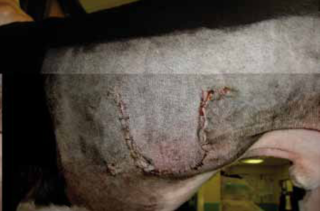
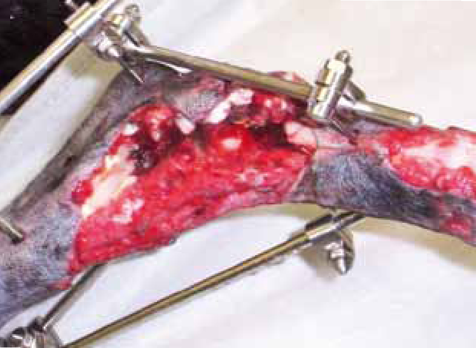
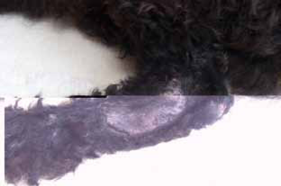
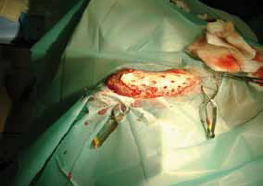
Free skin grafts are a versatile and accessible method of closing small to large sized wounds, usually following a period of open wound healing especially in the distal limb. They involve the transfer of variable thicknesses of dermis, with the epidermis (White, 2009). In difficult areas such as the distal limbs (Figures 2 and 3), free skin grafts can be utilized to achieve durable epithelial cover. Full thickness grafts have the advantage of including the adnexal structures which improves cosmesis and are by far the most commonly used grafts in dogs and cats.
Free skin grafts lack a vascular attachment on transfer to the wound and must depend on imbibition of wound fluid from the wound bed for the first 48 hours. It follows, therefore, that having a healthy vascular bed plays a key role in the initial preparation for a free skin graft. The most common recipient bed is a clean healthy granulation tissue, a freshly created wound bed, i.e. healthy muscle and periosteum. Chronic granulation tissue (pale, poorly vascularized) is unsuitable but can be converted back to good tissue by serial debridement and bandaging for a few days prior to grafting (McGlennon and White, 1989).
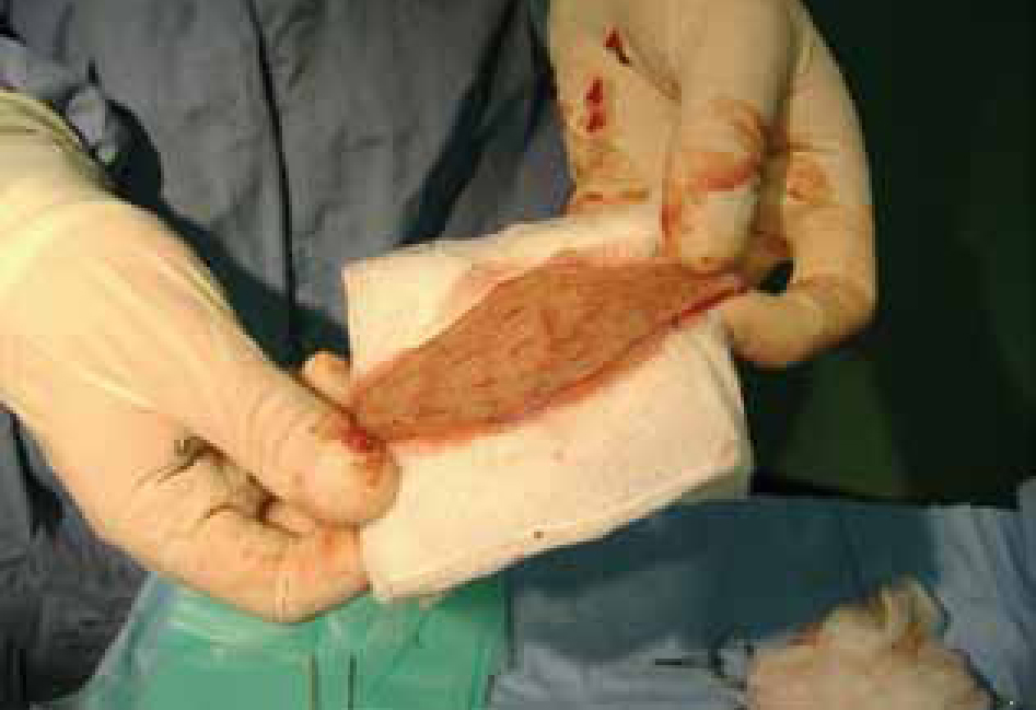
The graft is harvested from an area where there is plenty of free skin, the most common sites being the flank and the lateral aspect of the cervical region. In these two sites there is normally plenty of mobile skin which can be removed while allowing the resulting deficits to be closed easily without creating excessive tension (McGlennon and White, 1989). This is then prepared by removing all subcutaneous fat or muscle. The graft is then meshed by placing small incisions at staggered intervals (Figures 4 and 5). This is to allow effective drainage of fluid that may collect beneath the graft. The graft is orientated in the correct direction of hair growth, secured to the recipient site with sutures, overlapping the edge by 2-5 mm.
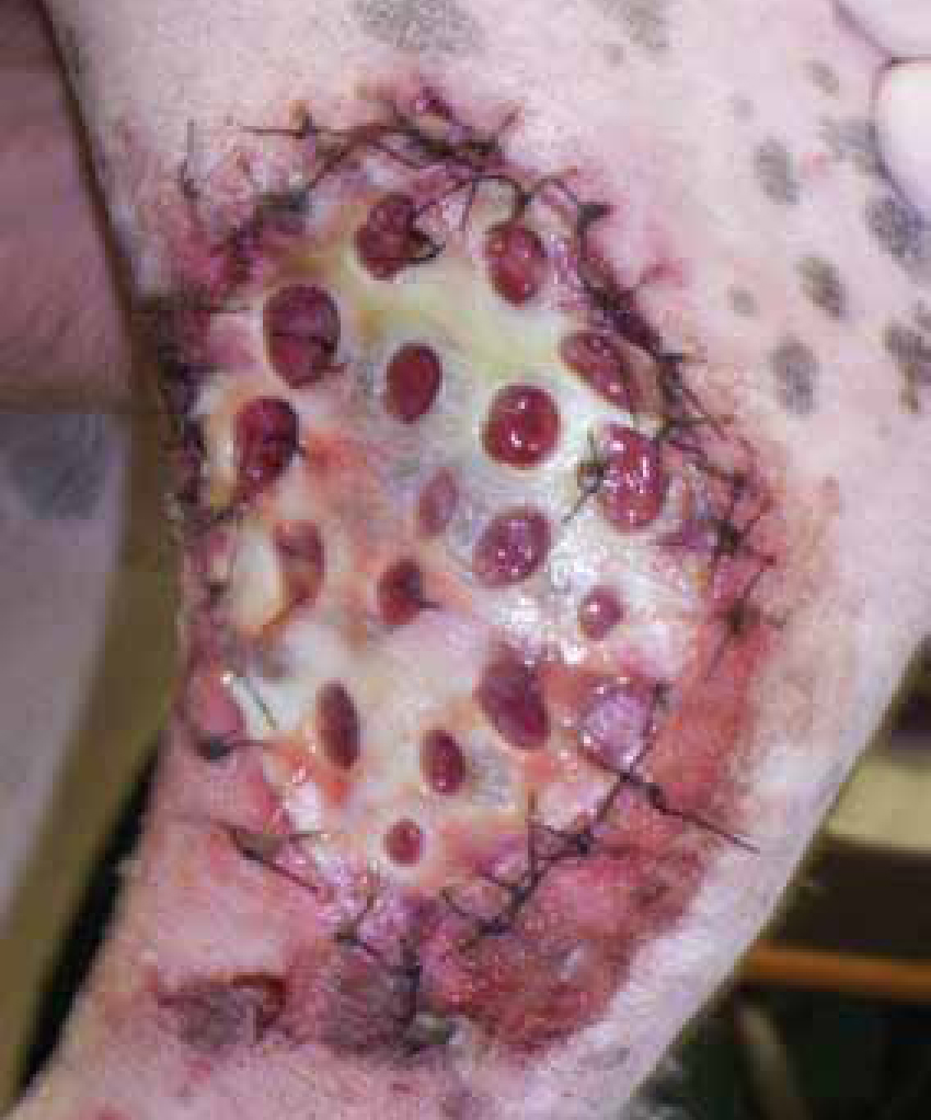
Any accumulation of debris, blood, purulent material or foreign material between the graft and the recipient site will impair plasmatic imbibition (absorption of recipient site wound fluid) and will lead to ischaemic necrosis of the graft. Therefore the recipient bed should be smooth, clean, free of infection and have all haemorrhaging controlled. For the first 48–72 hours the graft will appear swollen and slightly cyanotic (Figure 6). All subcutaneous fat must be removed to allow imbibition to occur. Vascularization of the graft occurs via entry of capillaries of old vascular channels (inosculation) or creation of new vessels (revascularization). These vessels are fragile and any movement of the graft will prevent a successful ‘take’–— therefore immobilization of the graft to the recipient bed by carefully suturing in position and subsequent bandaging to immobilize the area is very important.
The long-term viability of the graft depends on the development of new blood vessels between the recipient bed and the graft. Anything that interferes with this process during the first week after grafting will therefore jeopardize the success of the graft (McGlennon and White, 1989).
Post-operative and nursing considerations
Separation of the graft from the wound bed can occur for a number of reasons.
Accumulation of fluid below the graft
A seroma/haematoma will tend to separate the skin graft from the graft bed and impair the process of imbibition or hinder the development of revascularization (White, 2009). Haemostasis of the graft bed should, therefore, be thorough prior to graft placement. Where fluid accumulation can be expected (e.g. grafting a recent wound or a very haemorrhagic granulation tissue) some provision for drainage should be made. This is most easily achieved by using a mesh graft but can also be achieved using closed suction drainage systems or the use of vacuum assisted closure systems (see later).
Movement of the graft over the bed
- Suturing — the graft should not only be anchored at the periphery of the wound but at strategic points through its surface to maintain contact with the graft bed
- Bandaging — this is particularly important when grafting the extremities and a bandage incorporating a gutter splint should be used to aid immobilization. Attention to detail when bandaging is important and the following points should be considered. The initial bandage is usually left in place for at least 72 hours. Sedation is essential for the first bandage change to prevent excessive movement. Extreme care is required when lifting off the bandage material in case there is some adherence to the graft. The bandage is then changed every 2–3 days for approximately 10–14 days. A light dressing, such as Melolin, Smith and Nephew, London, is then placed for a further 2 weeks to protect the initially delicate epithelium.
- Construction — a low or non-adherent dressing is placed directly in contact with the graft to minimize disturbance of the graft when the dressing is changed. This may be either a non-adherent pad, e.g. Melolin (Smith and Nephew) or if copious exudation is expected an absorbent foam dressing can be used. Some surgeons recommend the use of non-adherent silicone dressings, e.g. Mepitel. The intermediate layer of the dressing should be absorbent, e.g. cotton wool or Soffban (Smith and Nephew), and combined with a conforming bandage. The outer layer should be strong, supporting and protective, e.g. Vetrap (3M) or Elastoplast (Smith and Nephew).
- Frequency of change — the graft should be disturbed as infrequently as possible during the first 7 days but accumulation of exudate at the wound surface should be kept to a minimum. Thus the frequency of changing will vary. In general, if a fresh wound is grafted copious exudate can be anticipated and the dressing may initially need to be changed daily. If, however, an established granulation tissue bed is grafted the dressing may be left in place for 3 to 4 days before a change.
- Duration of bandaging — this is a matter of judgement and experience but bandaging is usually maintained for 2 to 3 weeks until the graft has satisfactorily taken and, in the case of a mesh graft, re-epithelialization is complete.
- Self-mutilation — steps should be taken to prevent the patient from mutilating the graft by means of adequate bandaging and, where necessary, Elizabethan collars, as patients have been known to remove the dressing and consume the graft.
Alternative techniques
Should the graft appear to undergo necrosis, all may not be lost as the deeper dermal and adnexal layers may survive and epithelialization can occur from these areas. Therefore avoid debridement of the graft for at least 5–6 days even if it looks like full necrosis has occurred.
An alternative to full thickness meshed grafts is the use of punch grafts. These are harvested with a biopsy punch and placed in holes created in the granulation tissue bed. This is a very useful and fast technique for closure of small wounds, especially if more movement is anticipated.
There is currently research into the use of negative pressure therapy wound therapy or vacuum-assisted closure (VAC) systems for use with the placement of free skin grafts with anecdotal reports indicating an improved and more consistent graft take (Guille et al, 2007).
- Removal of fluid from the extravascular space
- Increased local circulation
- Improved formation of granulation tissue
- Increased bacterial clearance (Kirkby, 2010).
Graft survival depends on several factors. Complicating issues, such as graft movement, haematoma or seroma formation, and infection, can all lead to graft failure (Blackburn et al, 1998). VAC reduces or eliminates all of these potential problems (Welch and Lewis, 1999).
Conclusion
Properly applied and managed skin flaps and free skin grafts can significantly reduce the time and expense that might be spent in managing a wound that is otherwise allowed to heal by epithelialization and contraction. In cats and dogs free skin grafts are usually full thickness and they can be applied immediately to clean wound beds with a good blood supply, e.g. regions of the face or on muscular surfaces, or to mature granulation tissue. New technology such as the use of VAC may improve the outcome of skin graft take in small animal patients but obviously its use is still in its infancy in veterinary medicine.
The nursing care of these patients is vitally important in the success of the graft take, and the dressing care and post-operative nursing need to be carried out meticulously in order to achieve a successful outcome.
Key Points
- The ultimate success of the skin graft take will depend greatly upon surgical technique as well as post-operative care.
- It is important to ensure that traumatic wounds are free of infection, devitalized or necrotic tissue, or contamination before closure is performed.
- Preparation of the wound is vitally important in order to achieve optimal healing.
- Local flaps have significant limitations as they rely on stretching the skin close to the deficit.
- Free skin grafts are a versatile and accessible method of closing small to large sized wounds.
- Free skin grafts may be selected where closure of skin deficits is not achievable using local skin flap techniques.
- www.theveterinarynurse.com