
Osteoarthritis (OA), also termed degenerative joint disease or osteoarthrosis, is one of the most commonly seen, chronically painful conditions in dogs (Lindley and Taylor, 2010) and, if left unmanaged often leads to debilitating, painful lameness. In geriatric dogs, the incapacity caused by osteoarthritis may be the major contributing factor in a decision for euthanasia (May, 1994). This highlights the importance of appropriate, multi-modal management of the disease and good nursing management to facilitate the best quality of life possible for the dog.
Pathophysiology
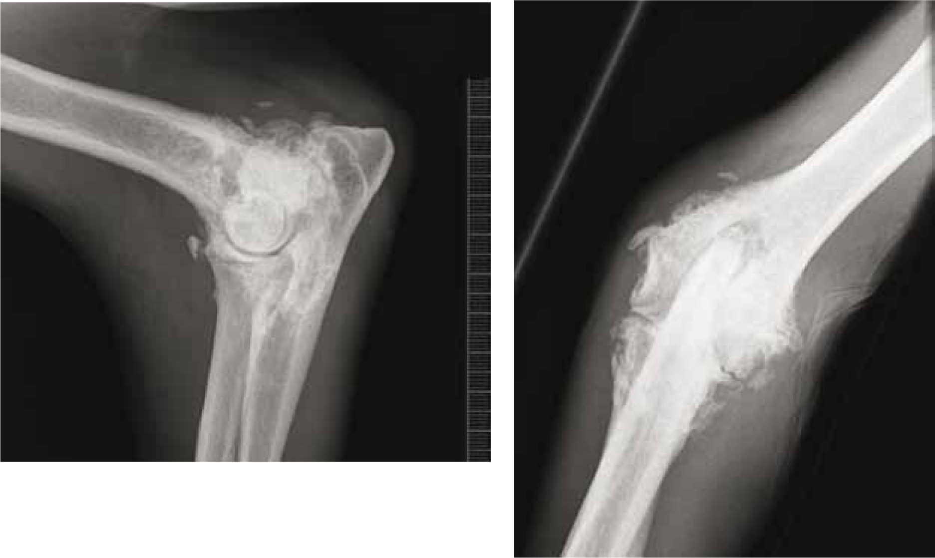
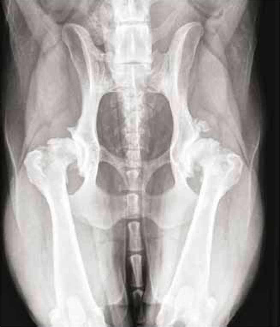
OA occurs when there is a loss of articular cartilage (Abercromby et al, 2006) as a result of both biological and mechanical events. These events destabilise the normal coupling of degradation and synthesis of articular cartilage chrondrocytes and extracellular matrix, and subchondral bone (Sharma and Kapoor, 2007). These changes lead to a softening, fibrillation (ulceration and loss of articular cartilage), sclerosis (thickening) and, eburnation (hardening) of subchondral bone, and osteophyte and subchondral cyst (fluid-filled sac in the bone just below the cartilage) formation (Figures 1 and 2) (Sharma and Kapoor, 2007). There are repair mechanisms present within cartilage, although it is likely that they become less efficient with age, which may partly explain the association between OA and ageing (Abercromby et al, 2006).
OA can be categorised as primary OA or secondary OA. Primary OA is idiopathic, with no clear cause, whereas secondary OA results following trauma, metabolic disorders, anatomic abnormalities or inflammatory arthritis (Fox and Millis, 2010). Secondary OA is far more commonly seen than primary OA (Abercromby et al, 2006). Patients with OA may experience joint pain and tenderness, reduced mobility, stiffness, reduced range of motion and reluctance to exercise, crepitus, muscle atrophy, joint effusion and inflammation. The pain is associated with direct stimulation of the joint capsule and bone receptors by cytokines and the degradative process (Fox and Millis, 2010). Along with effusion and consequent distension of the joint capsule, abnormal articulation of the joint, stimulation of subchondral bone from abnormal loading such as obesity or poor joint congruity, and irritation of muscles, tendons and ligaments occurs (Fox and Millis, 2010). If pain is left unmanaged it can cause a cascade of problems including depression, fear, anxiety and decreased food intake (Flaherty, 2009).
Management of OA
Diagnosis of OA will be carried out by the veterinary surgeon through physical examination, radiography and synovial fluid analysis. If it is considered necessary and funds are available the veterinary surgeon may proceed with arthroscopy, computerised tomography or magnetic resonance imaging to establish a more accurate diagnosis of cartilage loss.
The main objectives when treating OA is to slow down the progression of the disease, reduce pain and inflammation and, address aggravating factors. Carmichael (2006) has devised the ABCDE (analgesia, bodyweight, control, disease modification, exercise) approach to OA, which may be helpful when formulating a holistic treatment plan for patients with the disease.
Analgesia
Pain management should be a priority with these patients to reduce discomfort and improve quality of life. Non-steroidal anti-inflammatory drugs (NSAIDs) are immensely beneficial for the treatment of pain and inflammation associated with OA and include deracoxib, meloxicam, etodolac, firocoxib, carprofen (Abercromby et al, 2006) and maxacoxib.
NSAIDs act by inhibiting the cyclooxygenase (COX) enzymes, and reduce inflammation and discomfort, and allow the animal increased mobility. COX-2 triggers the conversion of arachidonic acid into prostaglandins, prostacyclin and thromboxanes (Kerr, 2007). These compounds are metabolites of arachidonic acid that originate from the phospholipids found in cell membranes. They cause vasodilation and neutrophil chemotaxis, resulting in increased vascular permeability and cellular influx (Kerr, 2007). NSAIDs are the most popular analgesic agents used in veterinary practice for chronic and acute pain (Gaynor and Muir, 2009), however, problems can arise with their use such as gastrointestinal mucosal damage, renal toxicity and clotting abnormalities (Gaynor and Muir, 2009) due to the inhibition of COX 1. If NSAIDs are contraindicated, such as in patients with gastrointestinal or renal disease, alternative analgesics should be utilised.
Paracetamol may be prescribed especially if NSAIDs are not tolerated by the patient. Although paracetamol has been used for more than a century in humans (Zhang et al, 2004), it is not currently licensed for use in animals (Roberts, 2011) therefore, there is very little literature detailing the use of this drug in dogs. There have however, been several human studies (Pavelka, 2005; Sinatra et al, 2005; Arici et al, 2009) concluding that paracetamol is an effective alternative to NSAIDs, where NSAIDs are not tolerated. Further studies are required to evaluate the effectiveness of paracetamol in the treatment of pain in dogs.
Gabapentin is a drug used as an adjunct anticonvulsant and analgesic for treating chronic, neuropathic pain (pain from injury to the nervous system) in dogs. Gabapentin may be used together with opioids during an acute pain phase (Kerr, 2007) especially if the patient is unable to take NSAIDs. the mechanism of analgesia for gabapentin is still not fully understood (Kerr, 2007) but it appears to be mediated through blockade of calcium channels in neurons decreasing the release of excitatory neurotransmitters such as substance P, glutamine and norepinephrine, reducing the depolarization of nerves in the spinal cord involved in conduction of pain perception. There are currently no controlled veterinary clinical studies available that evaluate the analgesic properties of gabapentin, although a number of human studies have suggested that gabapentin can be used as part of a multimodal regimen. Gabapentin may cause hypersomnia at higher doses and has been reported to cause seizures in humans if the drug is discontinued suddenly so, gradual weaning off gabapentin is recommended in these circumstances (Posner, 2012).
Amantadine is an antiviral drug with analgesic properties and may be used as an adjunct drug with NSAIDs and opioids, in the clinical management of canine osteoarthritic pain (Lascelles et al, 2008). Amantadine is a NMDA receptor antagonist preventing and/or attenuating central sensitisation at the dorsal horn of the spinal cord (Posner, 2012). Used at sub-anaesthesia doses there are very few behavioural side effects seen with amantadine (Posner, 2012).
Other analgesic drugs such as tramadol may also be prescribed as part of a multi-modal approach to pain associated with OA. Tramadol is classified as an atypical, centrally acting opioid analgesic (Kerr, 2007) and has mild mu opioid action. Side effects are rare but may include vomiting, diarrhoea, anorexia and constipation.
Acupuncture has been advocated as having beneficial effects for some patients with OA (Langley-Hobbs, 2010). Acupuncture is a form of Chinese traditional medicine based on the meridian system, where 28–35 gauge needles are inserted into different points along this system (Cantwell, 2010). Electrical stimulus of the needles may also be used to provide a greater effect. The effects of acupuncture may be mediated through the release of endorphins and beta adrenoreceptor activity (Cantwell, 2010). Acupuncture may also contribute to inhibitory pain pathways by stimulating Aβ fibres thus reducing nociception via the gate control theory of pain (Cantwell, 2010).
Bodyweight
Obesity can significantly increase the pain associated with OA and may increase the progression of the disease due to a higher load the joints need to withstand. Body condition scoring is an effective way to assess the level of obesity in dogs and should be regularly reviewed. Nurse-led weight management clinics are a useful way to educate and encourage owners to aid weight loss of their dog. Owners should be advised that the dog is likely to require fewer calories (Langley-Hobbs, 2010) due to a reduction in activity as a result of the pain associated with OA.
Control (client education/clinical monitoring)
The owner will play an integral role in reducing the risk of their dog developing complications associated with OA and providing palliative care. The most prominent clinical symptom of OA is chronic pain which often results in a deterioration in the patient's wellbeing. An important part of managing chronic pain in dogs is to allow owners to feel in control and empowered by providing them with the required knowledge and understanding of the condition. An understanding of the disease keeps the owner motivated and ‘onside’ (Abercromby et al, 2006). The veterinary nurse should make it a priority to provide the required information on OA to the owner and to be a source of support if the owner has any concerns. The owner should understand that the disease is a life-long condition and cannot be cured, but managed if specific strategies are in place.
Pain relief can be provided via the use of analgesics and by using aids at home to improve the patient's quality of life, such as padded bedding, ramps and non-slip flooring. Nurse-led arthritis clinics can be extremely useful in providing support to the patient and the owner and, to track the patient's progress (Abercromby et al, 2006). Owner questionnaires can be used to determine and monitor how successful treatments are (Hudson et al, 2004). It is also useful to create an information leaflet along with some useful, current websites that the owner can be directed to at the stage of diagnosis.
Disease (chondroprotection)
The aim in treating the disease is to slow down the progression of the disease or, to perform salvage surgery such as joint replacement or arthrodesis. There are many nutraceuticals available for OA including glucosamine, chondroitin sulphate, extract of turmeric, methylsulphonylmethane (MSM) and extract of green-lipped mussel (Chan, 2010). Despite the wide use of these supplements currently published research has failed to find a significant benefit with their use for patients with OA (Vandeweerd et al, 2012). More research is required to evaluate these supplements further, and there are current trials that have significant results that have yet to reach the published literature (M. Pead, personal communication).
There has been promising evidence, documenting the positive effects of Omega-3 fatty acids. Roush et al (2010) completed a study of 127 dogs and concluded that dogs fed a diet high in omega-3 fatty acids had a significantly lower serum concentration of arachidonic acid at 6, 12, and 24 weeks. This suggests joint inflammation was reduced in patients fed a diet high in omega-3 fatty acids. Owners articulated that dogs fed the test food had a notably improved ability to rise from a resting position at 6 weeks and improved ability to walk at 12 and 24 weeks, compared with control dogs (Roush et al, 2010). Prescription diets may be beneficial, such as Hills j/d which has high levels of the omega-3 fatty acid, eicosapentaenoic acid.
Exercise
Exercise is vital in order to maintain cardiovascular function, improve muscle strength and prevent atrophy, maintain appropriate weight, and reduce joint stiffness. Despite this, veterinary practitioners should be careful not expect too much from these patients. Complete rest is important during the acute phase of the disease but, it is important that exercise is gradually increased once the dog shows improvement (Lindley and Taylor, 2010). The owner should be advised to begin with three, 5 minute lead walks a day, only increasing the length of the walks when the owner and veterinary surgeon are happy with his/her progress. Once an improvement is noted the walks should be increased weekly by 5 minutes until the patient is able to tolerate 30–40 minute walks without pain or significant stiffness. If the dog is unable to tolerate long walks the duration of the exercise should be decreased and the frequency increased (Abercromby et al, 2006). Exercise should be altered depending on the dog's progress, if the dog has a ‘flareup’, such as lameness, inflammation or pain, exercise should be reduced. Dogs with OA will cope better on flat ground with avoidance of hills and uneven terrain. During periods of acute pain and exacerbation of clinical signs veterinary advice should be sought and exercise regimens modified.
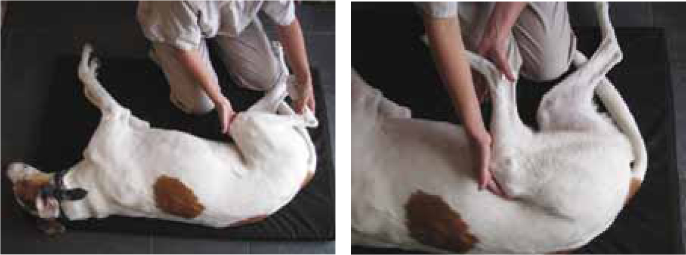
Physiotherapy may provide comfort, increase mobility and reduce swelling and, stiffness. The use of passive movements (Figure 3), stretches, heat therapy and massage prior to exercise, may prove beneficial (Langley-Hobbs, 2010). Heat therapy can include the use of hot water bottles and heat packs applied to the affected joint for 15–30 minutes, but the temperature of the pack should not exceed 75°C (Sharp, 2010).
Cold therapy (cryotherapy) following exercise or at the site of inflammation may also provide some relief (Langley-Hobbs, 2010). Cold compression units are available although the simplest method of cold application is with a commercial cold pack or a freezer bag filled with crushed ice and wrapped in a damp towel or cloth (Sharp, 2010). Cold therapy should be applied for 10–15 minutes every 2–4 hours during an acute phase of the disease.
Hydrotherapy may be indicated, especially with patients that are unable to walk long distances or find land-based exercise too painful. The buoyancy of the water will relieve the pressure on the joints temporarily and allow greater joint flexion. This increased joint movement may improve muscle strength, reduce swelling and stiffness (Lindley and Smith, 2010).
The owners should be fully informed of all the treatment options available to them so that they can make the right decision for their pet and their financial circumstances (Abercromby et al, 2006). Veterinary nurses can assist owners with this process.
Conclusion
For veterinary nurses involved in the provision of analgesia, it is important to understand the rationale behind using a combination of agents, rather than reliance on a single drug. Knowledge of the most appropriate analgesic agents for individual patients is essential in order to provide optimal pain relief and reduce the negative effects of specific drugs in high risk or ageing patients. Veterinary nurses play an integral role in the care of patients with OA and can be a huge source of support and guidance for the patient and owner. A good outcome and improved quality of life for these patients requires a multidisciplinary team approach that involves the entire pathway of care. Veterinary nurses can facilitate this care with careful assessment, planning, implementation and evaluation of the patient and continued support for the owner.